Molecular Human Reproduction, Vol.21, No.9 pp. 736–744, 2015 Advanced Access publication on June 26, 2015 doi:10.1093/molehr/gav037
ORIGINAL RESEARCH
Hypermethylation of the HLA-G promoter is associated with preeclampsia Yao Tang 1,2, Haiyan Liu 1, Han Li1, Ting Peng1, Weirong Gu 1,2,*, and Xiaotian Li 1,2 1 2
The Department of Obstetrics, Obstetrics and Gynecology Hospital, Fudan University, 419 FangXie Road, Shanghai 200011, PR China Shanghai Key Laboratory of Female Reproductive Endocrine Related Diseases, Shanghai 200011, PR China
*Correspondence address. Fax: +86-21-63455090. E-mail:
[email protected]
Submitted on December 19, 2014; resubmitted on June 3, 2015; accepted on June 22, 2015
abstract: Preeclampsia (PE) is a severe pregnancy-induced disorder characterized by hypertension and proteinuria and a leading cause of perinatal maternal –fetal mortality and morbidity in developing countries. Dysregulated human leukocyte antigen (HLA)-G was found in placentas as well as in maternal sera from PE patients; however, the reason for this difference is unknown. As accumulating evidence has confirmed that DNA methylation is an important mechanism for regulating gene expression, we sought to test the hypothesis that alteration in the DNA methylation of the HLA-G promoter region is responsible for decreased expression of HLA-G in PE. Bisulfite pyrosequencing showed that a series of CpG sites in the HLA-G promoter region were significantly more highly methylated in PE than in normal pregnancy (NP). Interestingly, the hypermethylated CpG sites were mostly reported to be binding sites of active transcription factors. To further investigate the regulation of HLA-G methylation, we also defined the expression patterns of DNA methyltransferases (DNMTs) in placental tissue using immunohistochemistry and quantitative polymerase chain reaction analyses. Here, we demonstrate that DNMT-1 is overexpressed and HLA-G expression is reduced in PE women when compared with NP. Furthermore, both treatment with the DNMT inhibitor 5-aza-2′ -deoxycytidine and specific knockdown of DNMT-1 using siRNAs can significantly increase the expression level of HLA-G in a trophoblastic cell line, indicating the potential mechanism of DNMT-1-mediated DNA methylation in HLA-G regulation. Taken together, our research confirms that DNMT-1-mediated promoter hypermethylation of HLA-G is associated with PE. These findings provide new insights into the diagnosis and treatment of PE. Key words: HLA-G / preeclampsia / DNA methylation / DNMT-1
Introduction Preeclampsia (PE) is a pregnancy-induced disorder characterized by hypertension, proteinuria and, sometimes, mild-to-severe edema. This multisystemic disorder can lead to severe clinical complications, such as HELLP (hemolysis, elevated liver enzymes and low platelets) when a hemolytic process is observed, and, more rarely, to eclampsia, when generalized seizures appear (Steegers et al., 2010). PE is a leading cause of perinatal maternal –fetal mortality and morbidity, especially in developing countries (Duley, 2009). While the etiology and pathogenesis of PE remain largely unclear, it is generally accepted that the syndrome consists of two successive processes that include poor placentation in early pregnancy followed by placental oxidative stress (Steegers et al., 2010). Genetic and epigenetic factors, as well as immunological and nutritional factors, are believed to contribute to the mechanism of PE (Serrano, 2006; Chelbi and Vaiman, 2008). In a successful pregnancy, human leukocyte antigen (HLA)-G is a fundamental molecule that induces maternal immune
tolerance and protects the fetal-derived placenta and fetal antigens from immune rejection by uterine natural killer cells and antigenprocessing cells (Chumbley et al., 1994). Dysregulated HLA-G has been found in most PE placentas, and HLA-G expression is reduced or absent in PE placentas compared with normotensive placentas (Hara et al., 1996; Goldman-Wohl et al., 2000; Zhu et al., 2012). Consistent with placental HLA-G expression, the serum HLA-G concentration is also decreased during the third trimester of PE pregnancies, as well as during the early gestational weeks in females who eventually develop PE, which strongly argues that soluble HLA-G may be an early predictor of PE (Yie et al., 2004, 2005). A low level of HLA-G expression in both the placenta and maternal serum might account for the disorder by mediating immune maladaptation at the maternal – fetal interface (Yie et al., 2004). In addition, our previous work demonstrated that alterations in HLA-G might directly interfere with the invasiveness function of trophoblast cell lines through different cell signaling pathways, confirming its significant role in the mechanism of PE (Li et al., 2011; Liu et al., 2013a, b).
& The Author 2015. Published by Oxford University Press on behalf of the European Society of Human Reproduction and Embryology. All rights reserved. For Permissions, please email:
[email protected]
737
HLAG promoter hypermethylation in preeclampsia
However, the regulation of HLA-G in PE remains unknown. Unlike other HLA class I molecules, the sequences involved in the transcriptional regulation of HLA class I genes are disrupted in the HLA-G gene (Moreau et al., 2009). The demethylation reagent 5-aza-2′ -deoxycytidine (5-Aza-dC) has been reported to reverse HLA-G gene repression in a number of tumor cell lines (Moreau et al., 2003). Spontaneous demethylation in the HLA-G promoter region has also been found in ovarian tumor cells compared with normal epithelial cells, and this demethylation is accompanied by an increased level of HLA-G protein in tumor cells (Menendez et al., 2008). Because promoter methylation is assumed to be a crucial mechanism that regulates gene expression (Deaton and Bird, 2011), the methylation status of the HLA-G promoter might account for its expression level, at least in part. However, whether the methylation status of the HLA-G promoter region is altered in PE patients, resulting in changes in its expression, has not been investigated. Our study aims to investigate whether elevated methylation of the HLA-G promoter is related to PE. In addition, we hypothesized that the key enzymes that regulate DNA methylation [DNA methyltransferases (DNMTs)] contribute to the altered methylation pattern of the HLA-G promoter region in trophoblastic cells. So far, we are the first group to use PE placentas to study HLA-G. As the traditional methods used by previous studies, such as methylation-specific polymerase chain reaction (PCR) (MSP) and bisulfate-sequencing PCR (BSP), cannot accurately reflect low levels of CpG methylation and most likely have a high level of false-negative results, we chose to perform bisulfite pyrosequencing, which is considered a gold standard for methylation quantification (Dupont et al., 2004), to determine the promoter methylation status of HLA-G in PE. Our findings increase our understanding of the role of epigenetics in HLA-G regulation in PE.
Materials and Methods Patients and samples collection Placentas and sera samples from 20 normal and 19 PE pregnancies were collected for this case – control study. The inclusion of PE patients followed the criteria of the American College of Obstetricians and Gynecologists [hypertension (blood pressure ^140/90 mmHg after 20 weeks’ gestation) and proteinuria (^300 mg/24 h or ^1+ dipstick)]. The normal pregnancies (NPs) consisted of healthy women undergoing selective cesarean. The
clinical characteristics of the recruited pregnancies are shown in Table I. Several tissue samples (0.5 cm × 0.5 cm) were obtained from the maternal side of the placenta soon after each selective cesarean section. After removal of the maternal blood cells by washing the tissue in sterile phosphate-buffered saline, a block of tissue was fixed in 4% formalin for immunohistochemistry (IHC), and the remaining tissue was aliquoted and snap frozen in liquid nitrogen and transferred to storage at 2808C for later use. Five milliliters of blood was taken from PE and NP by venipuncture in sterile conditions and collected in sterile tubes. Blood samples were centrifuged for 15 min at 600g at 48C. Serum was separated within 15 min after collection of blood and frozen at 2808C until assayed. Duration of the storage before measurement was up to 6 months.
Ethical approval This study was approved by the local ethics committees of Obstetrics and Gynecology Hospital of Fudan University (Shanghai, China), and written consent was obtained from all of the patients before the collection of the placenta and blood.
Genomic DNA extraction and quantification of DNA methylation Genomic DNA from placental tissue and cell lines was extracted using the TIANamp Genomic DNA Kit (TIANGEN, Beijing, China) according to the manufacturer’s instructions. The DNA concentration was evaluated using an Eon Microplate Spectrophotometer. Purified DNA with an optical density (OD) value between 1.8 and 2.0 was assumed to be of good quality. One thousand nanograms of DNA was modified using the EZ DNA Methylation-Gold Kit (ZYMO, USA). The modified DNA was stored at 2808C. To assess the HLA-G methylation status of the tissue and quantify the percentage of methylation of each individual CpG, bisulfite-modified DNA was amplified using bisulfite PCR primers with a biotin label on the 5′ end of the reverse primer, according to Menendez et al. (2008). The pyrosequencing primers were designed by Pyromark Assay Design software (QIAGEN, Germany) according to the HLA-G reference sequence GenBank J03027.1. All primers are shown in Table II. The PCR reactions were performed using HotStart PCR enzymes (TAKARA, Japan) following the manufacturer’s instructions. The optimized PCR conditions were 55 cycles of 958C for 30 s, 588C for 30 s, 728C for 1 min and a final step of 728C for 5 min. The PCR products were then processed and sequenced to measure the percentage of DNA methylation using the Pyromark Q96 MD Pyrosequencer system (QIAGEN).
Table I Characteristics of the study population. Characteristic
NP (N 5 20)
PE (N 5 19)
P-valuea
............................................................................................................................................................................................. Maternal age (years)
30.70 + 5.10
30.53 + 4.67
0.8104
Gestational age (weeks)
38.21 + 0.85
37.77 + 0.93
0.1178
Pregnancy body mass index (kg/m2)
28.36 + 3.38
29.88 + 4.23
0.3394
BP systolic (mmHg)
115.05 + 10.02
153.79 + 20.39
0.0001
BP diastolic (mmHg)
73.40 + 6.72
97.79 + 10.26
Proteinuria (g/24 h)
—
1.70 + 1.80
0.0001 —
Neonatal weight (g)
3286.75 + 488.58
3023.68 + 592.10
0.1031
Placenta weight (g)
510.00 + 82.10
470.26 + 74.81
0.0408
Gender ratio of newborns (male/female) All results are presented as mean + SD. a Obtained using the Mann –Whitney U test on Graph Pad version 5.0.
10/10
10/9
0.8695
738
Tang et al.
Table II Primer sequences (5′ –3′ ) used in this study. Bisulfite PCR primers
Quantitative PCR primers
............................................................................................................................................................................................. HLA-G
FW
AAGAGTATAGGAGGATAGGTAAGG
RVa
AACACCATAACCACCATCCTTAAC
Pyrosequencing primers Array-1
HLA-G DNMT-1
AAGATTTAGGGAGATATTGAGATAG
Array-2
ATAGGYGGTGTATGGGTTGGGGAGG
Array-3
TATTAGGTGATAGGTTTTTAGAGAAG
GAPDH
FW
CTGGTTGTCCTTGCAGCTGTAG
RV
CCTTTTCAATCTGAGCTCTTCTTTCT
FW
GGAGGGCTACCTGGCTAAAGTC
RV
CCCGTTTTCTAGACGTCCATTC
FW
TTCGACAGTCAGCCGCATCTT
RV
CCCAATACGACCAAATCCGTT
FW, forward; RV, reverse. a This reverse primer should be labeled with biotin in its 5′ region when the PCR products are used for pyrosequencing.
Immunohistochemistry (IHC) For IHC staining, the placentas were harvest and fixed in 4% paraformaldehyde, embedded in paraffin and processed into 4-mm-thick sections. After incubation at 608C for 2 h, the slides were deparaffinized in xylene, rehydrated in a graded alcohol series and washed in running water. Endogenous peroxidase activity was blocked with 3% H2O2 in methanol for 15 min. For antigen retrieval, the slides were incubated in 0.01 M citric acid buffer at 1008C for 20 min. The sections were incubated with rabbit monoclonal antibodies directed against DNMT-1 (Epitomics, USA; dilution 1:100) and HLA-G (Abcam, USA; dilution 1:200) at 48C overnight in a humidified chamber. A horseradish peroxidase (HRP)-labeled (Gene Company, Hong Kong) secondary antibody was incubated with the slides for 30 min at room temperature followed by diaminobenzidine staining and hematoxylin counterstaining. Negative controls were created by omitting the primary antibody. All images were captured by an Olympus BX51 microscope. The original magnification was 200× for all the panels. Estimation of HLA-G and DNMT-1 expression was performed according to the German ImmunoReactive Score (Soslow et al., 2000). The staining intensity (0, no staining; 1, weak; 2, moderate; 3, strong) and staining quantity (0, no staining; 1 – 1 to 10%; 2 – 11 to 50%; 3 – 51 to 80%; and 4 – 81 to 100%) were evaluated successively by two researchers without any knowledge of the patients’ clinical data. The intensity and quantity of the staining were documented initially by Y.T. and verified by Haiyan Liu. The overall score was expressed as the summation of the intensity and quantity scores and defined as the quantity-intensity product with a range of 0 – 12.
Enzyme-linked immunosorbent assay (ELISA) The concentration of sHLA-G protein in sera samples from PE and NP patients was determined by a specific double monoclonal sandwich enzyme immunoassay ELISA technique (BioVendor Laboratory Medicine, Heidelberg, Germany) according to the manufacturer’s instructions. Briefly, standard and analyzed samples were incubated in microplate wells pre-coated with monoclonal anti-sHLA-G antibody. After 20 h incubation and washing, monoclonal anti-human b2-microglobulin antibody labeled with HRP was added to the wells and incubated for 60 min with captured sHLA-G. Following another washing step, the remaining HRP conjugate was allowed to react with the substrate solution. The reaction was stopped by addition of acidic solution, and absorbance of the resulting products was measured at wavelengths 450 and 630 nm. Standard curve was used to establish the concentrations of sHLA-G protein in analyzed samples.
complementary DNA was synthesized using the RevertAidTM First Strand cDNA Synthesis Kit (THERMO, USA). Quantitative real-time PCR (qRT-PCR) was conducted to detect the relative mRNA levels of genes using SYBRw Premix Ex TaqTM II (TAKARA) in a 7900HT Fast Real-time PCR System. All qRT-PCR reactions were performed in triplicate in a final volume of 15 ml according to the manufacturer’s protocol. Glyceraldehyde-3-phosphate dehydrogenase (GAPDH) was used as an internal control. The primers for qRT-PCR are shown in Table II. The relative expression of target genes in the samples was expressed as the averaged, normalized Ct value of each sample compared with the GAPDH Ct value of the corresponding sample based on the 2-DDCt method.
Cell culture and treatment The trophoblast-like cell line HTR-8/SVneo was a gift from Professor Graham at the University of Toronto, Canada. The cells were maintained in RPMI-1640 medium supplemented with 10% fetal bovine serum at 378C in a 5% CO2 humidified incubator. The culture medium was replaced every 24 h. Lipofectamine 2000 transfection reagent (Life Technologies, Invitrogen, USA) was used to transfect the HTR-8/SVneo cell line with siRNAs that targeted DNMT-1 mRNA according to the manufacturer’s protocol. The medium was replaced with fresh growth medium 6 h after transfection. The efficiency of cell transfection was evaluated by performing real-time PCR analysis. The cells were collected for the appropriate experiments 48 – 72 h after transfection. Demethylating treatment was carried out for 72 h with 5-Aza-dC at different concentrations of 2, 4 and 8 mM. The trophoblastic-like cell line HTR-8/ SVneo was cultured for 12 h before the treatment at a confluency of 70%. The cells were collected for the following experiments.
Statistical analysis Statistical calculations were performed using GraphPad Prism 5.1 (GraphPad Software, Inc., USA). Quantitative data are presented as the mean + SEM. The statistical significance of the variances was determined by the nonparametric Mann – Whitney U test, chi-square test or Student’s t-test for comparisons. A P-value of ,0.05 was considered statistically significant.
Results
RNA isolation and quantitative real-time PCR
Promoter methylation patterns of the HLA-G promoter in placentas from PE patients
Total RNA was extracted from cells and placenta samples using the TRIZOL reagent (Invitrogen, USA) according to the manufacturer’s instructions. Two micrograms of total RNA was then reverse transcribed, and first-strand
To obtain the methylation pattern of the HLA-G promoter region, we performed bisulfite pyrosequencing in placentas from PE and NPs and used three sequencing primers to detect the average % methylation
739
HLAG promoter hypermethylation in preeclampsia
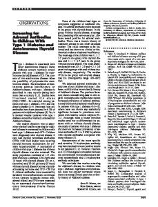
for CpG sites located from 28 to 2394 bp 5′ to the HLA-G transcriptional start site, which were reported to be regulated by DNA methylation in trophoblastic cell lines (Fig. 1A). The HLA-G promoter displays a different methylation profile in PE placentas compared with NP placentas. The average methylation levels across the promoter region are shown in order according to the location (Fig. 1B– D). Among all of the CpG sites examined, the 2188, 268 and 265 sites of the HLA-G promoter region have significantly higher methylation levels in PE than in NP tissues (P ¼ 0.0165, 0.0222 and 0.0037, respectively, Fig. 1C and D). The methylation percentage of the 2188 CpG site was 15.6 + 1.2% in PE placentas and 11.8 + 0.9% in NP placentas (Fig. 1C). The maximum difference was found at the 265 CpG site, with a methylation percentage of 8.6 + 1.3% in PE placentas versus 3.8 + 0.9% in NP placentas, while the minimum difference was found at the 268 site, with a methylation percentage of 9.5 + 1.1% in PE placentas versus 6.3 + 0.8% in NP placentas (Fig. 1D). As the region from
2290 to 2211 is reported to be a hypoxia-response element (HRE) that functions by binding the transcription activation factor HIF1-alpha (Menendez et al., 2008), we assayed the overall methylation level of this region except for 2211 CpG sites, which was overlapped by sequencing primer. Interestingly, although we did not find any significant difference in the percentage of methylation in any of the four CpG sites in the HRE (Fig. 1B), we found overall hypermethylation in the HRE in PE versus NP placentas (Fig. 1E, 46.0 + 0.6% in NP versus 48.5 + 1.0% in PE; P ¼ 0.0399).
HLA-G is down-regulated in PE and is accompanied by altered expression of DNMT-1 Because a high methylation level is believed to be closely associated with gene silencing and DNMTs are considered key enzymes that regulate
Figure 1 Bisulfite pyrosequencing to reveal the methylation patterns in the promoter region of HLA-G in placentas from PE and NP patients. (A) Schematic diagram of the promoter of the HLA-G gene. Each CpG site is indicated by vertical lines. The locations of the primers used for bisulfite PCR and pyrosequencing are shown by black and red arrows, respectively. The transcription start sites are indicated by boldface arrows. (B– D) The percentages of methylation at each CpG site in the promoter region of HLA-G in placentas from PE (n ¼ 19) and NP (n ¼ 20) patients using three sequencing primers. E shows the overall methylation level of the HRE element (from 2290 to 2211). Several CpG sites in the promoter region and HRE element are relatively hypermethylated in PE compared with NP placentas. Data are shown as the mean + SEM (*P , 0.05; **P , 0.001). PE refers to preeclampsia. NP refers to normal pregnancy.
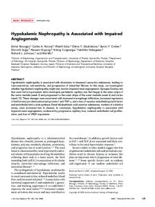
740 DNA methylation, we attempted to examine the expression levels of DNMTs and HLA-G. Immunostaining experiments on slides of placental tissue indicated that HLA-G was specifically expressed on the membranes of extravillous cytotrophoblasts (EVTs) (Fig. 2A), while DNMT-1 was highly expressed in the nuclei of both EVTs (Fig. 2A) and syncytiotrophoblasts (data not shown). The semi-quantitation of immunostaining for DNMT-1 and HLA-G was examined according to the German ImmunoReactive Score (Soslow et al., 2000). The immunostaining of DNMT-1 was significantly overexpressed in PE placentas than that in NP placentas (6.2 + 0.9 in PE versus 2.6 + 0.4 in NP, P ¼ 0.0069, shown in Fig. 2B), while the immunostaining of HLA-G showed us an opposite trends between the two population (3.8 + 0.4 in PE versus 9.5 + 0.5 in NP, P ¼ 0.0001, shown in Fig. 2C). However, we failed to detect and locate the DNMT3a/3b proteins in slides of placental tissue from
Tang et al.
both PE and NP in our IHC experiments (data not shown). We supposed that the DNMT3a/3b expression levels may be too low to detect, or they may be completely silenced in human placentas. As it was reported that HLA-G could be secreted or shed from trophoblastic cells of the placenta into maternal blood and soluble HLA-G was believed to be an early predictor of PE, we also determined the serum concentrations of sHLA-G in our study by performing ELISA. The sera concentration level of sHLA-G in PE was found to be significantly depressed when compared with that in NP (10.9 + 2.3 in PE versus 39.0 + 17.9 in NP, P ¼ 0.0348, shown in Fig. 2D). Furthermore, we also performed a qRT-PCR analysis to determine the mRNA expression levels of DNMT-1 and HLA-G in our study population of placentas. DNMT-1 was expressed at approximately 3- to 4-fold higher levels in placentas from PE placentas compared with NP placentas (3.5 + 0.7 in PE
Figure 2 DNMT-1 and HLA-G localization and expression in PE versus NP patients. (A) IHC and localization of HLA-G and DNMT-1 in placentas. Tissue sections were deparaffinized, rehydrated and stained with DNMT-1 and HLA-G antibodies. Biopsies from all PE and NPs were analyzed. HLA-G and DNMT-1 are co-expressed in extravillous trophoblastic cells. HLA-G is a specific marker for extravillous trophoblastic cells. O indicates extravillous trophoblastic cells. The original magnification in the representative results is 200 fold. Scale bars (10 mm) are shown on right side of each image. (B and C) The IHC quantifications of DNMT-1 and HLA-G in placenta tissues were estimated according to the German ImmunoReactive Score. DNMT-1 was significantly overexpressed in PE placentas, while HLA-G expression was decreased in PE compared with NP placentas. (D) The concentrations of sHLA-G (units/ ml) in sera samples from PE and NP patients were measured by ELISA. sHLA-G were significantly decreased in PE patients. (E and F) The fold change of the transcription levels of DNMT-1 and HLA-G in placentas from PE versus NP patients was evaluated using qRT-PCR analysis. The results indicate that HLA-G expression is significantly decreased, whereas DNMT-1 is significantly increased in PE placentas compared with NP placentas. The data are the mean + SEM that were acquired from 19 PE and 20 NPs (**P , 0.001).
741
HLAG promoter hypermethylation in preeclampsia
versus 1.0 + 0.2 in NP, P ¼ 0.0022, shown in Fig. 2E), while HLA-G was decreased by nearly half in PE placentas compared with placentas from NP patients (0.5 + 0.1 in PE versus 1.0 + 0.1 in NP, P ¼ 0.0033, shown in Fig. 2F). Interestingly, the results of qRT-PCR analysis were consistent with the quantifications of the IHC staining. We did not perform a correlation analysis because the statistical relationship of the two molecules did not indicate a biological relationship, and the placental tissues might be composed of multiple cell types.
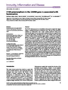
HLA-G is regulated by DNA methylation and DNMT-1 To further investigate the role of promoter DNA methylation in the deregulation of HLA-G in trophoblastic cells, we evaluated the effect of the methylation inhibitor 5-Aza-dC on HLA-G expression in HTR-8/ SVneo cells, which are human immortal trophoblastic cells that express a low level of the HLA-G molecule. The data indicated that the HLA-G transcripts were robustly increased after treatment with 2–8 mM 5-Aza-dC for 72 h (Fig. 3A). Bisulfite pyrosequence analysis of HLA-G promoter region in HTR-8/SVneo cell line before and after being incubated with 2 mM 5-Aza-dC indicated that there was an overall promoter demethylation in most of its CpG sites including 2188 site (Fig. 3C, 28.7 + 0.9 before versus 10.9 + 0.9 after demethylation treatment, P ¼ 0.0001) and 268 site (Fig. 3D, 30.5 + 1.8 before versus 1.4 + 1.4 after demethylation treatment, P ¼ 0.0002) as well as HRE region (Fig. 3E, 76.6 + 2.7 before versus 44.7 + 0.7 after demethylation
treatment, P ¼ 0.0003), which were hypermethylated in PE placenta. However, 265 CpG site did not show a significant demethylation after 5-Aza-dC treatment as it was initially hypomethylated in HTR-8/SVneo cell line (Fig. 3D, 1.3 + 1.3 before versus 0.9 + 0.9 after demethylation treatment, P ¼ 0.8). To better understand the methylation-dependent mechanism of the change in HLA-G, we further manipulated the expression of the primary DNMT, DNMT-1, by RNA interference. After the cells were incubated with two different siRNAs targeting DNMT-1 for 72 h, an increase in HLA-G expression was observed, although the increase was less than that observed following the 5-Aza-dC treatment (Fig. 3F). We hypothesize that this effect may be due to a different effect of the siRNA on DNMT activity. We did not evaluate the influence of DNMT3a/3b on HLA-G expression as these proteins were hardly detected in our placenta samples.
Discussion Recent evidence has indicated that multiple epigenetic mechanisms contribute to PE (Chelbi and Vaiman, 2008). Meanwhile, a wide range of PE-specific genes with aberrant DNA methylation and gene expression patterns have been identified, including apoptosis-related genes, cytokine receptor genes and obesity-related genes (Reimer et al., 2002; Tsoi et al., 2003; Yuen et al., 2010). In the current study, we have shown that a series of CpG sites in the HLA-G promoter region are hypermethylated in placentas from PE pregnancies compared with
Figure 3 HLA-G expression level is regulated by DNA methylation. (A) The fold changes in gene expression of HLA-G in HTR-8/SVneo cells treated with different concentrations of 5-Aza-dC for 3 days. The HLA-G expression level increased significantly as the concentration of 5-Aza-dC increased. (B– D) The percentages of methylation at each CpG site in the promoter region of HLA-G in HTR-8/SVneo cells before and after being incubated with 2 mM 5-Aza-dC for 3 days. (E) The overall methylation level of the HRE region (from 2290 to 2211) in HTR-8/SVneo cells before and after being incubated with 2 mM 5-Aza-dC for 3 days. (F) The fold changes in gene expression of HLA-G in HTR-8/SVneo cells transfection with two different siRNAs targeting DNMT-1. The knockdown efficiency of DNMT-1 is shown on the right, and HLA-G expression was significantly elevated. Each experiment was independently repeated three times. Each point represents the mean + SEM values from three independent experiments (*P , 0.05; **P , 0.001; ***P , 0.0001).
742 NPs. The altered methylation in placentas is accompanying by decreased expression of HLA-G as well as the overexpression of DNMT-1. Furthermore, both DNA demethylation reagent and the specific inhibition of DNMT-1 in vitro can lead to increased HLA-G expression, confirming the epigenetic regulation of the HLA-G gene in PE. In the present study, the percentage of HLA-G methylation was elevated in PE. Moreover, the expression level of HLA-G in both placenta tissues and maternal blood is decreased in PE patients when compared with those from NPs, which is consistent with previous studies by other research groups (Darmochwal-Kolarz et al., 2012). A hypermethylated promoter correlates with decreased gene expression because the methylated cytosine hinders the binding of transcription factors to the chromosome. Chelbi et al. (2007, 2012) have demonstrated that specific CpG demethylation in the SERPINA3 promoter region might help render the chromatin accessible to transcription factors, thus promoting its expression and participating in the onset of PE. Hence, we analyzed the particular CpG sites that exhibit different methylation patterns in our study population. Interestingly, the HRE (2211 to 2290) in the HLA-G promoter had a significantly higher overall methylation level in PE than in NP. HRE is a binding site for the transcription factor HIF-1, which plays a critical role in the cellular response to hypoxia and is reported to induce HLA-G expression in the HLA-G-negative M8 melanoma cell line (Mouillot et al., 2007). Thus, we supposed that the hypermethylated HRE might become less sensitive to HIF-1, which is induced by placental physiology and/or pathology caused by hypoxia during pregnancy, and thus fail to induce HLA-G expression and eventually disrupt the fetal–maternal immune tolerance or influence the trophoblastic cell functions that are mediated by HLA-G. Similarly, other CpG sites displaying a significantly higher level of methylation are located close to intact binding sites of regulatory proteins known to facilitate gene expression (CCAAT box at 276 and enhancer kB1 at 2188) (Menendez et al., 2008). The hypermethylation of these sites is likely to inhibit the activation of HLA-G expression through a cis-acting mechanism in PE. DNMTs are key enzymes that regulate DNA methylation. DNMT-1 acts as a ‘maintenance’ methyltransferase and is the primary DNMT in mammals, while DNMT3a/3b act as ‘de novo’ methyltransferases (Bestor, 2000). We demonstrate an up-regulation of DNMT-1 in PE placentas in our research. High levels of DNMT-1 expression were usually believed to be responsible for global hypermethylation. Accordingly, a global hypermethylation was shown PE placentas in Gao’s and Kulkarni’s studies (Gao et al., 2011; Kulkarni et al., 2011). However, this up-regulation of DNMT-1 contrasts with recent findings from several microarrays analysis in which no significant alteration of DNMT-1 was observed (Herse et al., 2007; Nishizawa et al., 2007). The discrepancy between our work and earlier work probably arises from the differences in methodologies used to detect DNMT-1 levels. The fact that some microarray designs might not distinguish all the isoforms of DNMTs separately as we did by performing real-time PCR would probably account for differences in the results. Moreover, the gestational week and the patient ethnicity may also have effects on it. DNMT-1 is closely correlated with the methylated CpG island phenotype (Teodoridis et al., 2008). Robert had shown us that DNMT-1 is required to maintain CpG methylation and aberrant gene silencing in human cancer cells, although the mechanisms of which DNMT1 involves with locus-specific methylation are uncertain (Robert et al., 2003). Consistently, our research indicated that DNMT-1 participated in HLA-G gene silencing through DNA methylation in HTR-8/SVneo. It was
Tang et al.
reported that HLA-G gene transcription is inhibited by DNA methylation in a series of tumor cell lines and reversal of methylation-mediated repression by DNA demethylation reagent 5-Aza-dC could directly induce HLA-G expression (Moreau et al., 2003). We find a similar phenomenon in HTR-8/SVneo. Furthermore, although to a lesser extent than 5-Aza-dC treatment, a reversion in HLA-G expression was also observed following specific DNMT-1 knockdown, which supports the critical role of DNMT1 in the epigenetic regulation of HLA-G expression. The aberrant DNMT-1 expression observed in PE in our study was consistent with Gao’s study (Gao et al., 2011). As a putative HRE was discovered in the 5′ UTR of DNMT-1 and HIF-1alpha-mediated hypoxia regulation of DNMT enzymes eventually alters the DNA methylation pattern (Watson et al., 2014), we guess that the pathophysiology due to hypoxia in the placenta from PE patients may have induced abnormal DNMT-1 expression. Combined with other transcription factors, increased levels of DNMT-1 might cause the hypermethylation of HLA-G promoter region and suppress HLA-G transcription. However, further research should be done to testify this hypothesis in the future. As we did not find clear expression of DNMT3a/3b in placentas at either the mRNA or protein levels, DNMT3a/3b knockdowns were not performed, and their impact on HLA-G expression was not evaluated in our in vitro experiment. However, these proteins may still contribute to DNA methylation. A possible reason for the low expression of these proteins is that de novo methylation mainly occurs during epigenetic reprogramming in the gametes and preimplantation embryo (Yin et al., 2012). After embryo implantation, methylation is maintained by DNMT-1, and no noticeable changes in de novo methylation occur in the post-differentiated human villous (Yin et al., 2012). Consistent with previous studies, our results indicate that the epigenetic regulation by promoter methylation is central for HLA-G expression as well (Moreau et al., 2003; Menendez et al., 2008; Holling et al., 2009). Our results represent a slightly different but more accurate assessment of the methylation percentage of each CpG site than previous studies due to the advanced methylation detection system that we employed. There are several limitations to our study. First, the purity and cell proportion of placental tissues may have an influence on the results. Repeating the experiment with primary cells might be a more preferable and less contrived approach to experiments in an immortalized trophoblast cell line that expresses a low level of HLA-G as primary trophoblast culture is reported to retain the PE phenotype for a few days (Zhou et al., 2013). Second, the study population was mostly in the late stages of pregnancy, while the onset of PE begins during early pregnancy; thus, we cannot clearly conclude whether the dysfunctional regulation of HLA-G is a cause or a consequence of PE. Thirdly, it is controversial to use GAPDH as a reference gene as some studies indicate that it was unstably expressed in the placenta across pregnancy. However, Kaitu’uLino proved that GAPDH did not vary in PE compared with normotensive pre-term and term placentas (Kaitu’u-Lino et al., 2014). In summary, we have characterized the expression of DNMTs in PE placentas and have demonstrated the involvement of DNMT-1mediated promoter hypermethylation in the regulation of HLA-G in PE. Further research is needed to illustrate the underlying mechanisms that regulate gene-specific methylation dysfunction in PE, which will help us identify specific epigenetic markers for PE, find new strategies to treat PE and thus improve the outcomes of affected women and their children.
HLAG promoter hypermethylation in preeclampsia
Authors’ roles Y.T. formed the study design, collected clinical samples, obtained results, performed data analysis and wrote this manuscript. Haiyan Liu and Han Li also participated in clinical samples collection and performed data analysis. W.G. edited the manuscript, and she is the corresponding author; he formed the study idea and study design and also participated in critical discussion. X.L. participated in the formation of study design, researched the data, reviewed and edited the manuscript. T.P. participated in the formation of study design and also in critical discussion.
Funding This research was supported by the Science Foundation of Shanghai Municipal Health Bureau (Grant No. 20134031 to W.G) and Natural Science Foundation of China (Grant Nos 81471470 to W.G., 81200440 to T.P. and 81270712 to X.L.).
Conflict of interest None declared.
References Bestor TH. The DNA methyltransferases of mammals. Hum Mol Genet 2000; 9:2395 – 2402. Chelbi ST, Vaiman D. Genetic and epigenetic factors contribute to the onset of preeclampsia. Mol Cell Endocrinol 2008;282:120 – 129. Chelbi ST, Mondon F, Jammes H, Buffat C, Mignot TM, Tost J, Busato F, Gut I, Rebourcet R, Laissue P et al. Expressional and epigenetic alterations of placental serine protease inhibitors: SERPINA3 is a potential marker of preeclampsia. Hypertension 2007;49:76 – 83. Chelbi ST, Wilson ML, Veillard AC, Ingles SA, Zhang J, Mondon F, GascoinLachambre G, Doridot L, Mignot TM, Rebourcet R et al. Genetic and epigenetic mechanisms collaborate to control SERPINA3 expression and its association with placental diseases. Hum Mol Genet 2012;21:1968–1978. Chumbley G, King A, Robertson K, Holmes N, Loke YW. Resistance of HLA-G and HLA-A2 transfectants to lysis by decidual NK cells. Cell Immunol 1994;155:312 – 322. Darmochwal-Kolarz D, Kolarz B, Rolinski J, Leszczynska-Gorzelak B, Oleszczuk J. The concentrations of soluble HLA-G protein are elevated during mid-gestation and decreased in pre-eclampsia. Folia Histochem Cytobiol 2012;50:286 – 291. Deaton AM, Bird A. CpG islands and the regulation of transcription. Genes Dev 2011;25:1010 – 1022. Duley L. The global impact of pre-eclampsia and eclampsia. Semin Perinatol 2009;33:130 – 137. Dupont JM, Tost J, Jammes H, Gut NG. De novo quantitative bisulfite sequencing using the pyrosequencing technology. Anal Biochem 2004; 333:119– 127. Gao WL, Li D, Xiao ZX, Liao QP, Yang HX, Li YX, Ji L, Wang YL. Detection of global DNA methylation and paternally imprinted H19 gene methylation in preeclamptic placentas. Hypertens Res 2011;34:655 – 661. Goldman-Wohl DS, Ariel I, Greenfield C, Hochner-Celnikier D, Cross J, Fisher S, Yagel S. Lack of human leukocyte antigen-G expression in extravillous trophoblasts is associated with pre-eclampsia. Mol Hum Reprod 2000;6:88 – 95. Hara N, Fujii T, Yamashita T, Kozuma S, Okai T, Taketani Y. Altered expression of human leukocyte antigen G (HLA-G) on extravillous trophoblasts in preeclampsia: immunohistological demonstration with
743 anti-HLA-G specific antibody “87G” and anti-cytokeratin antibody “CAM5.2”. Am J Reprod Immunol 1996;36:349– 358. Herse F, Dechend R, Harsem NK, Wallukat G, Janke J, Qadri F, Hering L, Muller DN, Luft FC, Staff AC. Dysregulation of the circulating and tissue-based renin-angiotensin system in preeclampsia. Hypertension 2007;49:604 – 611. Holling TM, Bergevoet MWT, Wierda RJ, van Eggermond M, van den Elsen PJ. Genetic and epigenetic control of the major histocompatibility complex class Ib gene HLA-G in trophoblast cell lines. In: Shoenfeld Y, Gershwin ME, (ed). Contemporary Challenges in Autoimmunity. Oxford: Blackwell Publishing, 2009, 538 – 544. Kaitu’u-Lino TJ, Hastie R, Cannon P, Lee S, Stock O, Hannan NJ, Hiscock R, Tong S. Stability of absolute copy number of housekeeping genes in preeclamptic and normal placentas, as measured by digital PCR. Placenta 2014;35:1106 – 1109. Kulkarni A, Chavan-Gautam P, Mehendale S, Yadav H, Joshi S. Global DNA methylation patterns in placenta and its association with maternal hypertension in pre-eclampsia. DNA Cell Biol 2011;30:79 – 84. Li HJ, Gu WR, Li XT. Effects of human leukocyte antigen-G on p38 mitogen-activated protein kinase signaling pathway in HTR-8/SVneo cell line. Zhonghua Fu Chan Ke Za Zhi 2011;46:271 – 276. Liu HY, Liu XY, Jin H, Yang FY, Gu WR, Li XT. Proteomic analysis of knock-down HLA-G in invasion of human trophoblast cell line JEG-3. Int J Clin Exp Pathol 2013a;6:2451– 2459. Liu X, Gu W, Li X. HLA-G regulates the invasive properties of JEG-3 choriocarcinoma cells by controlling STAT3 activation. Placenta 2013b; 34:1044– 1052. Menendez L, Walker LD, Matyunina LV, Totten KA, Benigno BB, McDonald JF. Epigenetic changes within the promoter region of the HLA-G gene in ovarian tumors. Mol Cancer 2008;7:11. Moreau P, Flajollet S, Carosella ED. Non-classical transcriptional regulation of HLA-G: an update. J Cell Mol Med 2009;13:2973 –2989. Moreau P, Mouillot G, Rousseau P, Marcou C, Dausset J, Carosella ED. HLA-G gene repression is reversed by demethylation. Proc Natl Acad Sci U S A 2003;100:1191 – 1196. Mouillot G, Marcou C, Zidi I, Guillard C, Sangrouber D, Carosella ED, Moreau P. Hypoxia modulates HLA-G gene expression in tumor cells. Hum Immunol 2007;68:277 – 285. Nishizawa H, Pryor-Koishi K, Kato T, Kowa H, Kurahashi H, Udagawa Y. Microarray analysis of differentially expressed fetal genes in placental tissue derived from early and late onset severe pre-eclampsia. Placenta 2007;28:487 – 497. Reimer T, Koczan D, Gerber B, Richter D, Thiesen HJ, Friese K. Microarray analysis of differentially expressed genes in placental tissue of pre-eclampsia: up-regulation of obesity-related genes. Mol Hum Reprod 2002;8:674 – 680. Robert MF, Morin S, Beaulieu N, Gauthier F, Chute IC, Barsalou A, MacLeod AR. DNMT1 is required to maintain CpG methylation and aberrant gene silencing in human cancer cells. Nature Genet 2003;33:61–65. Serrano NC. Immunology and genetic of preeclampsia. Clin Dev Immunol 2006;13:197 – 201. Soslow RA, Dannenberg AJ, Rush D, Woerner BM, Khan KN, Masferrer J, Koki AT. COX-2 is expressed in human pulmonary, colonic, and mammary tumors. Cancer 2000;89:2637– 2645. Steegers EAP, von Dadelszen P, Duvekot JJ, Pijnenborg R. Pre-eclampsia. Lancet 2010;376:631– 644. Teodoridis JM, Hardie C, Brown R. CpG island methylator phenotype (CIMP) in cancer: causes and implications. Cancer Lett 2008;268:177– 186. Tsoi SM, Cale JM, Bird IM, Kay HH. cDNA microarray analysis of gene expression profiles in human placenta: up-regulation of the transcript encoding muscle subunit of glycogen phosphorylase in preeclampsia. J Soc Gynecol Invest 2003;10:496 – 502.
744 Watson CJ, Collier P, Tea I, Neary R, Watson JA, Robinson C, Phelan D, Ledwidge MT, McDonald KM, McCann A et al. Hypoxia-induced epigenetic modifications are associated with cardiac tissue fibrosis and the development of a myofibroblast-like phenotype. Hum Mol Genet 2014;23:2176 – 2188. Yie SM, Li LH, Li YM, Librach C. HLA-G protein concentrations in maternal serum and placental tissue are decreased in preeclampsia. Am J Obstet Gynecol 2004;191:525 – 529. Yie SM, Taylor RN, Librach C. Low plasma HLA-G protein concentrations in early gestation indicate the development of preeclampsia later in.pregnancy. Am J Obstet Gynecol 2005;193:204 – 208. Yin LJ, Zhang Y, Lv PP, He WH, Wu YT, Liu AX, Ding GL, Dong MY, Qu F, Xu CM et al. Insufficient maintenance DNA methylation is
Tang et al.
associated with abnormal embryonic development. BMC Med 2012; 10:10. Yuen RKC, Penaherrera MS, von Dadelszen P, McFadden DE, Robinson WP. DNA methylation profiling of human placentas reveals promoter hypomethylation of multiple genes in early-onset preeclampsia. Eur J Hum Genet 2010;18:1006– 1012. Zhou Y, Gormley MJ, Hunkapiller NM, Kapidzic M, Stolyarov Y, Feng V, Nishida M, Drake PM, Bianco K, Wang F et al. Reversal of gene dysregulation in cultured cytotrophoblasts reveals possible causes of preeclampsia. J Clin Invest 2013;123:2862– 2872. Zhu X, Han T, Yin G, Wang X, Yao Y. Expression of human leukocyte antigen-G during normal placentation and in preeclamptic pregnancies. Hypertens Pregnancy 2012;31:252 – 260.