Differentiation, Survival, and Function of Embryonic Stem Cell–Derived Endothelial Cells for Ischemic Heart Disease Zongjin Li, MD, PhD; Jenny C. Wu, PhD; Ahmad Y. Sheikh, MD; Daniel Kraft, MD; Feng Cao, MD, PhD; Xiaoyan Xie, PhD; Manishkumar Patel, PhD; Sanjiv S. Gambhir, MD, PhD; Robert C. Robbins, MD; John P. Cooke, MD, PhD; Joseph C. Wu, MD, PhD
Downloaded from http://circ.ahajournals.org/ by guest on June 8, 2018
Background—Embryonic stem (ES) cells are distinguished by their capacity for self-renewal and pluripotency. Here we characterize the differentiation of ES cell– derived endothelial cells (ESC-ECs), use molecular imaging techniques to examine their survival in vivo, and determine the therapeutic efficacy of ESC-ECs for restoration of cardiac function after ischemic injury. Methods and Results—Murine ES cells were transfected with a construct composed of a vascular endothelial cadherin promoter driving enhanced green fluorescence protein (pVE-cadherin-eGFP). Differentiation of ES cells to ECs was detected by FACS analysis using Flk-1 (early EC marker at day 4) and VE-cadherin (late EC marker at day 8). After isolation, these ESC-ECs express endothelial cell markers similar to adult mouse lung endothelial cells, form vascular-like channels, and incorporate DiI-labeled acetylated low-density lipoprotein (DiI-Ac-LDL). For in vivo imaging, ES cells were transduced with an ubiquitin promoter driving firefly luciferase and monomeric red fluorescence protein (pUb-Fluc-mRFP). A robust correlation exists between Fluc signals and cell numbers by ex vivo imaging analysis (R2⫽0.98) and by in vitro enzyme assay (R2⫽0.94). Afterward, 5⫻105 ESC-ECs or PBS (as control) was injected into the hearts of mice undergoing LAD ligation (n⫽15 per group). Bioluminescence imaging showed longitudinal survival of transplanted ESC-ECs for ⬇8 weeks. Echocardiogram demonstrated significant functional improvement in the ESC-EC group compared with control (P⫽0.04). Finally, postmortem analysis confirmed increased presence of small capillaries and venules in the infarcted zones by CD31 staining. Conclusions—This is the first study to track the fate and function of transplanted ESC-ECs in the heart. With further validation, these ESC-ECs could become a valuable source of cell therapy for induction of angiogenesis in the treatment of myocardial ischemia. (Circulation. 2007;116[suppl I]:I-46–I-54.) Key Words: embryonic stem cells 䡲 differentiation 䡲 endothelial cells 䡲 heart disease 䡲 molecular imaging
C
oronary artery disease (CAD) is the leading cause of morbidity and mortality in the US. Current treatments fail to address the underlying cause of heart failure, which are cell loss and tissue scarring.1 Stem cell therapy might ameliorate heart failure by promoting neovascularization, inhibiting cardiomyocyte apoptosis, and recruiting resident stem cells.2 Several elegant studies demonstrate that endothelial progenitor cells harvested from the bone marrow3,4 or circulating peripheral blood5,6 can contribute to angiogenesis and functional regeneration of ischemic or infarcted myocardium. These exciting results have led to a pilot clinical trial showing that injection of circulating endothelial progenitor cells can improve left ventricular ejection function (LVEF) in patients.7 In the TOPCARE-AMI trial, patients who received circulating endothelial progenitor cells after myocardial infarction manifested an improvement in LVEF from 51⫾10% to 59⫾10% at 4 months (P⬍0.001).7
However, a recent larger study from the same group showed that patients who received circulating endothelial progenitor cells had no improvement in LVEF at 3 months (⫺0.4⫾2.2; P⫽0.60).8 Although the study design and patient population are different between the 2 trials (which may partially explain the inconsistent results), the discrepancy suggests that a detailed mechanistic understanding of how these transplanted cells can engraft and improve cardiac function is still lacking. In addition, patients who have CAD as well as other comorbidities such as diabetes, hypertension, and hypercholesterolemia may have endothelial progenitor cells with impaired function and regenerative capacity.9,10 For this reason, the beneficial results observed in preclinical studies using progenitor cells from young healthy adult animals may not be achievable in patients with comorbid conditions.6,9,10 Therefore, evaluation of another source of cell therapy may be warranted.
From the Department of Radiology and Molecular Imaging Program at Stanford (Z.L., F.C., X.X., M.P., S.S.G., Jo.C.W.), the Department of Medicine, Division of Cardiology (Je.C.W., J.P.C., Jo.C.W.), the Department of Cardiothoracic Surgery (A.Y.S., R.C.R.), and the Department of Medicine, Division of Hematology (D.K.), Stanford University School of Medicine, Stanford, Calif. Presented at the American Heart Association Scientific Sessions, Chicago, Ill, November 12–15, 2006. Correspondence to Joseph C. Wu, MD, PhD, Stanford University School of Medicine, Edwards Building R354, Stanford, CA 94305-5344. E-mail
[email protected] © 2007 American Heart Association, Inc. Circulation is available at http://circ.ahajournals.org
DOI: 10.1161/CIRCULATIONAHA.106.680561
I-46
Li et al
Transplantation of ES Cell–Derived Endothelial Cells
Downloaded from http://circ.ahajournals.org/ by guest on June 8, 2018
With their unlimited self-renewal and pluripotency capacity, embryonic stem (ES) cells represent a new and exciting avenue of stem cell therapy. In cell culture, mouse and human ES cells can differentiate to endothelial cells through successive maturation steps as manifested by endothelial-specific markers such as fetal liver kinase 1 (Flk-1), platelet endothelial cell adhesion molecule (PECAM), VE-cadherin, von Willebrand Factor (vWF), and endothelial nitric oxide synthase (eNOS).11,12 In addition, these cells can participate in vasculogenesis by matrigel assay13 and incorporate DiI-acLDL,14 2 hallmarks of mature endothelial cell function. However, no study has addressed the behavior of these cells after transplantation into the heart. In this study, we hypothesize that ES cell– derived endothelial cells (ESC-ECs) can be a viable source of stem cell therapy for improving cardiac function after myocardial infarction. Importantly, fate of transplanted ES cells and their ESC-EC derivatives can also be tracked longitudinally and quantitatively using molecular imaging techniques.
Methods Culture and Maintenance of Mouse ES Cells The murine ES-D3 cell line (CRL-1394) derived from SV129 mice was obtained from the American Type Culture Collection (ATCC; Manassas, VA). The ES cells were electroporated with linearized pVEcadherin-eGFP-IE plasmid carrying pVEcadherin-eGFP-5⬘Int1 enhancer (gift from S. Nishikawa, Kyoto University, Japan) with neomycin resistant gene for selection of stable clones.15
Differentiation and Isolation of ESC-ECs ES cell differentiation was performed with minor modifications from previous protocols.11,12 Briefly, 3⫻104 cells per well were transferred to collagen IV-coated 6-well plates (Becton-Dickinson), cultured for 4 days, and sorted for Flk-1 (early EC marker). Flk-1⫹ cells were replated with the same medium supplemented with 50 ng/mL of VEGF (R&D Systems). After another 4 days of culturing, Flk-1⫹ cells were stained with VE-cadherin antibody (BD Pharmingen). Next, VE-cadherin (late EC marker) and eGFP double positive cells were isolated using FACScan (Becton Dickinson). The cells were maintained on EGM-2 medium (Clonetics) for 1 to 2 passages on fibronectin-coated plates (Invitrogen) for subsequent experiments. Control untransfected ES cells were differentiated under same conditions.
In Vitro Characterization of ESC-ECs Antibody staining, DiI-Ac-LDL uptake assay, matrigel assay, and RT-PCR were used to confirm endothelial cell phenotype of these Flk-1⫹/VE-cadherin⫹ purified cells. Antibodies used in this study were phycoerythrin (PE)-, Alex-594 –, or allophycocyanin (APC)conjugated anti-CD31, anti–Flk-1, anti-CD34, and APC-conjugated anti-rat IgG2a, rat anti-mouse VE-cadherin (BD Pharmingen). The stained cells were analyzed using FACS Vantage (BectonDickinson). Dead cells stained by propidium-iodide were excluded from the analysis. Isotype-identical antibodies served as controls (BD Pharmingen). Approximately 5⫻105 ESCs and 2⫻105 ESC-ECs were used to run the FACS. Cell sorting was performed as described.16 For uptake assay, ESC-ECs were incubated with 10 g/mL of DiI-Ac-LDL (Molecular Probes) at 37°C for 6 hours before detection with fluorescence microscopy as described.14 The formation of endothelial tubes was assessed by seeding cells in 6-well plates coated with Matrigel (BD Pharmingen) and incubating them at 37° for 24 hours as described.12 For RT-PCR analysis, total RNA was isolated using Trizol (Invitrogen) from undifferentiated ES cells at day 0, differentiated ES cells at day 4 (after Flk⫹ sort), differentiated ES cells at day 8 (after VE-cadherin⫹/eGFP⫹ double sort), and adult mouse microvascular endothelial cells (obtained
I-47
from lung lavage) as positive control. Primer sequences for these specific genes (CD31, Flk-1, eNOS, vWF, Oct-4, -actin) are listed in supplemental Table I (available online at http://circ.ahajournals.org). Unless stated otherwise, all experiments were performed in triplicate.
Transduction of ES Cells With Novel Double Fusion Reporter Gene To track transplanted cells in vivo, ES cells were also transduced at multiplicity of infection (MOI) of 10 with self-inactivating lentiviral vector carrying an ubiquitin promoter driving Fluc and mRFP. Stable clones were isolated using FACS for mRFP expression. Afterward, Fluc activity within different cell numbers (4⫻104 to 1⫻106) was confirmed ex vivo using Xenogen IVIS 200 system (Xenogen) and in vitro using enzyme assay as described.17 In vitro Fluc enzyme activity was expressed as relative light unit per microgram protein (RLU/g). Next, several assays were performed to assess the effects of double fusion (DF) reporter gene on ES cell differentiation, proliferation, and viability. Control nontransduced ES cells and ES cells with DF (ESC-DF) were stained for alkaline phosphatase (marker for undifferentiated cells) using BCIP (5-bromo-4-chloro3⬘-indolyphosphate p-toluidine salt)/NBT (nitro-blue tetrazolium chloride) dyes (Sigma). Images were obtained with a Zeiss Axiovert microscopy (Sutter Instrument Co). RT-PCR was performed comparing expression of Oct-4 (stem cell marker), Fluc (reporter gene), and -actin (housekeeping gene) in control ES cells and ESC-DF. Cell proliferation was measured by counting cells at days 0, 1, 2, and 3 time points. Finally, the trypan blue exclusion assay was used to assess viability and cytotoxicity under the same conditions. Six samples were performed and averaged.
Animal Surgery and ESC-EC Transplantation Ligation of the mid left anterior descending (LAD) artery was performed in adult female SV129 mice (Charles River Laboratories, Wilmington, Mass) by a single experienced surgeon (AYS). Myocardial infarction was confirmed by myocardial blanching and EKG changes. After waiting for 10 minutes, animals were then injected intramyocardially with 5.0⫻105 ESC-ECs at the peri-infarct zone (n⫽15), 5.0⫻105 undifferentiated ESCs (n⫽15), or PBS as control (n⫽15). In all groups, the volume of injection was 25 L using a 31-gauge Hamilton syringe. Study protocols were approved by the Stanford Animal Research Committee.
Optical Bioluminescence Imaging of Transplanted Cell Survival Cardiac bioluminescence imaging was performed using the Xenogen IVIS 200 system. After intraperitoneal injection of reporter probe D-Luciferin (150 mg luciferin/kg), animals were imaged for 1 to 10 minutes. The same mice were scanned for 4 to 8 weeks. Imaging signals were quantified in units of maximum photons per second per centimeter square per steridian (photons/sec/cm2/sr) as described.17
Left Ventricular Functional Analysis With Echocardiogram Echocardiography was performed using the Siemens-Acuson Sequioa C512 system equipped with a multi-frequency (8 to 14 MHz) 15L8 transducer by an investigator (F.C.) blinded to group designation. Analysis of M-mode images was performed using Siemens built-in software. Left ventricular end diastolic diameter (EDD) and end-systolic diameter (ESD) were measured and used to calculate fractional shortening (FS) by the following formula: FS⫽[EDD⫺ESD]/EDD.18
Histological Examination Explanted hearts from study and control groups were embedded into OCT compound (Miles Scientific). Frozen sections (5 m thick) were processed for immunostaining. To detect microvascular density (MVD) in the peri-infarct area, a rat anti-CD31 (BD Pharmingen) was used. The number of capillary vessels was counted by a blinded
I-48
A
Circulation
September 11, 2007
pVE-cadherin
eGFP
SV40
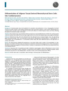
neor Figure 1. ES cell differentiation into endothelial-like cells. a, Schema of the endothelial cell lineage-specific promoter with VE-cadherin promoter driving eGFP and SV40 promoter driving neomycin resistant gene for antibiotic selection of stable clones. b, After 4 days on collagen IV plates, Flk-1⫹–positive cells can be identified by FACS. The outgrowth from these Flk-1⫹ cells exhibit predominantly 2 morphologies: cobblestone-like endothelial colony and striated smooth muscle-like cells as shown on brightfield microscopy (100⫻ magnification). c, After another 4 days of culturing on differentiation medium, ⬇36% of cells express both VE-cadherin surface marker and intracellular eGFP. These double-positive cells show typical endothelial cell morphology and can be expanded for additional 5 to 8 passages (100⫻ magnification).
B
C
Downloaded from http://circ.ahajournals.org/ by guest on June 8, 2018
investigator (X.X.) in 10 randomly selected areas using a light microscope (⫻200 magnification). Additional samples were used to examine whether transplanted ESC-EC differentiated into functional vascular and other cell lineages processed for hematoxylin and eosin (H&E) and fluorescence staining. To trace the ESC-EC in the ischemia heart, the slices were double stained with anti-RFP antibody (Chemicon International), anti-CD31 antibody, and DAPI (4, 6-diamidino-2-phenylindole) nuclear counterstain. For undifferentiated ES cell transplantation group, teratoma formation at 4 weeks was also evaluated by H&E and immunofluorescence staining.
Statistical Analysis ANOVA and repeated measures ANOVA with post-hoc testing as well as the 2-tailed Student t test were used. Differences were considered significant at probability values of ⬍0.05. Unless specified, data are expressed as mean⫾SD.
Statement of Responsibility The authors had full access to and take full responsibility for the integrity of the data. All authors have read and agree to the manuscript as written.
Results Differentiation of Embryonic Stem Cells to Endothelial Cells In mouse ES cells, there is a distinct expression pattern of endothelial specific genes: Flk-1 starting at day 2 to 3, PECAM at day 4, and VE-cadherin at day 5.12,19 To develop a reliable system of purifying mature ECs from ES cells, we used the VE-cadherin promoter driving eGFP construct for selection marker as shown in Figure 1a.15 After 4 days of culturing on collagen IV– coated plates and in differentiation medium, ⬇20% of ES cells express early EC marker (Flk-1) but not late EC marker (VE-cadherin). The outgrowth from Flk-1 ⫹ cells exhibit predominantly 2 morphologies: cobblestone-like endothelial colony and striated smooth muscle-like cells (Figure 1b). The Flk-1⫹–positive cells were subcultured with VEGF supplementation for another 4 days. Afterward, ⬇36% cells were isolated by FACS using both
VE-cadherin surface antibody as well as intracellular eGFP expression driven by the VE-cadherin promoter. After expansion in cell culture, these VE-cadherin⫹/eGFP⫹ cells have morphology typical of mature endothelial cells (Figure 1c).
In Vitro Characterization of ESC-ECs To further characterize these differentiated cells, flow cytometry was performed using antibodies directed against endothelial markers such as VE-cadherin, Flk-1, CD31, and CD34 (Figure 2a). Isolated cells at day 8 typically express robust levels of VE-cadherin and modest levels of Flk-1, CD31, and CD34. By contrast, undifferentiated ES cells typically express low levels of VE-cadherin, Flk-1, and CD34 but high levels of CD31. Uptake of DiI-ac-LDL has been used to characterize mature endothelial cells.14 As shown in Figure 2b, these VE-cadherin⫹/eGFP⫹ cells avidly incorporate DiIac-LDL. The characteristics of ESC-ECs were also assessed by culturing on matrigel, an extracellular matrix basement membrane that can be used to promote vascular morphogenesis of endothelial cells.13 In contrast to undifferentiated ES cells, ESC-ECs were able to form cord-like structures (Figure 2c). To understand the temporal kinetics of endothelial marker expression in these cells, we performed RT-PCR analysis of common endothelial markers. As shown in Figure 2d, the levels of Flk-1, eNOS, and vWF increased during ES cell differentiation from day 0 to day 4 (Flk-1⫹ sort) to day 8 (VE-cadherin⫹/eGFP⫹ double sort). The ES cell marker Oct-4 was expressed in high levels at day 0, but progressively decreased on ES cell differentiation into ESC-ECs at days 4 and 8. Adult mouse microvascular endothelial cells showed robust endothelial marker expression but no Oct-4 expression as expected. Finally, for both FACS and RT-PCR analysis, the CD31 expression was present in both undifferentiated mouse ES cells and their differentiated derivatives, a pattern that has been described by others.19 Thus, CD31 alone cannot be used as a marker for distinguishing mouse ES cells from ECs.
Li et al
VE-cadherin
A
CD31
Transplantation of ES Cell–Derived Endothelial Cells
Flk-1
I-49
CD34
Counts
ESC
ESC-EC
B
Downloaded from http://circ.ahajournals.org/ by guest on June 8, 2018
C
DiI-ac-LDL
ESC (24h)
eGFP
DiI-ac-LDL/eGFP
ESC-EC (24h)
D
Expression of DF Reporter Genes To develop an imaging assay for tracking transplanted ESCECs, we used a DF reporter gene consisting of Fluc-mRFP (Figure 3a). The efficiency of self-inactivating lentiviral transduction of mouse ES cells was ⬇21% (Figure 3b). On culturing onto 6-well plates, we observed a strong correlation (r2⫽0.98) between Fluc activity and cell numbers ex vivo using the Xenogen IVIS system. Similarly, there was a strong correlation (r2⫽0.94) between Fluc activity and cell numbers in vitro using luminometer enzyme assays (Figure 3c). Both control nontransduced ES cells and ESC-DF showed similar expression pattern of stem cell markers ALP and Oct4 on immunostaining and RT-PCR, respectively (Figure 3d and 3e). The cell proliferation and cell viability data are also
ESC-EC (48h)
Figure 2. In vitro characterization of murine ESC-ECs. a, FACS analysis shows increased expression of common endothelial cell markers (VE-cadherin, Flk-1, CD34) in differentiated ESC-ECs compared with undifferentiated ES cells. CD31 marker is highly expressed in both cell types. Y-axis represents cell number and x-axis represents fluorescence intensity. b, Uptake of DiI-ac-LDL by ESCECs. These cells are stably transfected with pVEcadherin-eGFP-SV40-neo construct and express eGFP on differentiation into endothelial cells. c, Endothelial tube formation by ESC-ECs after 24 and 48 hours of plating. Undifferentiated ES cells do not form cord-like structures (100⫻ magnification). d, Expression profile of endothelial cell-specific genes at different time points: undifferentiated ES cells (day 0), Flk-1⫹ cells (day 4), and VE-cadherin⫹/eGFP⫹ cells (day 8). The positive control is adult mouse endothelial cells (mEC) from the lung microvasculature. The negative control has no PCR templates.
similar between control ES cells and ESC-DF (Figure 3f). Finally, ESC-DF retained their ability to differentiate and express endothelial markers such as Flk-1⫹ (on day 4) and VE-cadherin⫹/eGFP⫹ (on day 8; data not shown). Taken together, these results are consistent with our previous studies showing minimal effects of reporter genes on ES cell survival, proliferation, and differentiation.17,20,21
Molecular Imaging of ESC-EC Survival in Living Animals To noninvasively assess the engraftment of ESC-ECs, bioluminescence imaging was performed longitudinally for 8 weeks (Figure 4a). Cell signal was most robust immediately after transplantation, which gradually decreased from day 2 (2.95⫻106⫾1.21⫻105 photons/sec/cm2/sr) to day 7 (3.56⫻105⫾2.36⫻104) to week 2
I-50
A
Circulation
September 11, 2007
5’ LVLTR pUbiquitin
Fluc
Fluorescence
RFP
FSC
B
mRFP SIN LTR 3’
C
Downloaded from http://circ.ahajournals.org/ by guest on June 8, 2018
D
Control
4×10 4
1×10⁵
4×10⁵
8×10 4
1×10 6
ESC
ESC-DF
E
ESC
Oct-4 Fluc
β-actin F
Cell proliferation
Cell Viability
(1.45⫻105⫾4.94⫻104) to week 4 (3.87⫻104⫾4.50⫻102) to week 6 (3.68⫻104⫾3.65⫻102) and to week 8 (3.60⫻104⫾2.57⫻103; P⫽0.001 versus control for all time points; Figure 4b). Control animals injected with PBS showed no imaging signals as expected. When cell signals were normalized to day 2, the quantified cell survival activity was 12.2⫾0.8% at day 7, 5.1⫾1.6% at week 2, 1.3⫾0.1% at week 4, 1.2⫾0.1% at week 6, and 1.1⫾0.1% at week 8 (Figure 4c). Transplanted ESC-ECs can improve left ventricular function: echocardiography performed 2 days before and 2 days after the LAD ligation showed comparable FS and LVEF between the control PBS group and ESC-EC group. However, 8 weeks after LAD ligation, the ESC-EC group had significantly higher FS (P⫽0.03) and LVEF (P⫽0.04) compared with the control group (supplemental Table I). At week 2, immunohistochemistry
ESC-DF
Figure 3. Transduction of ES cells with double fusion (DF) reporter gene. a, Schema of the DF reporter gene with ubiquitin promoter driving Fluc and mRFP joined by a 14-amino acid linker (LENSHASAGYQAST). b, FACS analysis showing lentiviral transduction efficiency (⬇21%) after 48 hours. Isolated cells are strongly positive for mRFP on fluorescence microscopy. c, Ex vivo imaging analysis of stably transduced cells show increasing bioluminescence signals with cell numbers (r2⫽0.98). In vitro enzyme assays also show increasing Fluc enzyme activity with cell numbers (r2⫽0.94). d, In situ immunostaining of control nontransduced ES cells and ESC-DF show similar expression pattern of alkaline phosphatase (purple and black) in 24-well plate (200⫻ magnification). e, RT-PCR analysis shows similar expression of stem cell marker (Oct4) in both cell types. Fluc is present only within ESC-DF cells as expected. -actin is used as housekeeping gene. f, Cell proliferation assay and trypan blue cell viability assay both show no significant difference between control ES cells and ESC-DF at various time points.
showed presence of ESC-ECs at the peri-infarct regions. These cells can be identified by double-staining for CD31 (endothelial marker) and mRFP (DF reporter gene) as shown in Figure 5a. However, relatively few surviving ESC-ECs surrounding the existing vasculature in the myocardium were identified by week 8, consistent with in vivo imaging data showing weak cell signal activity at later time points. Despite the poor cell survival, myocardial neovascularization as assessed by capillary density was enhanced in mice receiving ESC-EC transplantation compared with mice receiving PBS (P⫽0.01) at week 8 (Figure 5b).
Survival of Undifferentiated ES Cells in Ischemic Hearts Several groups have recently reported that transplantation of murine undifferentiated ES cells can improve cardiac func-
Li et al
A
Control
Transplantation of ES Cell–Derived Endothelial Cells
I-51
ESC-EC
D2
D7
W2
W4
W6
W8
B
Downloaded from http://circ.ahajournals.org/ by guest on June 8, 2018
tion in mice and rats after myocardial infarction without any evidence of graft rejection or teratoma formation.22–25 To compare the survival kinetics of undifferentiated ES cells versus differentiated ESC-ECs, we also injected 5⫻105 undifferentiated ES cells into mice with LAD infarction (n⫽15) and tracked their survival by bioluminescence imaging (Figure 6a). Quantitative analysis showed that there was acute donor cell loss from day 2 to day 7 followed by a drastic rebound of cell survival and proliferation from week 1 to week 4 (Figure 6b and 6c). This survival pattern is markedly different compared with differentiated ESC-ECs as shown in Figure 4b. Furthermore, the animal mortality was ⬇20%
Figure 4. Molecular imaging of ESC-EC fate after transplantation. a, A representative animal injected with 5⫻105 ESCECs shows significant bioluminescence activity at day 2, which decreases progressively over the following 8 weeks. A representative control animal injected with PBS shows no imaging signals as expected. b, Detailed quantitative analysis of signals from all animals transplanted with ESC-ECs (signal activity is expressed as photons/sec/cm2/sr). c, Donor cell survival plotted as % signal activity from day 2 to week 8.
(2/15) by week 4, ⬇46% (6/15) by week 6, and ⬇67% (10/15) by week 8. Postmortem analysis of explanted hearts at 4 weeks confirmed presence of teratoma formation with ectoderm, mesoderm, and endoderm cell lineages (data not shown). Persistence of mRFP expression in these ES cells carrying the DF reporter gene was confirmed by immunofluorescence staining as shown in Figure 6d.
Discussion In this study, we describe the differentiation, survival, and function of ES cell– derived endothelial cells in a murine model of myocardial infarction. The major findings can be
A
200µm
1 mm
CD31
mRFP
50µm
Merge
20µm m
B
Figure 5. Engraftment of ESC-ECs can improve neovascularization. a, i-iii, Representative histology of a heart transplanted with ESC-ECs and harvested at week 2. H&E stains of the periinfarct area demonstrate presence of inflammatory cells surrounding the necrotic region at low (50⫻) and high (200⫻) power magnification. Immunofluorescence staining of CD31 endothelial marker (green, arrow) and mRFP (red, arrow) demonstrate surviving ESC-ECs can incorporate into vasculature of myocardium. Nuclear staining is identified by DAPI (blue; 400⫻ magnification). b, Quantitative analysis of capillary density was significantly higher in the ESC-EC group compared with the control PBS group at week 8 (20⫻ magnification).
I-52
A
Circulation
Control
September 11, 2007
ESC
D2
D7
W2
W4
B
Downloaded from http://circ.ahajournals.org/ by guest on June 8, 2018
C
100µm
1 mm
cTnT
Figure 6. Teratoma formation after transplantation with undifferentiated ES cells. a, A representative SV129 mouse injected with 5⫻105 undifferentiated ES cells shows the lowest bioluminescence signals at day 7 which increase dramatically between week 2 and week 4. Control animal injected with PBS show no imaging signals as expected. b, Detailed quantitative analysis of signals from all animals transplanted with undifferentiated ES cells (signal activity is expressed as photons/sec/cm2/ sr). c, Donor cell survival plotted as % signal activity from day 2 to week 4. d, Representative histology of an explanted heart shows teratoma formation at week 4 (H&E staining). Immunofluorescence staining demonstrate transplanted ES cells (carrying DF reporter gene) have mRFP expression (red). The host myocardium was counter stained with cardiac troponin T (cTnT) (green) and nuclei were stained with DAPI (blue; 200 to 400⫻ magnification).
mRFP
Merge
50µm m
summarized as follows: (1) ES cells can be differentiated into endothelial-like cells using a sequential combination of Flk-1⫹ (early EC marker) and VE-cadherin⫹ (late EC marker) sorting; (2) these ESC-ECs express typical endothelial markers (Flk-1, eNOS, vWF) and can incorporate DiI-ac-LDL as well as form endothelial tubes on matrigel assay; (3) transplanted ESC-ECs can improve ventricular function and enhance neoangiogenesis in a mouse model of myocardial infarction; (4) importantly, transplanted cell fate can be monitored noninvasively within the same animals for up to 8 weeks using bioluminescence imaging; (5) however, a timedependent decrease of cell signal activity was observed
20µm m
within this time period, indicating significant donor cell loss; (6) by contrast, engraftment of undifferentiated ES cells lead to teratoma formation and early mortality in transplanted animals. Compared with adult stem cells, ES cells are unique in their ability to differentiate into virtually all cell types, including neurons, cardiomyocytes, hepatocytes, islet cells, skeletal muscle cells, and endothelial cells.26 Nevertheless, an obstacle to their therapeutic use is the lack of reliable methodology to purify cells of interest from other unwanted cell populations. One common approach is to select cell population with the use of lineage-specific promoters driving
Li et al
Transplantation of ES Cell–Derived Endothelial Cells
Downloaded from http://circ.ahajournals.org/ by guest on June 8, 2018
GFP or drug-resistant marker genes. In the case of endothelial cell isolation, this approach has been plagued by nonspecific activation of promoters in other cell types. For instance, the vWF promoter is active in megakaryocytes, the PECAM-1 promoter is active in hematopoietic cells, and the VEGF receptor 2 promoter is active in undifferentiated ES cells.15 By contrast, the VE-cadherin promoter is known to be constitutively expressed specifically by endothelial cells.27 Building on the experience from these studies, we used a combinatorial approach of both early (Flk-1⫹)12 and late endothelial cell marker (VE-cadherin⫹).19 We were able to differentiate mouse ES cells into endothelial-like cells that express surface markers similar to adult mouse endothelial cells. On isolation, these ESC-ECs can form tube-like structures when cultured on matrigel and can incorporate DiI-acLDL, similar to mature endothelial cells. Overall, our data concur with other studies describing the functionality of endothelial cells isolated from mouse,12,28 primate,28,29 and human11 ES cells. In this study, we were able to determine the kinetics of cell survival over time within the same individual. We used a novel imaging technique, which avoids the sampling biases and errors that may occur when groups of animals are euthanized at different time points.30 Our imaging data suggest that by week 8, ⬍2% of the transplanted ESC-ECs are still alive. This observation conforms with other studies showing poor donor cell survival using serial histology, TUNEL apoptosis assay, or Taqman Sry PCR techniques.31 Indeed, our imaging and histological analysis provide no definitive proof that cardiac myocytes are being regenerated after transplantation with ESC-ECs. These findings indicate that other mechanisms such as activation of paracrine pathways may play a role,2 but additional studies will be needed in the future to test this hypothesis. Interestingly, Levenberg et al showed that ESC-ECs, when seeded in biodegradable polymer scaffolds, can lead to long-term engraftment and formation of blood-carrying microvessels.11 Thus, tissue engineering techniques, rather than direct stem cell transplantation, may prove to be a more viable approach in the future.32 One of the limitations of our study is that we focused on murine ESC-ECs, which have different phenotype and functionality compared with human endothelial cells. Thus, future studies will require a head-to-head comparison of human ESC-ECs versus human endothelial progenitor cells, looking in particular at the issues of cell number, cell immunogenicity, cell engraftment, and functional effects. Another limitation is that we used a DF reporter gene to track cell fate. The Fluc can be used for high-throughput bioluminescence imaging of stem cell survival, proliferation, and migration in small animals whereas the mRFP can be used for single cell fluorescence microscopy and isolation of stable clones by FACS. However, both fluorescence and bioluminescence imaging rely on low energy light photons which become attenuated within deep tissues and thus are not applicable for large animal and human studies.30 In the future, positron emission tomography (PET) reporter such as the herpes simplex virus type 1 thymidine kinase (HSV1-tk) will need to be evaluated for tracking human ESC-ECs and other cell types. Although the feasibility of imaging viral-mediated PET
I-53
reporter gene expression has been demonstrated in patients with recurrent glioblastoma33 and hepatocellular carcinoma,34 the same approach will need to be rigorously validated and refined as well for tracking stem cells. In summary, stem cell therapy is an exciting area of investigation. With further validation, the Flk⫹/VE-cadherin⫹ isolated ESC-ECs described here could provide a continual source of endothelial cells for treatment of myocardial ischemia and peripheral vascular disease. Furthermore, we believe molecular imaging will likely play a critical role in monitoring the localization and viability of these transplanted cells (or engineered tissues) for cardiovascular diseases. The in vivo information gathered will provide greater insight into stem cell physiology in living subjects and lay the framework for more complex, refined studies in the future.
Sources of Funding This work was supported in part by grants from the National Heart, Lung, and Blood Institute (to J.C.W., J.P.C., S.S.G.), American Heart Association (to J.C.W.), Atorvastatin Research Award (to J.C.W.), American College of Cardiology Foundation & General Electrics (to J.C.W.), Small Animal Imaging Resource Program (to S.S.G.), In Vivo Cellular and Molecular Imaging Center (to S.S.G.), and the Stanford Dean’s Fellowship (to Z.L.).
Disclosures None.
References 1. Laflamme MA, Murry CE. Regenerating the heart. Nat Biotechnol. 2005; 23:845– 856. 2. Wollert KC, Drexler H. Clinical applications of stem cells for the heart. Circ Res. 2005;96:151–163. 3. Kocher AA, Schuster MD, Szabolcs MJ, Takuma S, Burkhoff D, Wang J, Homma S, Edwards NM, Itescu S. Neovascularization of ischemic myocardium by human bone-marrow-derived angioblasts prevents cardiomyocyte apoptosis, reduces remodeling and improves cardiac function. Nat Med. 2001;7:430 – 436. 4. Takahashi T, Kalka C, Masuda H, Chen D, Silver M, Kearney M, Magner M, Isner JM, Asahara T. Ischemia- and cytokine-induced mobilization of bone marrow-derived endothelial progenitor cells for neovascularization. Nat Med. 1999;5:434 – 438. 5. Asahara T, Murohara T, Sullivan A, Silver M, van der Zee R, Li T, Witzenbichler B, Schatteman G, Isner JM. Isolation of putative progenitor endothelial cells for angiogenesis. Science. 1997;275:964 –967. 6. Urbich C, Dimmeler S. Endothelial progenitor cells: characterization and role in vascular biology. Circ Res. 2004;95:343–353. 7. Schachinger V, Assmus B, Britten MB, Honold J, Lehmann R, Teupe C, Abolmaali ND, Vogl TJ, Hofmann WK, Martin H, Dimmeler S, Zeiher AM. Transplantation of progenitor cells and regeneration enhancement in acute myocardial infarction: final one-year results of the TOPCARE-AMI Trial. J Am Coll Cardiol. 2004;44:1690 –1699. 8. Assmus B, Honold J, Schachinger V, Britten MB, Fischer-Rasokat U, Lehmann R, Teupe C, Pistorius K, Martin H, Abolmaali ND, Tonn T, Dimmeler S, Zeiher AM. Transcoronary transplantation of progenitor cells after myocardial infarction. N Engl J Med. 2006;355:1222–1232. 9. Hill JM, Zalos G, Halcox JP, Schenke WH, Waclawiw MA, Quyyumi AA, Finkel T. Circulating endothelial progenitor cells, vascular function, and cardiovascular risk. N Engl J Med. 2003;348:593– 600. 10. Vasa M, Fichtlscherer S, Aicher A, Adler K, Urbich C, Martin H, Zeiher AM, Dimmeler S. Number and migratory activity of circulating endothelial progenitor cells inversely correlate with risk factors for coronary artery disease. Circ Res. 2001;89:e1– e7. 11. Levenberg S, Golub JS, Amit M, Itskovitz-Eldor J, Langer R. Endothelial cells derived from human embryonic stem cells. Proc Natl Acad Sci U S A. 2002;99:4391– 4396. 12. Yamashita J, Itoh H, Hirashima M, Ogawa M, Nishikawa S, Yurugi T, Naito M, Nakao K, Nishikawa S. Flk1-positive cells derived from
I-54
13.
14.
15.
16.
17.
18.
Downloaded from http://circ.ahajournals.org/ by guest on June 8, 2018
19.
20.
21.
22.
Circulation
September 11, 2007
embryonic stem cells serve as vascular progenitors. Nature. 2000;408: 92–96. Doetschman T, Shull M, Kier A, Coffin JD. Embryonic stem cell model systems for vascular morphogenesis and cardiac disorders. Hypertension. 1993;22:618 – 629. Voyta JC, Via DP, Butterfield CE, Zetter BR. Identification and isolation of endothelial cells based on their increased uptake of acetylated-low density lipoprotein. J Cell Biol. 1984;99:2034 –2040. Hisatsune H, Matsumura K, Ogawa M, Uemura A, Kondo N, Yamashita JK, Katsuta H, Nishikawa S, Chiba T, Nishikawa S. High level of endothelial cell-specific gene expression by a combination of the 5⬘ flanking region and the 5⬘ half of the first intron of the VE-cadherin gene. Blood. 2005;105:4657– 4663. Nishikawa SI, Nishikawa S, Hirashima M, Matsuyoshi N, Kodama H. Progressive lineage analysis by cell sorting and culture identifies FLK1⫹VE-cadherin⫹ cells at a diverging point of endothelial and hemopoietic lineages. Development. 1998;125:1747–1757. Cao F, Lin S, Xie X, Ray P, Patel M, Zhang X, Drukker M, Dylla SJ, Connolly AJ, Chen X, Weissman IL, Gambhir SS, Wu JC. In vivo visualization of embryonic stem cell survival, proliferation, and migration after cardiac delivery. Circulation. 2006;113:1005–1014. Collins KA, Korcarz CE, Lang RM. Use of echocardiography for the phenotypic assessment of genetically altered mice. Physiol Genomics. 2003;13:227–239. Vittet D, Prandini MH, Berthier R, Schweitzer A, Martin-Sisteron H, Uzan G, Dejana E. Embryonic stem cells differentiate in vitro to endothelial cells through successive maturation steps. Blood. 1996;88: 3424 –3431. Wu JC, Cao F, Dutta S, Xie X, Kim E, Chungfat N, Gambhir S, Mathewson S, Connolly AJ, Brown M, Wang EW. Proteomic analysis of reporter genes for molecular imaging of transplanted embryonic stem cells. Proteomics. 2006;6:6234 – 6249. Wu JC, Spin JM, Cao F, Lin S, Xie X, Gheysens O, Chen IY, Sheikh AY, Robbins RC, Tsalenko A, Gambhir SS, Quertermous T. Transcriptional profiling of reporter genes used for molecular imaging of embryonic stem cell transplantation. Physiol Genomics. 2006;25:29 –38. Hodgson DM, Behfar A, Zingman LV, Kane GC, Perez-Terzic C, Alekseev AE, Puceat M, Terzic A. Stable benefit of embryonic stem cell therapy in myocardial infarction. Am J Physiol Heart Circ Physiol. 2004;287:H471–H479.
23. Min JY, Yang Y, Converso KL, Liu L, Huang Q, Morgan JP, Xiao YF. Transplantation of embryonic stem cells improves cardiac function in postinfarcted rats. J Appl Physiol. 2002;92:288 –296. 24. Min JY, Yang Y, Sullivan MF, Ke Q, Converso KL, Chen Y, Morgan JP, Xiao YF. Long-term improvement of cardiac function in rats after infarction by transplantation of embryonic stem cells. J Thorac Cardiovasc Surg. 2003;125:361–369. 25. Behfar A, Zingman LV, Hodgson DM, Rauzier JM, Kane GC, Terzic A, Puceat M. Stem cell differentiation requires a paracrine pathway in the heart. Faseb J. 2002;16:1558 –1566. 26. Wobus AM, Boheler KR. Embryonic stem cells: prospects for developmental biology and cell therapy. Physiol Rev. 2005;85:635– 678. 27. Gory S, Vernet M, Laurent M, Dejana E, Dalmon J, Huber P. The vascular endothelial-cadherin promoter directs endothelial-specific expression in transgenic mice. Blood. 1999;93:184 –192. 28. Sone M, Itoh H, Yamashita J, Yurugi-Kobayashi T, Suzuki Y, Kondo Y, Nonoguchi A, Sawada N, Yamahara K, Miyashita K, Park K, Shibuya M, Nito S, Nishikawa S, Nakao K. Different differentiation kinetics of vascular progenitor cells in primate and mouse embryonic stem cells. Circulation. 2003;107:2085–2088. 29. Kaufman DS, Lewis RL, Hanson ET, Auerbach R, Plendl J, Thomson JA. Functional endothelial cells derived from rhesus monkey embryonic stem cells. Blood. 2004;103:1325–1332. 30. Chang GY, Xie X, Wu JC. Overview of stem cells and imaging modalities for cardiovascular diseases. J Nucl Cardiol. 2006;13:554 –569. 31. Reinecke H, Murry CE. Taking the death toll after cardiomyocyte grafting: a reminder of the importance of quantitative biology. J Mol Cell Cardiol. 2002;34:251–253. 32. Levenberg S, Rouwkema J, Macdonald M, Garfein ES, Kohane DS, Darland DC, Marini R, van Blitterswijk CA, Mulligan RC, D’Amore PA, Langer R. Engineering vascularized skeletal muscle tissue. Nat Biotechnol. 2005;23:879 – 884. 33. Jacobs A, Voges J, Reszka R, Lercher M, Gossmann A, Kracht L, Kaestle C, Wagner R, Wienhard K, Heiss WD. Positron-emission tomography of vector-mediated gene expression in gene therapy for gliomas. Lancet. 2001;358:727–729. 34. Penuelas I, Mazzolini G, Boan JF, Sangro B, Marti-Climent J, Ruiz M, Ruiz J, Satyamurthy N, Qian C, Barrio JR, Phelps ME, Richter JA, Gambhir SS, Prieto J. Positron emission tomography imaging of adenoviral-mediated transgene expression in liver cancer patients. Gastroenterology. 2005;128:1787–1795.
Differentiation, Survival, and Function of Embryonic Stem Cell−Derived Endothelial Cells for Ischemic Heart Disease Zongjin Li, Jenny C. Wu, Ahmad Y. Sheikh, Daniel Kraft, Feng Cao, Xiaoyan Xie, Manishkumar Patel, Sanjiv S. Gambhir, Robert C. Robbins, John P. Cooke and Joseph C. Wu
Downloaded from http://circ.ahajournals.org/ by guest on June 8, 2018
Circulation. 2007;116:I-46-I-54 doi: 10.1161/CIRCULATIONAHA.106.680561 Circulation is published by the American Heart Association, 7272 Greenville Avenue, Dallas, TX 75231 Copyright © 2007 American Heart Association, Inc. All rights reserved. Print ISSN: 0009-7322. Online ISSN: 1524-4539
The online version of this article, along with updated information and services, is located on the World Wide Web at: http://circ.ahajournals.org/content/116/11_suppl/I-46
Data Supplement (unedited) at: http://circ.ahajournals.org/content/suppl/2007/09/10/116.11_suppl.I-46.DC1
Permissions: Requests for permissions to reproduce figures, tables, or portions of articles originally published in Circulation can be obtained via RightsLink, a service of the Copyright Clearance Center, not the Editorial Office. Once the online version of the published article for which permission is being requested is located, click Request Permissions in the middle column of the Web page under Services. Further information about this process is available in the Permissions and Rights Question and Answer document. Reprints: Information about reprints can be found online at: http://www.lww.com/reprints Subscriptions: Information about subscribing to Circulation is online at: http://circ.ahajournals.org//subscriptions/