Published in IVIS with the permission of the editor
Close window to return to IVIS
Diaphragmatic Hernia in Horses in Israel: A Case Series Efraim, G.* and Kelmer, G.*
Large Animal Department, Veterinary Teaching Hospital, Koret School of Veterinary Medicine, The Robert H. Smith Faculty of Agriculture, Food and Environment, The Hebrew University of Jerusalem, Israel. * Corresponding Author: Dr. Gal Kelmer, Large Animal Department, Koret School of Veterinary Medicine, The Robert H. Smith Faculty of Agriculture, Food and Environment, The Hebrew University of Jerusalem, PO Box 12, 76100, Rehovot, Israel.Tel: +972-(3)-9688588, Fax: +972-(3)-9688525, Email:
[email protected]
AB ST RAC T
Diaphragmatic hernia (DH) is possibly more frequent in the horse than typically reported in the literature. Since DH is not as rare as perceived, reporting on its occurrence is critical in order to increase awareness of this problem. DH typically presents as an emergency situation, whose prompt treatment influences the prognosis. Thus, it should be included in the differential diagnosis of horses presenting with signs of colic or of respiratory distress, or with a combination of the two. The aims of this study were to determine the prevalence of DH cases at the Veterinary Teaching Hospital of the Koret School of Veterinary Medicine and to describe the clinical signs, case management and outcome of these cases. The equine medical records of our hospital for January 2008 – August 2012 were reviewed. The information retrieved included chief complaints, mode of diagnosis, physical characteristics of the hernia, organs found in the thorax, treatment, results and, where relevant and pathology report. The number of DHs was compared to the hospital’s caseload and colic surgeries during the reference period. Four horses were presented with DH in the reference period constituting 0.46% of all surgeries and 1.29 % of all colic surgeries in the reference period, and 0.14% of the annual referral figure. Three of the four cases showed signs of colic with respiratory symptoms, whereas the fourth presented only acute respiratory distress. The survival rate was 25%, one patient being successfully treated. The prevalence of DH was found to be more frequent at the KVSM-VTH than previously reported. Early diagnosis and a suitable surgical approach proved essential to the successful surgical repair of DH, but the overall survival rate remained low. Keywords: Diaphragmatic Hernia; Equine; Prevalence; Respiratory Distress; Colic.
INTRODUCTION
Diaphragmatic hernias (DH) are classified as true, having a hernia sac, or false where this is a lack of a hernia sac. False hernias can probably be better defined as a diaphragmatic defect, rupture or tear (1). Nevertheless, in order to conform to the current literature the term DH will be used throughout the manuscript for consistency. A further classification takes into account the etiology of DH, namely congenital and acquired. The congenital form is mostly associated with an abnormal development of the various parts of the diaphragm, which do not fuse together, while the acquired form is usuIsrael Journal of Veterinary Medicine Vol. 70 (1) March 2015
ally caused by trauma or increased intra-abdominal pressure (e.g. parturition (1, 2)). Congenital as well as acquired DH are typically left-sided (1, 3), though a right sided congenital, Morgagni type DH has been also reported (4). Interestingly, there is an in-between form that can be defined both as traumatic-acquired and as congenital. This form of DH occurs upon parturition in which the foal’s ribs fracture and tear its diaphragm upon passing through the birth canal (5). The location of the defect in the diaphragm varies, but it most commonly occurs at the junction between the tendineous and the muscular parts of the diaphragm. The location of Diaphragmatic Hernia in Horses
37
Published in IVIS with the permission of the editor
Close window to return to IVIS
Research Articles
the lesion is probably affected by the nature of the etiologic factor (1). The organs that are usually incarcerated in DH are the small and large intestines (1, 2, 6), but the spleen, the stomach and the liver can also be involved (2). All types of DH can remain subclinical for prolonged periods and clinical signs typically appear acutely upon incarceration or strangulation of intestine. Clinical signs are typically signs of colic, but may include and limited to respiratory distress (7). Symptoms however may be mild, such as lethargy and exercise intolerance alone, up to a stage when organs migrate into the thoracic cavity. Initial suspicion of DH can be established following physical examination, when signs of colic are accompanied by respiratory distress. Other, inconsistent, non-specific, signs include resistance upon nasogastric intubation and a sensation of emptiness upon rectal palpation. Useful diagnostic techniques include thoracic radiographs and ultrasonographic evaluation (1). Notwithstanding, diagnosis is unfortunately often reached only by exploratory celiotomy or post-mortem examination (2). Treatment is exclusively surgical and aims at removing the herniated bowel from the thorax, with or without resection, and repairing the diaphragmatic defect (8). Post-operative treatment should comprise management of pneumothorax and pleuritis in addition to the typical post-operative colic treatment regimen. This report describes the diagnostic procedures and the treatment of four cases of DH which were recorded at the Veterinary Teaching Hospital of the Koret School of Veterinary Medicine (KVSM-VTH) during January 2008 – August 2012. The study was carried out in order to better understand DH prevalence and possibly assist improve future treatment of similar cases. MATERIALS AND METHODS
This article is a retrospective cohort study that was carried out in the KVSM-VTH between January 2008 and August 2012. The clinical and clinical pathology records of the DH cases recorded in the reference period were reviewed. The clinicians involved in the cases also assisted in the retrieval of information regarding the clinical history, signalment, medical and surgical treatment, and outcome for their respective cases. Records were found for two fillies, a mare and a stallion, aged 10 days, 7 months, 18 years and 17 years respectively.
38
Efraim, G.
The main equine population seen in the hospital comprised riding and breeding horses. The total number of cases for each year was retrieved and an average case load per year was computed. The same calculation was performed for the number of surgeries and colic surgeries at the hospital. The prevalence of DH was calculated in percentage terms, along with the percentage of DH in total surgeries and colic surgeries. RESULTS
During the reference period, the KSVM-VTH received an average of 67 surgical colics per year, and DH comprised 1.29% of our surgical colic caseload. The KVSM-VTH, with an average of 637 referrals per year over the reference period ( January 2008 –August 2012), received four horses with DH during this time which amounted to 0.14% of the total referrals. Only one of the four cases with DH survived (25%) to discharge with a good long-term outcome. CLINICAL CASES
CASE 1 Clinical History
A 10-day-old Arabian filly was presented to the KVSMVTH, after uneventful pregnancy and parturition. The owners reported having noticed signs of weakness and carpal swelling on the right forelimb the following day. On arrival to the hospital the complete blood count (CBC) revealed, WBC of 6.3x10⁹/L, Reference Range (RI): 5.6-12.1x10⁹/L), packed cell volume (PCV 37%, (RI): 27-43%) and total solids (TS) of 6g/dl, (RI): 6-8g/dl) were within the reference ranges. Sample of synovial fluid taken from the right inter-carpal joint was consistent with synovial infection (TS 7g/dL, (RI): 2-3.5g/dl; lactate 8 mmol/L, (RI): 0-2 mmol/L; glucose 19 mg/dL, (RI): 76-130 mg/dL).
Case Management
Amikacin (20 mg/kg s.i.d., Vetmarket, Shoaham, Israel), ampicillin (20 mg/kg q.i.d., Penibrin, Sandoz GmbH, Kundl, Austria), flunixin meglumine (0.5 mg/kg b.i.d., Norbrook laboratories Ltd, Newry, N .Ireland) and LRS (Teva Medical, Petah Tikva, Israel) were administered intravenously and ranitidine (7 mg/kg t.i.d., Dexcel Pharma, Jerusalem, Israel) was administered orally. The next day the filly underwent Israel Journal of Veterinary Medicine Vol. 70 (1) March 2015
Published in IVIS with the permission of the editor
Close window to return to IVIS Research Articles
arthroscopic surgery under general anaesthesia. In both the inter-carpal and the radio-carpal joints, pannus, synovial edema and discoloration were found, consistent with synovial sepsis. In both joints, pannus was removed, partial synovectomy was performed, followed by generous lavage (12 liters per joint) and amikacin (1g) was injected. The joints were lavaged daily under heavy sedation (butorphanol (3 mg, Morphasol, Animedica GmbH, Senden, Germany), diazepam (10 mg, Teva Medical, Petah Tikva, Israel) and xylazine (10 mg, Sedazine, AST Farma, Oudewater, The Netherlands)) using teat cannulas, for 10 days. Since clinical signs persisted despite the aggressive management, joint lavage was replaced by regional limb perfusion (RLP) with imipenem (Merck Sharp & Dohme, Chibret, France) alternating with intra-articular injections with imipenem. The same day the filly exhibited difficulty in expiration subsequent to tranquilization. Dyspnea recurred the following day and lateral thoracic radiographs taken in recumbent position showed no evidence of a respiratory pathology. After another RLP procedure carried out the same week, the patient was kept under observation, with daily bandage changes, as there was improvement in the condition of carpus. During the third week of hospitalization, the filly’s lameness and general condition improved, whereas acute respiratory distress continued to flare up occasionally. On day 23 of hospitalization, the filly developed severe respiratory distress accompanied by tachycardia and tachypnea (heart rate 120 beats/min and respiratory rate 56 breaths/ min). At that stage, the filly had a severe bout of coughing and then collapsed in agonal breathing. An attempt was made to insert an endotracheal tube, but the filly collapsed and stopped breathing. Intubation was successfully performed but resuscitation efforts were attempted to no avail, until death ensued.
Post-mortem examination
The post-mortem examination revealed: yellowish liquid filling the thoracic cavity, the lung lobes were collapsed, and discoloured, with consolidation in ~30% of the lung field. In addition, there was a 15 cm long defect in the right crus of the mid-diaphragm at the border of the muscular and fibrous portions of the diaphragm. The margins of the opening were thick. Multiple loops of the small intestine were found in the thoracic cavity. The dorsal loops appeared thickened and compromised (Figure 1). Israel Journal of Veterinary Medicine Vol. 70 (1) March 2015
Figure 1: Necropsy photograph of case 1, depicting diaphragmatic hernia in a 10 days old Arabian filly. Vertical white arrow points to the herniated small intestine while the horizontal white arrow points to the torn diaphragm. Vertical black arrow points to the stomach while the horizontal black arrow points to the collapsed lungs.
CASE 2 Clinical History
A 7 months old Arabian filly had had a history of severe abdominal pain. Two days earlier the owner called the referring veterinarian to treat the filly for a ventral abdominal swelling. The veterinarian detected that the swelling included the right-side thorax, and administered anti-inflammatory medication. Several hours before arriving to the hospital, the filly showed severe signs of abdominal pain, which were unresponsive to analgesics, and was referred to the hospital. On arrival to the hospital The filly was severely painful and required potent analgesia in order to tolerate the initial evaluation. A swelling was detected on the right hemithorax and on the ventral part of the abdomen. Blood tests included CBC: ( WBC of 4.2x10⁹/L, Reference Range (RI): 5.6-12.1x10⁹/L), packed cell volume (PCV 45%, (RI): 2743%) and total solids (TS 8.2g/dl, (RI): 6-8g/dl) were consisted with mild dehydration and leukopenia. On ultrasound examination, a portion of the small intestine with peristaltic movement, and fractured ribs were visible in the swelling on the right hemi-thorax. At that stage, the filly was submitted for urgent abdominal exploration under general anaesthesia.
Case Management
Preoperative medication was administered intravenously as follows: benzylpenicillin sodium (20,000 IU/kg, Norbrook laboratories Ltd, Newry, N. Ireland), gentamicin (6.6 mg/kg, Diaphragmatic Hernia in Horses
39
Published in IVIS with the permission of the editor
Close window to return to IVIS
Research Articles
Figure 2: Intra-operative photograph of case 2, depicting a diaphragmatic tear in a 7 months old Arabian filly. The image is viewed through an abdominal approach for exploratory celiotomy.
Figure 3: Intra-operative photograph of case 2, depicting torn mesentery and damaged small intestine in a 7 months old Arabian filly suffering from diaphragmatic hernia. The arrows points to the extensive tear in the small intestine mesentery.
Gentaveto, Eurovet, Netherlands) and flunixin meglumine (1.1 mg/kg). The patient was then premedicated intravenously with 100 mg xylazine, and induction was performed with 220 mg of ketamine (Clorketam, Vetquinol, Paris, France) and 10 mg of diazepam. The filly was positioned in dorsal recumbency and isoflurane (Piramal Critical Care, Inc, Bethlehem, Pennsylvania, USA) was used to maintain anaesthesia. A ventral abdominal midline approach was performed, and small intestine entrapped in a diaphragmatic tear, were observed. The diaphragmatic tear was about 10 cm long, located on the right ventral muscular portion of the diaphragm (Figure 2). After carefully reducing the incarcerated intestine into the abdominal cavity, it was found to be non-viable. Over 70% of small intestine was discolored and had no pulse or motility with several tears in the mesentery (Figure 3). In addition, the right hemi-thorax had a large defect that contained some of the damaged intestine, and four sharp-edged fractured ribs 12-15. The ribs were fractured at the costo-chondral junction. There was no external wound, neither at the thorax nor caudally, but the ribs lacerated the thoracic wall allowing several loops of small intestine to migrate subcutaneously. Due to poor prognosis the filly was euthanized at the owner’s consent.
to the initial analgesic therapy in the field. Nevertheless, a few hours later the mare showed severe signs of colic and was referred to the hospital. On arrival at the hospital the mare had severely compromised cardiovascular status with marked tachycardia (100 beats per minute), hyperemic mucous membranes and cold extremities. Auscultation revealed decreased borborygmus and nasogastric intubation produced 13 liters of reflux. On rectal examination, dry feces and a gas-distended large colon were felt. A CBC revealed a high WBC count (15.4x10⁹/L, RI: 5.6-12.1x10⁹/L) and elevated PCV, indicative of hemoconcentration (PCV 48%, RI: 27-43%), while TS was low (4.7 mg/dL, RI: 6-8mg/dL), and lactate concentration was elevated (5.1 mmol/L, RI: < 2 mmol/L). During examination, the mare showed signs of uncontrollable pain and immediate exploratory celiotomy was therefore uninitiated.
CASE 3 Clinical history
An 18 year-old mixed breed mare suffered from colic which had begun about twelve hours earlier and had responded well
40
Efraim, G.
Case Management
Preoperatively the mare was given sodium penicillin G (20,000 IU/kg) and gentamicin (6.6 mg/kg) intravenously. The mare underwent induction and isoflurane and anaesthesia. With the mare in dorsal recumbency, a ventral midline abdominal approach was performed. Exploration revealed a defect in the diaphragm, about 22-cm long, situated in the left dorsal part of the diaphragm, at the musculo-tendinous junction. The stomach, several meters of the small intestine, the pelvic flexure and a left lobe of the liver were incarcerated in the thorax. Furthermore, a large colon volvulus was found (360° counter-clockwise). The cecum and the large colon were edematous and purple. Part of the omentum was adhered Israel Journal of Veterinary Medicine Vol. 70 (1) March 2015
Published in IVIS with the permission of the editor
Close window to return to IVIS Research Articles
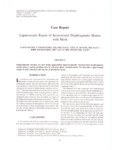
Figure 4: Intra-operative photograph of case 3, depicting a polypropylene mesh stapled to a tear in the diaphragm in an 18 yearold mixed breed mare. Long vertical arrow points to the finochietto rib retractors, short vertical arrow points to the polypropylene mesh and the oblique arrow depicts the skin staples used to attach the mesh to the diaphragm.
to the lateral border of the hernia. The abdominal organs were retrieved and returned in their normal position in the abdominal cavity. The volvulus was manually corrected, which led to an improvement in color and motility of the large colon. The omentum was ligated and resected. A blood gas analysis performed an hour after induction showed respiratory acidosis (with values of 102.9 mm Hg for PaO2, (RI: 100-500 mm Hg) 50 mm Hg for PaCO2 (RI: 30-45 mm Hg) and 7.26 for blood pH (RI: 7.35-7.45), which was resolved by having the mare tilted 30 degrees head up (reverse trendelenburg position) for an hour in order to decrease the pressure exerted on the lungs by the abdominal organs. Attempts to correct the hernia through the abdominal cavity failed. The deep dorsal location and considerable tension on the sutures, due to the large gap in the diaphragm; combined with the friable diaphragmatic muscular tissue; hindered the repair. The abdomen was lavaged and closed in a routine fashion. At that stage the mare was positioned in right lateral recumbency as the left hemithorax was prepared for surgery. An incision was made in the left hemithorax over the 11th rib, approximately 30cm length of rib was removed using a gigli wire saw (Narang Medical Limited, Delhi, India) and the space between the 10th and the 12th rib was expanded using a Finochietto rib spreader. The tear was located and an atelectatic left lung lobe was detected. Suturing proved difficult using this approach. A polypropylene mesh was attached to cover the diaphragmatic defect using surgical skin staples Israel Journal of Veterinary Medicine Vol. 70 (1) March 2015
(Figure 4). At termination of the procedure two 32 French drains (Well Lead Medical, Panyu district, Guangzhou city, China) were inserted, one dorsally at the 13th intercostals space and one ventrally at the 8th intercostal space. Postoperatively the mare received intensive treatment including: intravenous fluid therapy consisting of crystalloids (LRS) and colloids (Hetastarch, Fresenius Kabi AG, Badhomburg, Germany)), fresh frozen plasma, antibiotic therapy (penicillin 22.000 IU/kg q.i.d. IV and gentamicin 6.6 mg/kg s.i.d. IV), analgesics (flunixin 1 mg/kg b.i.d. IV) and anti-endotoxin therapy (polymyxin B 6000 IU/kg b.i.d., IV, X-GEN Pharmaceuticals Inc, Northport, NY). Ice therapy applied on all four extremities to prevent for laminitis. Initially the horse received a nasal oxygen supply (15 liters/min). Post-operative radiographs confirmed good lung inflation and verified that the mesh was intact at its location. The drains were removed the day after surgery following a significant decrease in the amount of blood and air that was drained. Two days after surgery significant edema developed at both incisions but both remained dry. At that stage, oral antimicrobials (enrofloxacin (7.5 mg/kg s.i.d., Phibro Animal Health Corporation, Petah Tikva, Isreal) and metronidazole (25 mg/kg t.i.d., Vetmarket, Shoham, Israel)) and gastric mucosal protectants (omeprazole 4 mg/kg s.i.d., Nature Vet Pty Ltd., Glenorie, NSW, Australia) and ranitidine 6.6 mg/ kg q.i.d.) were added. The mare recovered smoothly and was discharged 12 days after surgery. The mare resumed her career as an endurance horse, with no further respiratory or colic problems in a five-year post-operative follow-up.
CASE 4 Clinical History
A 17-year-old male Quarter horse was referred to the KVSM-VTH with clinical signs of acute colic, which lasted for 12 hours. He was unresponsive to analgesics or sedatives and was referred to the hospital. The owners reported weight loss and occasional breathing difficulties in the last few months. The horse presented to the hospital with a heart rate of 40 beats/minute, a respiratory rate of 30 breaths/minute with the presence of a heart murmur. On rectal examination cecal impaction containing hard fecal material was palpated. The horse was treated with mineral oil (Vetmarket, Shoaham, Diaphragmatic Hernia in Horses
41
Published in IVIS with the permission of the editor
Close window to return to IVIS
Research Articles
Israel) and water through the nasogastric tube and LRS intravenously. Though the horse received potent analgesia several times, pain recurred and emergency celiotomy was recommended.
Case management
Preoperative medication was administered intravenously as follows: benzylpenicillin sodium (20,000 IU/kg), gentamicin (6.6 mg/kg), and flunixin meglumine (1.1 mg/kg). The horse was placed under general anesthesia, positioned in dorsal recumbency and isoflurane anesthesia was used. On exploratory celiotomy a 6 cm x 6 cm right ventral diaphragmatic tear was found. The cecum was incarcerated in the hernia and it was adhered to the pleura in several places. During manipulation of the cecum significant bleeding occurred in the thorax. The hernia was closed by direct suturing, and a drain was inserted into the mid-height, right hemi-thorax at the 12th inter-costal space. Following recovery the horse had respiratory distress and thoracic radiographs revealed large intestine in the thorax, along with an old fracture in the right fourteen rib. The horse was taken to surgery in an attempt to repeat the repair of the diaphragm, however death occurred during induction. DISCUSSION
Diaphragmatic hernia is commonly referred to as a rare lesion in the horse (9), however it is probably more common than previously considered (1). In one recent study, DH made up to 1% of all colic surgeries performed between 1998 and 2005 at a university referral hospital (1). This figure is comparable with the percentage of surgeries for ileocecal intussusception or gastrosplenic entrapment (1.3%, 0.3%, respectively) – two conditions that are not considered rare (10). The incidence of DH reported in the KVSM-VTH in this study (1.3 %) is similar to the results mentioned above. In another retrospective study from a single center, over two cases of DH per year were recorded (6). Thus, according to this and to our experience, DH is quite uncommon but definitely not rare. The four cases making up the current series presented exclusively diaphragmatic ruptures or defects, none of which had a hernia sac. This is in accordance with previous studies, confirming that true DHs, containing a hernia sac, are indeed rare (1). With regard to etiology, it is reasonable to assume that all our cases had a traumatic origin, in two cases, the
42
Efraim, G.
trauma was recent and in the other two, it occurred months prior to the acute presenting episode. In several reported cases, there was a history of trauma months to years prior to an acute episode of colic. It is likely that the damage to the mare’s diaphragm occurred at the time of the trauma/ parturition, however the acute episode was probably triggered by the sudden incarceration of viscera in the thoracic cavity. In case 1 (the 10-day old foal) the respiratory clinical signs were mild but evident since arrival and most likely had existed since parturition. It is likely that during parturition, while passing in the birth canal, the filly fractured her ribs, which tore the diaphragm. This case falls into the mixed category of congenital DH with a traumatic cause. In congenital DH, the defect is located mainly on the left side, owing to the incomplete fusion of the pleuroperiontal folds that is secondary to the slower development of the left lung (1). Acquired DH also seems to be predominantly left-sided, probably because the liver acts as a protection on the right side (1, 2). In contrast to the predominance of left-sided DH in the literature (1, 2, 6), in three of the four cases that make up our series, the lesion was located on the right side of the diaphragm. In three of the presented cases, the DH appeared on the ventral aspect, which is consistent with their traumatic history combined with the evidence of ipsilateral rib fractures. Overall, two cases in this series are presumed to be related to parturition associated trauma (cases # 1 and #3), while two were likely due to direct external thoracic trauma (cases # 2 and #4). The diagnosis of DH is typically difficult to make even during pre-operative evaluation. The clinical manifestations of DH typically include acute abdominal signs or colic; colic and dyspneic signs may be consecutive (11) or simultaneous, and only seldom do respiratory signs occur alone. Our first case presented with exclusive respiratory signs for several days prior to a fatal abdominal crisis episode. The respiratory signs can be attributed to the defect in the diaphragm leading to a loss of negative pressure in the thoracic cavity resulting in atelectatic lungs. This may be supported by a report describing a mare suffering from a diaphragmatic rent without any herniation and showing respiratory signs only (12). The fatal episode could be attributed to the migration of viscera into the thoracic cavity. It is interesting that case 4 had history of respiratory problems, demonstrated as episodes of dyspnea, months prior to the abdominal crises that led to the referral. Cases #2, #3 in our series did show signs of abdominal Israel Journal of Veterinary Medicine Vol. 70 (1) March 2015
Published in IVIS with the permission of the editor
Close window to return to IVIS Research Articles
pain, involving respiratory distress as well, however, at least initially, respiratory distress was interpreted as part of the abdominal crisis. This is consistent with the reported concern that respiratory signs in foals with DH, are often masked by the acute abdominal crises and attributed to abdominal pain and stress (11). Most commonly, DH is diagnosed either at abdominal exploration or at necropsy. Radiographs and ultrasonography are considered the most useful diagnostic aids for attaining ante-mortem or pre-operative diagnosis of DH (1). In the current series, ultrasonographic evaluation was useful though not definitive in the diagnosis of DH in one horse; in two cases DH was diagnosed by abdominal exploration, and in one case only at necropsy. Ultrasonographic evaluation can nevertheless easily lead to false positive diagnosis due to the bell-shaped diaphragm and the cranial migration of the small intestine. In our experience, when used judiciously, both ultrasound and radiograph can be helpful in the diagnosis of DH. An effort should be made to incorporate these modalities in the workup of any colic case where clinical signs are indicative of respiratory compromise, or where the history or initial evaluation may lead the clinician to suspect DH (1, 12). In case #1 in this series clinical signs were exclusively of a respiratory nature, radiographs however revealed no DH. The difficulty in sometimes detecting DH should be borne in mind and one should not rely on any single negative diagnostic test to rule out diagnosis of DH. The only definitive treatment for DH is surgical repair. The prognosis in general is not good and it largely depends on early diagnosis, on the location and length of the tear, and on the degree of intestinal damage (2, 6, 8). Romero and Rodgerson (2) reported that out of 25 horses which were operated on, only 6 survived more than three months following surgery, while 11 were euthanized during surgery. Similarly, 7 out of 44 survived to discharge in a recent retrospective study by Hart and Brown (6). Nevertheless, long term results were positive in the same study with 71 % of horses discharged surviving for over a year. Recently a DH diagnosed during abdominal exploration was successfully repaired, three weeks later, in the standing horse, using thoracoscopy (13). Although the thoracic approach to DH repair is described in the literature, the conventional approach is typically abdominal. Hart and Brown (6) described repairing nine DHs via an abdominal incision, and Romero and Rodgerson (2) reported repairing DH in 14 horses using the same approach. Israel Journal of Veterinary Medicine Vol. 70 (1) March 2015
In that study, five of the horses that underwent surgery had DH corrected by means of a polypropylene mesh applied by direct suturing or using a hand stapling device. Four of these horses did not survive to discharge. This may indicate low chances of success in correcting DH using a mesh through an abdominal approach. Case #4 in our series was treated by direct suturing through the abdomen and failed immediately after surgery. This defect was neither large nor dorsal but still failed. It may further support the inferiority of the abdominal approach for repairing DH. The thoracoscopy technique used by Röcken et al. (13) agrees with our elected thoracic approach, although the techniques used and the type of lesion was different. In that report, an endoscope was used, while in case #3; in the current report; an open approach was used performed by rib resection. Furthermore, in that study the defect was corrected using interrupted sutures; conversely, in our case the defect was corrected with mesh and staples. All the above supports our clinical impression that approaching the DH through the thorax is preferable. Thoracoscopy may prove the method of choice for repairing DH in the future since it provides the best access to the lesion and is minimally invasive. By and large the surgical success rate for equine DH repair remains low. In Romero and Rodgerson’s report (2), 25 horses with DH underwent surgery, but only eight survived to discharge. Similar results were reported by Hart and Brown (6), in a population of 26 horses that underwent surgery, seven were discharged from hospital, and two of them died within the first year. The same study showed that horses with dorsally located rents, which were 10 cm or more in diameter, had the worst prognosis with 92% of them being euthanized; while rents located ventrally and less than 10 cm in diameter were associated with a far better success rate (63%). In the current study, however the only survivor had a large and dorsal tear. No conclusions, with regards to the effect of lesion location and size on prognosis, can be drawn from this single case. Nevertheless, thoracic approach may enable repair of tears that are irreparable by the traditional abdominal approach and thus improve the overall survival rate. In this study, 25% of the horses with DH survived, which is a similar survival rate to other reports (2). Two cases were related to parturition, one in the dam and the other in the neonate emphasizing that Mares and foals should be monitored for clinical signs related to DH. The thoracic approach proved Diaphragmatic Hernia in Horses
43
Published in IVIS with the permission of the editor
Close window to return to IVIS
Research Articles
useful in the current report as well as in a recent study. In conclusion, earlier diagnosis and improved surgical techniques may improve the survival rate of horses suffering from DH. ACKNOWLED GEMENTS The authors are grateful to the referring veterinarians and the dedicated hospital staff without whom these difficult cases could not have been managed.
REFERENCES
1. Kelmer, G., Kramer, J. and Wilson, D. A.: Diaphragmatic hernia: etiology, clinical presentation, and diagnosis. Compend. Equine. 3:28-36, 2008. 2. Romero, A. E. and Rodgerson, D. H.: Diaphragmatic herniation in the horse: 31 cases from 2001-2006. Can. Vet. J. 51:1247-1250, 2010. 3. Barker, I. K.: The peritoeneum and retroperitoenum. In: Jubb, K. V. F, Kennedy, P. C. and Palmar, N. (Eds.): Pathophysiology of domestic animals. 4th ed. Academic Press, San Diego, pp. 425428, 1993. 4. Pauwels, F. F., Hawkins, J. F., McHarg, M.A., Rothenbuhler, R. D., Baird, D. K. and Moulton, J. S.: Congenital retrosternal (Mor-
44
Efraim, G.
gagni) diaphragmatic hernias in three horses. J. Am. Vet. Med. Assoc. 231:427-432, 2007 5. Schambourg, M. A., Laverty, S., Mullim, S., Fogarty, U.M. and Halley, J.: Thoracic trauma in foals: post mortem findings. Equine Vet. J. 35:78-81, 2003. 6. Hart, S. K. and Brown, J. A.: Diaphragmatic hernia in horses: 44 cases (1986-2006). J. Vet. Emerg. Crit. Care. 19:357-362, 2009. 7. Speirs, V. C. and Reynolds., W. T.: Successful repair of a diaphragmatic hernia in a foal. Equine Vet. J. 8:170-172, 1976. 8. Kelmer, G., Kramer, J. and Wilson, D. A.: Diaphragmatic hernia: treatment, complications, and prognosis. Compend. Equine 3:3746, 2008. 9. Collier D.S.: Comparative aspects of diaphragmatic hernia. Equine Vet. Edu. 31:358-359, 1999. 10. Mair T.S., Smith L.J.: Survival and complication rates in 300 horses undergoing surgical treatment of colic. Part 1: Short-term survival following a single laparotomy. Equine Vet. J. 37:296-302, 2005. 11. Tapio, H., Hewetson, M. and Sihvo, H. K.: An unusual cause of colic in a neonatal foal. Equine Vet. Edu. 24:334-339, 2012. 12. Goehring, L. S., Goodrich, L. R.and Murray, M.J.: Tachypnoea associated with a diaphragmatic tear in a horse. Equine Vet. J. 31:443-445, 1999. 13. Röcken, M., Mosel, G., Barske, K. and Witte, T. S.: Thoracoscopic diaphragmatic hernia repair in a warmblood mare. Vet. Surg. 42:591-594, 2013.
Israel Journal of Veterinary Medicine Vol. 70 (1) March 2015