Determination of water quality parameters in Indian ponds using remote sensing methods
Diploma thesis of Christian Gemperli
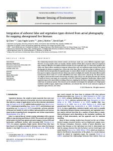
Department of Geography, University of Zurich Supervised by Prof. Dr. K.I. Itten Assisted by Dr. M. Kneubuehler (RSL) & Dr. R. Zah (EMPA) Zurich, 01.06.2004
ACKNOWLEDGMENTS
Many persons contributed to the success of this thesis. I would like to thank everybody who supported me during this time. Special acknowledgement deserve • Prof. Dr. K. I. Itten for his interest and the perusal of my manuscript, • Mathias Kneubuehler and Rainer Zah for the support and advices, • M. Schaepman for the interest and support in the initial phase, • the West Bengal Jute Project for the financial and logistical support, • Dominik Noger for the advices concerning sampling procedure and measurements, •
Daniel Schlaepfer for the valuable advices regarding data processing and statistics,
• the staff of Bidhan Chandra Krishi Viswavidyalaya University, Mohanpur, for the cooperation & infrastructure. Special thank goes to Prof. Dr. Hussein & Dr. Saha, • the Spektrolab Team for the patience to answer my questions, • Fieldwork Team for the cooperation & good time in India, • Mr. Uehlinger (EAWAG) for the contribution to the technical infrastructure, • Angela for the corrections, • Sibylle for the patience, • and finally my parents who enabled and supported my studies.
Zurich, May 2004
Christian Gemperli
ABSTRACT
In this thesis, the potential of existing algorithms and space borne imaging spectrometers for monitoring water quality were evaluated. In addition, the general state and the trophic conditions of the studied water bodies were assessed. The concept of eutrophication was used to describe water quality, using chlorophyll as an indicator for the presence of algae in the water. Among the several concepts of eutrophication, the Trophic State Index of Carlson was chosen to define the trophic state of the water bodies. Five small domestic ponds and one bayou of the Ganga river near the town of Kalyani, State of West Bengal, India, were selected as test-sites. During a field campaign in May 2003, reflectance spectra were measured 1m above the water surface using a handheld spectroradiometer. Concentration of TCHL and other limnological standard parameters such as Secchi Disk Depth, Nutrient content TOC and DOC were determined using laboratory analysis and a colorimetric testkit. A comparison of several existing semi-empirical algorithms to determine chlorophyll content was made by applying them on the spectra and chlorophyll measurements collected in-situ. Simple reflectance band ratio and Continuum Interpolated Band Ratio (CIBR) were tested at several wavelengths. Three tested algorithms rely on the reflectance peak at 700 nm whose shape and position depend strongly upon chlorophyll concentration. The area and magnitude of the peak above a baseline as well as the position of the peak show a linear relationship to chlorophyll-a concentration from laboratory spectrophotometric measurements. The algorithms were further applied onto spectra convolved to the bands of the hyperspectral sensors APEX, HyMap and Hyperion. A comparison of the spectral characteristics of the chosen sensors was made. The domestic ponds showed extremely high chlorophyll concentrations around 300 µg/l and very low Secchi Disk Depths around 0.1m indicating a hypertrophic situation. On the other hand, the nutrient concentrations were surprisingly low. The bajou showed almost no chlorophyll concentrations, as well as low nutrient concentrations and higher Secchi Disk Depths. All algorithms proved to be of value. Best results were obtained by using the algorithms Magnitude above a Baseline and CIBR, with yielded R 2 around 0.97 and mean deviations around 20 µg/ l. Given the large range of CHL concentrations, these results can be seen as very satisfactorily. However, certain presumptions had to be made and there still remain limitations concerning the used input data. Therefore, the results have to be treated with a certain caution. The differences of the outcome of the sensors were small, but the results indicate that an intermediate band width of around 10 nm seems appropriate. All sensors showed an adequate band positioning for the algorithms based on the peak near 700 nm. Using simple band ratios and CIBR, APEX and Hyperion perform better than HyMap.
ZUSAMMENFASSUNG
In dieser Diplomarbeit wurde das Potential von existierenden Algorithmen und Sensoren zum Monitoring von Wasserqualität evaluiert. Das Konzept der Eutrophierung wurde herangezogen, um die Qualität des Wassers zu beschreiben, wobei Chlorophyll als Indikator für den Algenwuchs diente. Mittels dem Trophic State Index (TSI) von Carlson wurde der trophische Status der Teiche bestimmt. Das Untersuchungsgebiet befindet sich in der Nähe der Kleinstadt Kalyani in West Bengalen, Indien. Während der Feldarbeit im Mai 2003 wurden in fünf kleinen Teichen anthropogenen Ursprungs und einem Altarm des heiligen Flusses Ganges Messungen vorgenommen. Die spektrale Reflektanz des Wassers wurde 1 m über der Wasseroberfläche mittels einem tragbaren Spektroradiometer gemessen. Die Konzentration von Chlorophyll und anderen limnologischen Standardparametern wie Nährstoffgehalt, TOC und DOC, wurden mittels Laboranalysen und einem kolorimetrischen Schnelltestkit bestimmt. Zusätzlich wurden Tiefe, Secchi Disk Tiefe und Temperatur gemessen. Verschiedene semi-empirischen Algorithmen zur Bestimmung des Chlorophyllgehalts wurden miteinander verglichen, indem sie auf die in-situ gesammelten Spektraldaten und Chlorophyllmessungen angewendet wurden. Einfache Band Ratio und Continuum Interpolated Band Ratio (CIBR) wurden bei verschiedenen Wellenlängen getestet. Drei weitere angewendete Algorithmen basieren auf dem Reflektanzmaximum bei 700 nm, dessen Form und Position stark von der Chlorophyllkonzentration abhängt. Die Fläche und die Magnitude des Peaks über einer Normalisierungslinie, wie auch die Position des Peaks auf der Wellenlängen-Achse, zeigen eine lineare Beziehung zu dem im Labor bestimmten Chlorophyllgehalt. Weiter wurden die Algorithmen auf Spektren angewendet, die auf die Bänder von drei ausgewählten hyperspektralen Sensoren (Hyperion, HyMap und APEX) gefaltet wurden. Dadurch konnten die ausgewählten Sensoren miteinander verglichen werden. Die kleinen, anthropogen entstandenen Teiche zeigten extrem hohe Chlorophyllgehalte um 300 µg/l und Secchi Disk Tiefen von weniger als 0.1m, was auf eine hypertrophische Situation hindeutete. Dennoch waren aber die gemessenen Nährstoffkonzentrationen überraschenderweise relativ klein. Im Altarm des Ganges konnte kein Chlorophyll nachgewiesen werden. Die Nährstoffkonzentrationen waren auch hier sehr klein, die Secchi Disk Tiefe grösser. Alle Algorithmen bewährten sich. Die besten Resultate wurden mit der Magnitude des 700 nm Peak und CIBR erzielt, mit R 2 von 0.97 und mittleren Abweichungen von rund 20 µg/l. In Hinsicht auf den sehr grossen Range von Chlorophyllgehalten innerhalb des Datensatzes, kann dieses Resultat als sehr befriedigend bezeichnet werden. Es mussten jedoch Annahmen getroffen werden und weiterhin bestehen einige Unsicherheiten bezüglich der verwendeten Datensätze. Deshalb sind die erzielten Resultate mit gewisser Vorsicht zu geniessen.
Die Unterschiede zwischen den einzelnen Sensoren waren sehr gering, die Resultate deuten jedoch an, dass für die verwendeten Algorithmen eine Bandbreite von etwa 10 nm ideal ist. Die Bänder aller Sensoren sind für die auf dem 700 nm Peak basierenden Algorithmen geeignet. Bei der Anwendung von einfachen Band Ratios und CIBR zeigten APEX und Hyperion bessere Resultate als HyMap.
TABLE OF CONTENTS
TABLE OF CONTENTS .........................................................................................1 LIST OF FIGURES .................................................................................................. 4 LIST OF TABLES .................................................................................................... 5 ABBREVIATIONS................................................................................................... 6
1. INTRODUCTION ......................................................................................... 7 1.1 Background ..................................................................................................................................... 7 1.2 Scope & aims ................................................................................................................................... 7 1.3 Outline.............................................................................................................................................. 8
2. THEORY ....................................................................................................... 9 2.1 Water quality................................................................................................................................... 9 2.1.1 Overview.................................................................................................................................................... 9 2.1.2 Eutrophication .......................................................................................................................................... 9 2.1.2.1 Trophic State Index (TSI) by Carlson [5] ................................................................................................................ 10
2.2 Water Optics.................................................................................................................................. 11 2.2.1 Inherent optical properties .................................................................................................................... 11 2.2.2 Apparent optical properties .................................................................................................................. 12 2.2.3 Interaction between Inherent and Apparent Optical Properties....................................................... 13 2.2.4 Optically active substances .................................................................................................................... 13 2.2.4.1 Water ........................................................................................................................................................................ 14 2.2.4.2 Yellow substances (Gelbstoff) ................................................................................................................................. 14 2.2.4.3 Seston ....................................................................................................................................................................... 14
2.2.5 Influence of the constituents onto the reflectance spectrum .............................................................. 15
2.3 Remote sensing of water quality parameters ............................................................................. 16 2.3.1 Approaches.............................................................................................................................................. 16 2.3.2 Sensors..................................................................................................................................................... 17 2.3.2.1 Selection of different sensors for convolution of spectral bands ............................................................................. 18
3. DATA ACQUISITION ............................................................................... 21 3.1 Study area ...................................................................................................................................... 21 3.2 Selection of water bodies .............................................................................................................. 22 3.3 Data ................................................................................................................................................ 23 3.3.1 Spectral data ........................................................................................................................................... 23 3.3.2 Water constituents data ......................................................................................................................... 24 3.3.2.1 Sampling scheme...................................................................................................................................................... 25 3.3.2.2 Determination methods ............................................................................................................................................ 25 3.3.2.3 Quality remarks concerning chlorophyll in-situ data............................................................................................... 26
4. SPECTRAL DATA PROCESSING & ANALYSIS ................................. 27 4.1 Pre-processing ............................................................................................................................... 27 4.1.1 Correction of sunglint effects ................................................................................................................ 28 4.1.2 Convolution............................................................................................................................................. 29
1
4.2 Description of the reflectance curve............................................................................................ 29
5. LIMNOLOGICAL ASSESSMENT........................................................... 31 5.1 General description of the water bodies ..................................................................................... 31 5.2 Analysis of retrieved limnological data....................................................................................... 32 5.2.1 Chlorophyll and assumed algae composition....................................................................................... 32 5.2.2 Nutrient content...................................................................................................................................... 34 5.2.3 Organic Carbon...................................................................................................................................... 34 5.2.4 Secchi disk depth .................................................................................................................................... 35 5.2.5 Other limnological parameters ............................................................................................................. 35 5.2.5.1 pH ............................................................................................................................................................................. 35 5.2.5.2 Total hardness .......................................................................................................................................................... 35 5.2.5.3 Heavy metals ............................................................................................................................................................ 35
5.3 Trophic state.................................................................................................................................. 36 5.4 Conclusions.................................................................................................................................... 37
6. METHODOLOGY OF PARAMETER RETRIEVAL ............................ 38 6.1 Methods used in this study........................................................................................................... 38 6.1.1 Selection of appropriate methods ......................................................................................................... 38 6.1.2 Chlorophyll determination algorithms................................................................................................. 38 6.1.2.1 6.1.2.2 6.1.2.3 6.1.2.4 6.1.2.5
Band ratio ................................................................................................................................................................. 38 Continuum Interpolated Band ratio (CIBR)............................................................................................................. 39 Area above baseline ................................................................................................................................................. 40 Peak Magnitude above baseline ............................................................................................................................... 41 Position of the Peak near 700 nm............................................................................................................................. 42
6.1.3 Statistical methods.................................................................................................................................. 43 6.1.3.1 Regression Analysis ................................................................................................................................................. 43
7. RESULTS OF PARAMETER DETERMINATION ............................... 45 7.1 Setup............................................................................................................................................... 45 7.2 Modelling using Fieldspec FR PRO bands ................................................................................. 46 7.2.1 Simple band ratios.................................................................................................................................. 46 7.2.2 CIBR ........................................................................................................................................................ 47 7.2.3 Area above base line............................................................................................................................... 48 7.2.4 Peak Magnitude above a baseline ......................................................................................................... 49 7.2.5 Position of peak near 700 nm ................................................................................................................ 49
7.3 Modeling using convolved spectra............................................................................................... 50
8. DISCUSSION............................................................................................... 52 8.1 Interpretation of results of spectral modelling........................................................................... 52 8.2 Quality assessment and source of errors .................................................................................... 54
9. CONCLUSIONS.......................................................................................... 55 9.1 Future Improvements................................................................................................................... 55 9.1.1 Sampling Procedure ............................................................................................................................... 55 9.1.2 Data Processing....................................................................................................................................... 55
9.2 Defiances for a future monitoring system................................................................................... 56 9.3 Outlook........................................................................................................................................... 56
2
REFERENCES............................................................................................ 58 APPENDIX ................................................................................................... 62 A. Additional Tables and Figures..................................................................................................... 62 Limnological classification schemes ............................................................................................................... 62 Sampling scheme and parameter determination........................................................................................... 63 Results of limnological measurements............................................................................................................ 64
B. Used methods for parameter extraction ..................................................................................... 65 B.I
Chlorophyll determination in water samples by Annon`s method.................................................... 65
B.II Dissolved organic carbon by dichromate oxidation method of Vance .............................................. 66 B.III Total organic carbon by Nelson Somers method................................................................................. 67
C. Description of additional limnological parameters.................................................................... 68 D. Field sheets..................................................................................................................................... 71
3
LIST OF FIGURES
Fig. 2.1:
Absorption coefficients of active constituents. ................................................. 14
Fig. 2.2:
Scattering coefficients of active constituents.................................................... 14
Fig. 2.3:
Absorbance peak of phycocyanin on spectrum of P2. ...................................... 16
Fig. 2.4:
Band positioning of Landsat ETM 7 on ASD spectroradiometer spectrum ..... 18
Fig. 3.1:
Map of West Bengal District ............................................................................ 21
Fig. 3.2:
Landsat ETM panchromatic image 22.04.2002 ................................................ 22
Fig. 3.3:
Selected water bodies and covered transects .................................................... 23
Fig. 4.1:
Averaged spectra of each water body ............................................................... 27
Fig. 4.2:
Effect of sunglint on reflectance spectra........................................................... 28
Fig. 4.3:
FWHM defined as spectral resolution .............................................................. 29
Fig. 5.1:
From left to right: P1, P2, P5 and P6 ................................................................ 31
Fig. 5.2:
Dominant phytoplankton in Indian reservoirs .................................................. 33
Fig. 6.1:
Band ratio algorithm ......................................................................................... 39
Fig. 6.2:
Continuum Interpolated Band ratio (CIBR)...................................................... 40
Fig. 6.3:
Base line algorithm ........................................................................................... 41
Fig. 6.4:
Peak Magnitude above Baseline algorithm by Gitelson ................................... 42
Fig. 6.5:
Peak Position algorithm by Gitelson................................................................. 42
Fig. 7.1:
Linear (left) and logarithmic (right) curve fit between CHL in-situ data and the normalized spectra ............................................................................................ 46
Fig. 7.2:
Linear curve fit for CIBR [651/675/713] (left) and its band positions on the reflectance spectra............................................................................................. 48
Fig. 7.3:
The range of the position of the peaks on the wavelength axis ........................ 49
Fig. 7.4:
Spectra convolved to the bands of the chosen sensors...................................... 51
Fig. 8.1:
Necessary bands for used algorithms on band positions of chosen sensors ..... 53
4
LIST OF TABLES
Tab. 2.1:
Comparison of TSI to Water Quality Parameters and Lake Productivity......... 11
Tab. 2.2:
Characteristics of chosen sensors...................................................................... 19
Tab. 3.1:
Recorded spectra ............................................................................................... 24
Tab. 3.2:
Determined limnological parameters ................................................................ 24
Tab. 3.3:
TCHL values for data set1 & 2 ......................................................................... 26
Tab. 5.1:
Characteristics of the selected ponds ................................................................ 32
Tab. 5.2:
Correlation between several limnological parameters ...................................... 35
Tab. 5.3:
Calculated TSI values ....................................................................................... 36
Tab. 7.1:
Results of ratios: linear fit; raw data ................................................................. 46
Tab. 7.2:
Results for ratios: linear fit; normalized at 900nm ........................................... 46
Tab. 7.3:
Results for ratios: logarithmic fit. ..................................................................... 47
Tab. 7.4:
Statistical coefficients for CIBR algorithm applied on Fieldspec FR PRO ...... 47
Tab. 7.5:
Results for area above baseline algorithm ........................................................ 48
Tab. 7.6:
Statistical coefficients for peak magnitude above a baseline............................ 49
Tab. 7.7:
Statistical coefficients for peak position ........................................................... 49
Tab. 7.8:
Results of simple band ratio; logarithmic fit..................................................... 50
Tab. 7.9:
Results of CIBR; linear fit ................................................................................ 50
Tab. 7.10:
Results of area a. baseline for convolved spectra ............................................. 50
Tab. 7.11:
Results of magnitude a. baseline for convolved spectra ................................... 50
Tab. 8.1:
Position of needed bands and availability of the sensors.................................. 52
Tab. A.1:
Classification scheme according to frequency of overturn by WHO ............... 62
Tab. A.2:
Characteristics of trophic states according Carlson. ......................................... 62
Tab. A.3:
Determined parameters for each sampling location.......................................... 63
Tab. A.4:
Measuring range and estimated error of colorimetric method (Aquanal)......... 63
Tab. A.5:
Results of limnological parameters determined in India .................................. 64
Tab. A.6:
Results of ICP-OES determination at EMPA ................................................... 64
5
ABBREVIATIONS
Spectrometric abbreviations a b c:
absorption coefficient [ m –1 ] scattering coefficient [ m –1 ] beam attenuation coefficient [ m –1 ]
RR : R ( –0 ) LW ( 0 ) ED( 0 )
Remote Sensing reflectance [%] Sub-surface irradiance reflectance [%] Water-leaving radiance [ Wm –2 nm –1 sr–1 ] Downwelling irradiance [ Wm –2 nm–1 ]
nm wvl
nanometer wavelength
Statistical abbreviations MD ℵ MD a R2
Std error
per cent Mean Deviation absolute Mean Deviation Correlation Coefficient Standard error
Limnological abbreviations SDD CHL-a TCHL TOC DOC ZN AS Th Integ. SDD ICP-OES
Secchi Disk Depth Chlorophyll a Total chlorophyll Total Organic Carbon Dissolved Organic Carbon Zinc Arsenic Total hardness Mixed sample from the water column between 0 m and SDD. Inductively Coupled Plasma Optical Emission Spectroscopy
n/d bdl
not determined below detection limit
Units µg/L mg/L
micrograms per Liter or ppb (parts per billion). milligrams per Liter or ppm (parts per million).
6
Chapter 1
INTRODUCTION
1 INTRODUCTION
1.1 Background The West Bengal Jute Project lead by IBF AG, Sustena IG and EMPA deals with the sustainable management of fiber crops in West-Bengal over the whole value chain. In jute production, retting is a very important step to the final quality of the fibre. After harvesting, the crops are immersed in rivers, canals, ponds and ditches and left there for about 2 to 3 weeks before the fibres can be extracted from the plant. An important condition for good retting is the quality of the used water, meaning, the water should be clear and oxygen-rich. Therefore, retting in rivers is preferred to retting in ponds and other water bodies without outlet. At the same time, retting influences the water quality. It can be assumed that the input of large amounts of organic material and nutrients affects the water quality at least temporarily. Classical techniques for determining indicators of water quality involve in-situ measurements and laboratory analysis. Although these approaches give accurate results, they are time-consuming, expensive and do not give a spatial view on the needed variables as they represent point-in-time measurements. Remote sensing of water quality parameters has the potential to determine these parameters at relatively low costs on a frequent basis offering a spatial view on the parameters. These advantages are especially interesting in remote areas of little or restricted access and with few analytical resources such as laboratories and trained staff. To get an overview of the current state of the various ponds within the wide and sometimes difficultly accessible terrain of West Bengal, India, remote sensing techniques seem to be appropriate due to the reasons mentioned above.
1.2 Scope & aims The scope of this work is to investigate the potential of algorithms in monitoring water quality parameters in West Bengal ponds. The concept of eutrophication was chosen to describe water quality, using chlorophyll as indicator for the presence of algae in the water. A semi-empirical approach was used to determine the parameter. The algorithms chosen from the literature had to perform well in other studies on water bodies with comparable limnological characteristics. The aims of the study are as follows • to asses the trophic state of the ponds, • to evaluate semi-empirical algorithms for determining chlorophyll-a content using spectral reflectance data collected with a handheld field-spectroradiometer, • to apply these algorithms to the spectra convolved to the bands of selected sensors, • and to evaluate the most suitable sensors and applied algorithms for determining chlorophylla content in the study area.
7
INTRODUCTION
Chapter 1
1.3 Outline Chapter 2 deals with the theoretical background of the field of study. It gives a description of the concept of eutrophication and of the optical properties of water and its constituents, such as phytoplankton, total suspended solids and colored dissolved organic matter. An overview of the approaches used for the determination of these constituents and the resulting advantages and disadvantages is presented as well as a short description of the technical characteristics of the sensors, of which the potential was investigated. Chapter 3 gives a detailed description of the campaign, which was carried out to gather spectral and limnolocigal data of the ponds. It describes the study area, where the field measurements took place, and it shows the schemes which were used in the sampling process and the methods to extract the desired parameters. A list of all gathered data and quality remarks is presented. Chapter 4 describes the process of data pre-processing and explains briefly the concept of convolution of spectral bands. The spectral reflectance curves of the water bodies are analyzed and discussed briefly. In Chapter 5 the results of the limnological assessment are discussed. A description of the limnological characteristics of the ponds are given and conclusions about its trophic state are drawn. Chapter 6 deals with the methods used for the retrieval of chlorophyll-a of the reflectance spectra and the statistical parameters having used to quantify the results. In Chapter 7, the results of the different methods for the determination of the parameters are presented for (1) the spectra measured with the handheld spectroradiometer, and (2) for the mentioned spectra which were convoluted to the spectral bands of the sensors described in Chapter 2. Chapter 8 discusses the results presented in Chapter 7. It shows the limitations and quality aspects of the data, which were used in the modeling process and gives explanations for the results. In Chapter 9, improvements for the future are proposed and general defiances for a water quality monitoring system based on Remote Sensing in West Bengal are considered.
8
Chapter 2
THEORY
2 THEORY
2.1 Water quality 2.1.1 Overview There is no single definition of water quality, as it depends on the respective processes and on the intended use of the water. In this work the focus is laid on the process of eutrophication. Trophic state is an absolute scale that describes the biological condition of a water body and does not imply a normative statement per se. It has to be stressed that trophic state is not the same thing as water quality but trophic state certainly is one aspect of water quality. To give a general impression of the state of the ponds, some additional limnological standard variables determined during the campaign will be discussed in Chapter 4.
2.1.2 Eutrophication The term ‘eutrophication’ means nutrient enrichment of lakes and describes the processes in water caused through over-enrichment by nutrients. Too much nutrient input causes a chain of events that may have undesirable effects on lakes, e.g. growth of algae and macrophytes. The enhanced rates of decomposition and attendant consumption of oxygen lead finally to oxygen depletion in the hypolimnion, decreasing the number of species. Eutrophication can occur under natural conditions, but often the process is induced by anthropological factors, such as fertilizers and sewage [5]. Organic matter introduced to surface waters (e.g. jute), leads to enhanced rates of decomposition and attendant consumption of oxygen. Numerous definitions of trophic state exist in the literature and there have been several attempts to classify lakes according to their trophic condition. They vary in the indices they use to assess the trophic state and in the classification schemes they use to separate lakes in different classes. Generally, one can distinguish between classification approaches which are based on productivity and biomass (e.g. chlorophyll-a concentration) or definitions based on the factors determining this production (e.g. phosphorus loadings). Other approaches focus on the effects that are caused by the process of eutrophication (e.g. hypolimnetic oxygen depletion, Secchi Disk Depth). Some contemporary classification schemes use only a single variable to define the trophic state of lakes. This use of a single variable simplifies the classification procedure considerably because only one variable has to be measured. At the same time, this approach reduces the concept of eutrophication, which rather describes a process, than a state or class, significantly. The early approach of Naumann (1919) divided the lakes into several classes based on biomass production, e.g oligotrophic lakes were low in production, eutrophic lakes were very productive. He then related the classes to several indicators, such as humic content, nitrogen and phosphorus, iron, pH, oxygen, and carbon dioxide [45].
9
THEORY
Chapter 2
Hutchinson (1969) and Odum (1969) emphasized the importance of the watershed to define the trophic state by the loading of nutrients to the lake. In this context, trophic state was a description of the potential for a lake to respond to nutrient loading rather than a description of the response. A eutrophic system would be a system in which the total potential concentration of nutrients was high, whether or not there was a correspondingly high algal or macrophyte density [5]. The OECD index of Vollenweider and Kerekes (1980) is a classification system based on the probability that a lake or reservoir will have a given trophic state. The index was generated by asking a group of scientists their opinion as to what was the average value of the indicator (e.g. Total phosphorus) for each trophic class. Therefore, lakes of the same concentrations may be in more than one trophic class. The assumed advantage of the probabilistic approach is that responses to a given variable will vary from lake to lake and therefore prediction of its trophic state is best stated in probabilistic terms [45]. Carlson (1977) suggested returning to the initial principles of trophic state: a quantifiable concept based on biomass. He used Naumanns (1929) original idea of classification according to plant biomass. But instead of the distinct typological classes, Carlson assumed algal biomass to be from a continuous range of values. This approach will be used in this work as it is relatively simple to use and only requires a minimum of data [51]. 2.1.2.1 Trophic State Index (TSI) by Carlson [5] According Carlson, trophic state is defined as the total weight of living biological material (biomass) in a water body at a specific location and time. It is estimated independently by three variables: chlorophyll-a pigments, Secchi depth, and total phosphorus (TP). For each variable the Trophic State Index (TSI) is calculated by a separate equation. The advantage of this index is that a similar TSI value can be obtained from each of the three variables. Although these three variables should co-vary, they should not be averaged, because neither transparency nor TP are independent estimators of trophic state. For the purpose of classification, priority is given to chlorophyll because this variable is the most accurate of the three at predicting algal biomass. Secchi depth and TP should be used as a surrogate and not as a co-variate of chlorophyll. The basic Secchi Disk index (Equation 2.1) was constructed from a doubling and halving of Secchi disk transparency, where the base index value is a Secchi depth of 1 m, the logarithm of which is 0 (Tab. 2.1). ln SD TSI ( SD ) = 10 6 – ------------- , ln 2
(2.1)
The index uses relationships between trophic variables to produce equations that allow the index to be calculated from variables other than Secchi depth. The indices for the chlorophyll and TP Equation 2.2 are obtained in a similar manner, but instead of a Secchi depth value in the numerator, the empirical relationship between chlorophyll or TP and Secchi depth is given. 2.04 – 0.68 ln CHL TSI ( CHL ) = 10 6 – --------------------------------------------- , ln 2
(2.2)
10
Chapter 2
THEORY
48 ln ------TP- , TSI ( TP ) = 10 6 – -----------ln 2
(2.3)
where TP and CHL-a are expressed in µg/l. SD is expressed in meters. TSI is unitless.
Trophic state
TSI Secchi Disk (m)
Oligotrophic
Mesotrophic Eutrophic
Total Phosphorus (µg/L)
Chlorophyll-a (µg/L)
0
64
0.75
0.04
10
32
1.50
0.12
20
16
3
0.34
30
8
6
0.94
40
4
12
2.60
50
2
24
6.40
60
1
48
20
70
0.5
96
56
80
0.25
192
154
90
0.12
384
427
100
0.06
768
1,183
Hypereutrophic
Tab. 2.1: Comparison of Trophic State Index to Water Quality Parameters and Lake Productivity [5].
The range of the index (Tab. 2.1) is from approximately zero to 100, although the index theoretically has no lower or upper bounds. Unlike Naumann’s typological classification of trophic state, the index reflects a continuum of states and there are no lake “types”.
2.2 Water Optics The optical properties of water are often divided into the Inherent Optical Properties (IOP) and the Apparent Optical Properties (AOP), which are defined by the first mentioned.
2.2.1 Inherent optical properties The Inherent Optical Properties are the properties of the medium itself (i.e., water plus constituents). They depend only upon the medium. Thus, regardless of the ambient light field the IOP are measured by active optical instruments. The main active constituents which will be described in (Chapter 2.2.4) are aquatic humus, phytoplankton and tripton. There are two main optical processes, absorption and elastic scattering, quantified by the absorption coefficient a and the volume-scattering function β, respectively. Other inherent optical properties, which can be obtained from the absorption coefficient and the volume-scattering function, are the scattering coefficient b and the beam attenuation coefficient c [8]. The absorption coefficient is defined as the sum of the individual absorption coefficients of these water constituents (Beers law). Assuming a linear relationship between absorption and concentration, it is given as
11
THEORY
Chapter 2
a ( total ) = C ( w ) × a ( w ) + C ( ah ) × a ( ah ) + C ( ph ) × a ( ph ) + C ( t ) × a ( t ) ,
(2.1)
where a ( w ) is the absorption by pure water, a ( ah ) by aquatic humus, a ( ph ) by phytoplankton, a ( t ) by tripton and C the corresponding concentration of the constituents. As scattering is mainly caused by water, phytoplankton and tripton the scattering coefficient b can be written as b ( total ) = C ( w ) × b ( w ) + C ( ph ) × b ( ph ) + C ( t ) × b ( t ) ,
(2.2)
The beam attenuation coefficient, representing the total loss of light due to absorption and scattering [7], is defined as c = a +b.
(2.3)
2.2.2 Apparent optical properties Apparent optical properties depend both on the medium and on the ambient light field. Therefore, in contrast to the IOP, they can only be determined in-situ. Commonly used apparent optical properties are the radiance, irradiance, reflectance and the vertical-attenuation coefficient. Dekker [7] and Frauenhofer [8] give a detailed overview. In remote sensing of water quality, the most important reflectance definitions are the sub-surface irradiance reflectance and the remote sensing reflectance. For the remote sensing reflectance two different definitions exist. Sometimes, the radiance reflectance is referred to remote sensing reflectance which is given through LW( 0 ) - [ sr –1 ] , R R = ------------ED( 0 )
(2.4)
where L W ( 0 ) is the water-leaving radiance influenced by the active constituents. E D ( 0 ) the downwelling irradiance.
is
If a white reflectance panel is used to ‘calibrate’ the sensor, a diffuse homogeneous mixture of all the full sky and sun radiance reflects at nearly 100 percent up to the fiber-optic input. Therefore, the irradiance becomes E(λ ) = π ⋅ L(λ ) ,
(2.5)
leading to the bi-directional reflectance, which will be used in this work. LW ( 0 ) - [ sr –1 ] R R = ------------LD( 0 )
(2.6)
12
Chapter 2
THEORY
The sub-surface irradiance reflectance which is often used in analytical models has to be measured below the water surface. It is defined as the ratio of the upwelling and the downwelling irradiance [12]. EW ( –0 ) R ( – 0 ) = -----------------. ED ( –0 )
(2.7)
2.2.3 Interaction between Inherent and Apparent Optical Properties Several models exist, which link the IOP to the AOP. Analytical and semi-analytical approaches use them to relate the different measured IOP to the AOP obtained by remote sensing methods. An overview of models mainly for ocean water is given by Gordon & Morel [18] and extended for inland water by Kirk [26]. A commonly used model links R ( – 0 ) to the backscattering and absorption coefficients with bb b - = r 1 ----b , R ( – 0 ) = r 1 -----------------( a + bb ) c
(2.8)
where a and b b are the absorption and backscattering coefficients given by Equation 2.1 and Equation 2.2. c defines the beam attenuation coefficient and r 1 is a coefficient dependent on solar zenith angle and the volume scattering function [7].
Monte Carlo simulations by Morel & Prieur [33] showed, that Equation 2.8 can be simplified to b R ( – 0 ) = r 1 ----b a
,
(2.9)
b –1 where r 1 is approximately 0.33 for ----b smaller than 0.25 m . a
Two very simple conclusions can be drawn from Equation 2.8: (1) The greater the backscattering, the more light is scattered up towards the water surface, the greater the radiance reflectance. (2) The greater the absorption, the greater the probability that a photon is absorbed prior to being scattered upward, the lower the radiance reflectance [30].
2.2.4 Optically active substances The main optically active constituents that influence the IOP are pure water, yellow substances, often referred to as Gelbstoff, and seston which can be further divided into tripton and phytoplankton. 2.2.4.1 Water Pure water does not absorb strongly in the blue-green region but increasingly at wavelengths beyond 550 nm. At 700 nm absorption increases exponentially with a peak at approximately 750 nm. Scattering by water is inversely proportional to wavelength and causes its blue color.
13
THEORY
Chapter 2
2.2.4.2 Yellow substances (Gelbstoff) Yellow substances, in the literature also referred to as Gelbstoff, aquatic humus, humic acid or gilvin, are decomposed organic substances originating from authochtonous production within the water or from allochthonous sources from organic breakdown products on land. Yellow substances mainly consist of dissolved organic carbon in form of dissolved fulvic and colloidal humic acids [7]. Absorption of yellow substances decreases exponentially with increasing wavelength. In open oceanic waters, its absorption can be neglected at wavelengths beyond 625 nm [28]. However, in inland waters with high content of humus concentration it is not negligible: according to Dekker [7], yellow substances can make up to 30% of the absorption by pure water at 700 nm and influence total absorption up to wavelengths beyond of 720 nm. As in productive lakes an increase in absorption of yellow substances tends to covary with an increase in lake trophic status [7]. Therefore, influence in the Indian ponds should be strong. The parameter is especially interesting for remote sensing, as it influences the determinability of other substances, but it has hardly been determined successfully by means of remote sensing yet [24]. 2.2.4.3 Seston Seston is the collective name for all particulate matter suspended in water which does not pass through a filter of 0.45 µm [7]. It consists of live organic material (phytoplankton) and dead organic (detrius) and inorganic matter. Tripton refers to the sum of all dead material, thus detrius and inorganic material1 [25].
Fig. 2.1: Absorption coefficients of active constituents after [12].
Fig. 2.2: Scattering coefficients of active constituents after [12].
Tripton As stated above, tripton is the sum of dead organic and non-organic material suspended in water. Absorption properties are similar to those of aquatic humus. It shows an exponentially increasing slope in the blue region. With increasing wavelength, absorption decreases; it is almost not existent in the red region [28]. It must be kept in mind, however, that the inorganic parts in high concentration will have their own color and will influence the absorption properties independently of the other constituents [24].
1.
Seston = phytoplankton + Tripton [dead organic (Detrius) + inorganic matter]
14
Chapter 2
THEORY
Phytoplankton Phytoplankton refers to all phototrophic active organisms in water. Phytoplankton influences the optical properties of the water by means of absorption and scattering. During absorption the pigments transform light (photons) into other forms of energy. The most relevant pigments for water optics are chlorophylls, phaeophytins, carotenoids and phycobiliproteins. The group of chlorophylls is comprised of CHL-a, -b, -c1, -c2, and -d, which vary in their chemical structure. Unlike all other chlorophyll pigments, CHL-a can be found in all photosynthetic active organisms. Therefore, it is the most important pigment serving as an indicator for the biological activities in inland water. The absorption spectrum of phytoplankton is mainly dominated by absorption of CHL-a; the influence of the accessory pigments -a and -b can be neglected in the red region of the spectrum. The absorption maxima of CHL-a can be found at 440 nm in the blue and at 675 nm in the red portion of the spectrum, whereby the position of the maxima can vary up to 5 nm. In the green region chlorophyll absorption is low which gives the pigment its characteristic color. Depending on the composition of algal species, the proportion of CHL-a to total chlorophyll (TCHL) varies. In the cyanophycea (blue-green algae), CHL-a occurs unaccompanied by other chlorophyll groups [24]. Pheophytin is a product of chl degradation. Its chemical structure and absorption properties are almost identical to those of CHL-a. Carotenoids occur much more often than chlorophylls, but their role in the photosynthetic process of algae is less crucial [24]. Red and blue algae contain phycobiliproteins, which absorb between 540 and 650 nm. The pigment phycocyanin, which occurs in cyanobacteria, belongs to this group and has its peak absorption at 630 nm [7].
2.2.5 Influence of the constituents onto the reflectance spectrum The range between 400- 500 nm is influenced by all optically active substances. Strong absorption in the blue is shown by dissolved organic matter as well as by chlorophyll and carotenoids. Scattering by particulate matter is also strong. The reflectance minimum at 440 nm represents the first CHL-a absorption peak and can be used for the estimation of CHL-a in ocean water (band ratio [440/550]). Due to the dominance of dissolved organic matter and particulate material in eutrophic water bodies, absorption of pigments becomes masked in this region. Another trough caused by absorption of carotenoids can be found at approximately 490 nm. As absorption of chlorophyll is low in the green region, algae-rich spectra show a peak in reflectance at around 550-570nm, which is called the green peak. The existence of blue-green algae in the water body is indicated by a trough in reflectance at approximately 630 nm, the phycocyanin absorbtion peak (Fig. 2.3). Increasing content of phycocyanin leads to increase in depth of the trough and shifts the green peak towards shorter wavelengths. Thus, in waters with blue-green algae, the green peak position depends on at least two factors: carotenoid and phycocyanin concentrations [16].
15
THEORY
Chapter 2
10
9
Phycocyanin absorption peak CHL-a absorption peaks CHL-b absorption peaks ‘Green Peak’
8
Reflectance (%)
‘Peak near 700nm’
7
6
O2 Absorption
5
4
3 400
450
500
550
600
650
700
750
800
850
Wavelength (nm)
Fig. 2.3: Absorbance peak of phycocyanin on spectrum of P2.
The reflectance peak near 700 nm, which was described by Gitelson [13], lies in between two reflectance minima. At lower wavelengths CHL-a has its peak absorption at 675 nm (Fig. 2.3). At this wavelength, absorption and scattering by cell walls are almost in equilibrium. Therefore, reflectance depends mainly on scattering of in-organic particulate matter. Beyond 750 nm, water absorbs increasingly and reflectance, depending mainly on both organic and in-organic suspended matter, is insensitive to algal pigments. The shape of the peak depends strongly on CHL-a concentration. With increasing content, its magnitude increases and the peak shifts towards longer wavelengths. There have been several attempts to explain the peak near 700 nm. It might be caused through: (1) fluorescence of phytoplankton pigments (Morel and Prieur [33]), (Gordon [17]), (2) anomalous scattering caused by the chlorophyll absorption peak at 675 nm (Morel and Prieur [33]) or (3) a minimum on the combined absorption curve of water and algae for high chlorophyll values (Vasilkov & Kopelevich [52]). [13]
2.3 Remote sensing of water quality parameters 2.3.1 Approaches Since the late 1970’s, bio-optical algorithms were developed to determine water quality parameters. Initially, algorithms were developed for oceans, where optical properties are determined solely by phytoplankton and its breakdown products. A few spectral bands in the blue to green spectral region are sufficient to determine CHL-a concentrations with adequate precision in these optically relatively simple waters known as Case 1 waters. In Case 2 waters (inland and coastal waters), the optical properties are determined, additionally to phytoplankton, by a composite of dissolved organic matter from terrestrial origin, dead particulate organic matter and particulate
16
Chapter 2
THEORY
inorganic matter. As the constituents are not statistically correlated, its determination is much more complex and less accurate. CHL-a algorithms developed for Case 1 waters are generally not applicable to Case 2 waters as the blue-green region is often masked by Gelbstoff absorption. Therefore, they concentrate on the second absorption maximum of CHL-a in the red and near infrared region of the spectrum [8]. Morel & Gordon [17] distinguished three different approaches to estimate concentrations of water constituents, the empirical, the semi-empirical and the physical approach. The empirical approach seeks statistical relationships between spectral bands or band combinations and the measured water parameters without including knowledge about spectral characteristics of the constituents or any physical explanation of the relationship. With the semi-empirical method, physical and spectral knowledge (e.g. absorption features) are used to develop the algorithms, which are then correlated to the measured constituents. The statistical coefficients are normally bound to the specific region and time of calibration. Analytical approaches derive the concentration of the constituents by modeling the way of the light flux in the water and the atmosphere by using the Inherent and Apparent Optical Properties. The semi-analytical approach mentioned by Keller [24] uses simplified analytical models. The advantage of the empirical approaches is its easy implementation as less mathematical skills and computation time is required. On the other hand, the method is not applicable if the parameter has a no distinctive absorption features as it is the case for Gelbstoff and to a certain extent for suspended matter [24]. The advantage of the analytical approach is that all constituents can be determined simultaneously, if the inherent properties of the parameters are well known and large amounts of in-situ data are given. The approach is further applicable to various water types and is not bound to the time the in-situ measurements were made [7]. The accuracy of the different approaches depends on the water type and is difficult to predict. Even if in recent years the development of analytical approaches has advanced, semi-empirical methods are still mostly used in this field [16],[46],[30],[29],[19].
2.3.2 Sensors Until today, remote sensing of water quality in inland waters has mainly relied on spaceborne multispectral sensors such as Landsat TM, SPOT-HRV and IRS-LISS and on airborne instruments varying from multi-spectral scanners to line spectrometers and imaging spectrometers, such as the CASI, AISA, AVIRIS, HyMap, and ROSIS [8].
17
THEORY
Chapter 2
A review of the literature on the application of empirical approaches using multispectral sensors shows ambiguous results: While some of the authors claim to achieve satisfying results using broadband sensors (Thiemann & Kaufmann) [46], others yielded poor results, especially in highly turbid and eutrophic waters (Oestlund et al, [38]), (Haermae et al, [19]). 10
Band1
Band2
Band3
Band4
9
Reflectance (%) Reflectance (%)
8
7
6
5
4
3
2 400
450
500
550
600
650
700
750
800
850
900
Wavelength (nm) Wavelength (nm) Fig. 2.4: Spectral band positioning of Landsat ETM 7 on ASD spectroradiometer spectrum
Most algorithms for the determination of CHL-a require a band near 675 nm and another near 700 nm (Chapter 6.1.2). (Fig. 2.4) shows the positioning of the spectral bands of Landsat ETM. It reveals that the bands are not only very broad but also not suitably positioned for the detection of CHL-a. The spectral positioning of other multispectral sensors such as ASTER, IRS-LISS III and SPOT-HRV is similar in the Red/NIR region and it can be assumed that neither of these sensors are suitable for the mentioned purpose. Spaceborne hyperspectral sensors with lower spatial resolution such as MERIS, MODIS and MOS have been used since several years for the monitoring of larger lakes and coastal waters [24]. But at the moment, few hyperspectral sensors for civil purposes are in space with a ground solution suitable for the monitoring of small lakes and ponds. Due to its spatial resolution of 30 m, the new hyperspectral imaging spectrometer Hyperion on the EO-1 platform, launched in November 2000, gives rise to new possibilities of operational monitoring [48]. The Compact High Resolution Imaging Spectrometer (CHRIS) on the platform PROBA with a ground resolution of 20 m complies with these requirements as well. But since it is a technology demonstrator, it is not in operational use. 2.3.2.1 Selection of different sensors for convolution of spectral bands Three sensors were evaluated in this study for their suitability for CHL-a monitoring. The sensors had to comply with the following criteria essential for an operational monitoring of small ponds: • the spatial resolution must be at least 30 m; • the spectral resolution must allow the appliance of the algorithms discussed in (Chapter 6). Two airborne and one spaceborne sensor were selected. (Tab. 2.2) gives an overview on their technical characteristics. In the comparison, only spectral characteristics were taken into account, radiometric and other constraints were neglected.
18
Chapter 2
THEORY
Hyperion was selected because of its unique spatial resolution of a spaceborne hyperspectral sensor. For a monitoring program of small-sized water bodies such as the case in West Bengal, Hyperion is still the only hyperspectral sensor which can fulfill this task at reasonable costs. HyMap was an interesting candidate as it has been in operational use since a long time already serving for this purpose. Last but not least, the choice fell on APEX, because this sensor is still in development and few studies have been done on it. Hyperion Hyperion was launched in November 2000 on board of the EO-1 platform positioned in orbit flying on an altitude of 705 km in formation with the Landsat 7 satellite. The three primary instruments currently carried by the EO-1 spacecraft are the Advanced Land Imager (ALI)1, the Linear Etalon Imaging Spectrometer Array Atmospheric Corrector (LAC)2 and Hyperion. Hyperion provides a high resolution hyperspectral imaging spectrometer capable of resolving 220 spectral bands (from 400 to 2500 nm) with a 30 m spatial resolution. Each image frame taken by the pushbroom scanner captures the spectrum of a stripe of 30 m length and 7.5 km width (swath with). The products length is either 42 or 187 km [48].
Hyperion Scanning mechanism Spectral range
HyMap
Push-broom
APEX Push-broom
400 - 2500 nm
450 - 2500 nm
Spatial resolution
30m
2-10 m
2-5m
Spectral resolution
10 nm
15 nm
1.5 - 10 nm
Spectral coverage
continuous
continuous
continuous
Number of bands
220
200
>300
Spaceborne
Airborne
Airborne
Platform
385 - 2500 nm
Tab. 2.2: Characteristics of chosen sensors
HyMap HyMap is an opto-mechanical scanner, developed by HyVista Corporation, which can be used on a light aircraft (e.g. Cessna 404), providing spectral coverage across the wavelength interval 450 - 2500 nm. Probe-1, the first sensor of the HyMap series was delivered as a 96 channel instrument [10]. Subsequently developed, the newest generation, referred to as ARES [34], has now up to 200 bands with a spectral resolution of 15nm. The sensor has a swath with of 2.3 km at 5 m IFOV along 1. 2.
19
ALI is a multispectral sensor producing images similar to those of the Enhanced Thematic Mapper Plus (ETM+) of Landsat 7 [48]. LAC is an imaging spectrometer covering the spectral range from 900 to 1600 nm which is well suited to monitor the atmospheric water absorption lines for correction of atmospheric effects in multispectral imagers such as ETM+ on Landsat [48].
THEORY
Chapter 2
track or 4.6 km at 10 m IFOV respectively, flying on a operational altitude of 2000 – 5000 m aGL. Cocks et al. [6] give further information over the characteristics and position of the bands of the 128-band series, which was used in this study.
APEX APEX is an airborne dispersive pushbroom imaging spectrometer which is currently developed by VITO (Belgium) and the RSL. At the time of this writing various parts were being finalized in design, breadboarding and performance analysis of the processing chain. The construction of the sensor is due to completion in early 2006. The system is optimized for land applications including limnology, especially the monitoring of water constituents such as chlorophyll, Gelbstoff and suspended matter. The sensor is operating in the wavelength range between 380 and 2500 nm. The spectral resolution is around 10 nm in the SWIR and 5 nm in VIS/NIR range of the spectrum. The total FOV is on the order of ± 14 deg, recording 1000 pixels across track, and max. 300 spectral bands simultaneously. A comprehensive overview of the characteristics of APEX is given by Schaepman et al [54].
20
Chapter 3
DATA ACQUISITION
3 DATA ACQUISITION
3.1 Study area The study area is situated in the state of West Bengal, India. Agriculture plays a pivotal role in the state's income, and nearly three out of four persons in the state are directly or indirectly involved in agriculture. The state accounted for 66.5 percent of the country's jute production including mesta in 1993-94. The summer months are from March to June. The monsoon season lasts from June to September and brings heavy rain. The monsoon brings respite to the parched plains but they often cause floods and landslides. The winter months are from October to February. Summer temperatures range from 24°C to 40°C and winter temperatures from 7°C to 26°C. The yearly average rainfall is around 175 cm [16].
Study area
Fig. 3.1: Map of West Bengal District; from [31]
Nadia District is located in the low-land basin of the Ganga river northeast of the state’s capital, Kolkata. As it can be seen in (Fig. 3.2) the rural area is scattered with small ponds and bayous (oxbow lakes) of the Hooghly river which crosses the district in the south. Their sizes vary between a few square meters to several square kilometers. Many of them are man made, serving as water reservoirs for various purposes, such as washing, bathing, irrigation, fishing and jute retting.
21
DATA ACQUISITION
Chapter 3
3.2 Selection of water bodies Between May, 23rd and June, 06th 2003, a field campaign being organized by the West Bengal Jute Project took place in the above mentioned area. The aims of the campaign were to collect physical-chemical parameters and spectral data on jute fields and water bodies of this region. The field work was conducted by Margarita Osses (EMPA), Mathias Kneubuehler (RSL), Gil Meienberger and Christian Gemperli. As there was very little knowledge about the water bodies and the general circumstances prior to the campaign, the selection of the water bodies had to be made during the campaign. The following criteria were considered: (1) (2) (3) (4) (5) (6)
All ponds must be suitable for jute retting. Minimum size must be 150 m x 100 m. Driving distance must be less then 1.5 h from Kalyani. The water bodies must be easily accessible. The permission of the owner must be obtained. A broad range of estimated reflectance spectra should be in the samples of the selected water bodies (estimation through water color and turbidity).
P2 - P4
P1 P6
P5
Fig. 3.2: Landsat ETM panchromatic image 22.04.2002
Due to time and logistical problems, physical-chemical water samples could only be taken from 4 ponds, radiometric measurements were made at 6 ponds. All selected water bodies (Fig. 3.2) are located near the rural town of Kalyani (24°37’59’’N, 89°28’59’’E) in Southern Nadia District. Five ponds (P1, P2, P3, P4, P6) are man made. For the remainder of this work, these ponds will be referred to as ‘domestic ponds’. One water body (P5) is of a larger size; it is a bayou of the Hooghly river.
22
Chapter 3
DATA ACQUISITION
P1
P2-P4
P5
P6
Fig. 3.3: Selected water bodies and covered transects
3.3 Data 3.3.1 Spectral data The radiometric characterization of the selected ponds was determined by using a FieldSpec Pro FR Spectroradiometer from Analytical Spectral Devices (ASD) [1]. Reflectance measurements were made at 400 to 2500 nm at a spectral resolution of 1nm, with a fore-optic FOV of 8°. Spectra were recorded using a white reference panel (spectralon) to obtain absolute reflectance values. Concerning the system configuration of the spectroradiometer, the spectrum averaging (number of averaged scans) of dark current was set to 25, the number of scans for white reference was set to 10. A fore-optic ‘bare fibre’ was chosen. The measurements were taken approximately 1m above the water surface from a boat, while holding the sensor perpendicular with a small inclination to prevent sunglint. The spectral scans were made following a biased systematic sampling scheme. The water bodies were covered with 1-2 line transects of approximately 50-120 m length using wooden fisher boats (Fig. 3.3). The shape and the position of the transects were defined in the field according to the characteristics of the pond. The radiometric scans were made at regular intervals of approximately 5 m. The exact position of each measurement was logged using a Garmin GPS, as well as attribute data such as sky conditions, wave size, water color and vegetation (see Appendix D).
23
DATA ACQUISITION
Chapter 3
The spectral measurements took place between 10 am and 14 pm. The sky conditions were for the most parts hazy with some high-level clouds, as it is typical before the arrival of the monsoon.
Pond
N° of spectral scans
N° of correspond. CHL samples (set1)
Boat
1
367
2
Yes
2
123
1
Yes
3
60
0
No
4
106
0
No
5
287
2
Yes
6
188
2
Yes
Tab. 3.1: Recorded spectra
Spectral data was taken from 6 ponds, four of them were covered with 1-2 transects using a boat (P1, P2, P5, P6). At two ponds (P3, P4), measurements were made only at the shoreline. At each pond, approximately 150 single spectra were recorded (Tab. 3.1).
3.3.2 Water constituents data It was the aim of the campaign to get a broad overview of the general state of the water bodies, upon which several limnological parameters were determined. They were analysed either at a local laboratory, using a portable testkit in the field or at the EMPA laboratories in Switzerland using the ICP-OES method.
Parameter
Abbr.
Determination method
Units
Collecting Depth
Chlorophyll-a
CHL-a
Laboratory; accord. Annon
mg/L
Integrated SDD
Dissolved Organic Content DOC
Laboratory; accord. Vance et al.
mg/L
Integrated SDD
Total Organic Content
TOC
Laboratory; accord. Nelson-Somer mg/L
Integrated SDD
Oxygen Saturation
OS
2 different Online Devices
% / mg/L Surface, bottom
Temperature
t
Online Device
°C
Depth
Depth
Secchi Disk
m
Secchi Disk Depth
SDD
Secchi Disk
m
Ortho-Phosphate
PO4
Colorimetric; Aquanal Testkit
mg/L
Integrated SDD
Total Phosphorus
TP
ICP-OES
mg/L
Integrated SDD
Nitrite
NO2
Colorimetric; Aquanal Testkit
mg/L
Integrated SDD
Nitrate
NO3
Colorimetric; Aquanal Testkit
mg/L
Integrated SDD
Ammonium
NH4
Colorimetric; Aquanal Testkit
mg/L
Integrated SDD
Total hardness
CA/MG Colorimetric; Aquanal Testkit
°d
Integrated SDD
pH
pH
Colorimetric; Aquanal Testkit
unitless Integrated SDD
Zinc
Zn
Aquanal Testkit / ICP-OES
mg/L
Integrated SDD
Arsenic
As
Aquanal Testkit / ICP-OES
mg/L
Integrated SDD
Surface, bottom
Tab. 3.2: Determined limnological parameters
24
Chapter 3
DATA ACQUISITION
Appendix C gives a short description of the main parameters. (Tab. 3.2) shows the complete list of all parameters. As not all of them are relevant for the subject of this study, they will not be discussed in detail. 3.3.2.1 Sampling scheme
The limnological sampling was performed on the same day as spectral characterization of the water (Tab. 3.1). In all ponds, one sample was taken at a location in the middle of the water body; in bigger ponds, an additional sample was taken nearer to the shoreline. Appendix A.4 gives an overview of the measurements for each sampling location. Due to the shallowness of the ponds and low SDD values, mixed samples were taken from the surface to SDD (Integrated SDD). Instead of using a water sampler, the samples were taken by hand filling 1l polyethylene bottles. Samples which were determined in Switzerland using ICP-OES were taken similarly using 0.1l bottles. The cap of the bottle was closed under water to avoid air bubbles in the sample. Storage of the samples determined in India was done according to the procedure described by Lindell et al. [30]. Immediately after sampling and during transport, all samples were stored cool using ice. Within 4 hours, they were either brought to the laboratory and deep frozen until parameter determination or analyzed in the field using a colorimetric testkit. The 0.1l bottles destined for Switzerland were stored cool in the fridge during the stay but not during transport to the EMPA laboratories. They were not acidulated. 3.3.2.2 Determination methods
The determination of Chl, DOC and TOC was done by the Department of Agricultural Chemicals, University of Bidhan Chandra Krishi Viswavidyalaya, Mohanpur. It was intended to use the standard method according ISO 10260 [30] for determination of CHL-a, but as the laboratory was not used to this method, Annon’s method B.I was applied. Chlorophyll content of the samples was assessed by spectrophotometric determination of the acetone extract of the sample at 645 and 663 nm. Dissolved organic carbon was determined by using the dichromate oxidation method of Vance et al B.II. Total organic carbon was determined using the Nelson Somer’s method B.III. Phosphate, nitrite, nitrate, ammonium, total hardness, pH, zinc and arsenic were determined within less than four hours after sampling by the campaign team using an Aquanal© Water Laboratory Testkit (37557) based on the colorimetric method. Every measurement was repeated and colorimetric comparison was done by two persons. Measuring range and uncertainty are given in Appendix A.4. Secchi Disk Depth was recorded using the procedure described by Lindell et al. [30]. The samples brought to Switzerland in closed 0.1l polyethylene bottles were determined using ICP-OES [21]. The following parameters were assessed: K, Na, Mg, Ca, TP, Cn, Cu, Cr, As. Every measurement was repeated 2 to 3 times; the measurement uncertainty (given as relative standard deviation) is lower than 10%. OS could not be determined as both online devices were damaged. Nevertheless, temperature could be measured.
25
DATA ACQUISITION
Chapter 3
3.3.2.3 Quality remarks concerning chlorophyll in-situ data
Unfortunately, instead of CHL-a, total chlorophyll (TCHL) was determined at the laboratory. Certain assumptions considering the assumed algal composition had to be made (see Chapter 5.1) to use this data-set together with algorithms developed for CHL-a. The first set of chlorophyll data, which was collected the same day as spectral measurements were done, was analyzed wrongly in the laboratory. For all ponds a chlorophyll concentration of 0 µg/ mL was determined. After change of the method, they were analyzed again approximately one week later. The results of this sample set are referred to as ‘set1’. During the time between the first and second determination the remaining water of the samples was stored frozen in a deep freeze storage.
Sample (set1) TCHL (µg/mL) Corresp. spectra Sample (set2) TCHL (µg/mL) Corresp. spectra P1S1
0.36
excluded
P1S2b
0.33
N
P1S2
0.3
N
P1S2b
n/d
N
P2S1
0.36
Y
P2S1b
0.28
N
P2S2
n/d
P2S2b
0.33
N
P5S1
0
Y
P5S1b
0
N
P5S2
0
N
P5S2b
0
N
P6S1
0.52
Y
P6S1b
0.66
N
P6S2
0.4
N
P6S2b
0.68
N
Tab. 3.3: TCHL values for data set1 & 2
A second set, referred to as ‘set2’, was collected the last day of the campaign. For set2 there is no temporally synchronous reflectance data and the samples were not taken exactly at the same location. Correlation between the two sets (averaged per pond) gives a correlation coefficient of 0.93 but the values differ significantly. Only set1 was used for modeling purposes. Any effects on the chlorophyll concentration produced through long storage in the deep freeze storage could not be estimated. Unfortunately, for one of the seven samples of set1 the corresponding spectra were erroneous due to hot spot influence. For three samples no corresponding spectrum exists because of logistical problems during the campaign. One sample was not determined (Tab. 3.1).
26
Chapter 4
SPECTRAL DATA PROCESSING & ANALYSIS
4 SPECTRAL DATA PROCESSING & ANALYSIS
4.1 Pre-processing The binary output spectra of the Fieldspec spectroradiometer were converted into ASCII format using the portspec.exe application in DOS mode. The binary files were then loaded into ENVI and converted to spectral libraries. All collected data were fed into a GIS using ArcGIS software. Geodatabases were produced for the spectral and the limnological data. Each measured spectrum is represented through a point having the information logged in the field as attributes (Appendix D). With the help of this information, defective spectra could be identified. Spectra, which were clearly defected by hot spots and instrument errors or influenced by adjacency effects of water plants or bottom effects, were excluded for further analysis. All remaining spectra were then within the range of 2 standard deviations calculated for each transect. A limitation of the acquired data is that only few samples exist where limnological and spectral measurements were collected at exactly the same time and place (see Tab. 3.1). To overcome this limitation, the nearest collected spectral measurements was determined for each water sample using GIS. In some cases the distance between the two locations was up to 20 m. Surprisingly, preliminary application of a simple band ratio on this newly compiled data-set showed very weak correlation. Therefore, it was decided to work with the mean spectra of each pond. Due to the above mentioned reason, the small size of the ponds and the few observations, all spectra within two standard deviation were averaged and used as mean spectra representing the ponds state (Fig. 4.1). 18
16
14
P1 P2 P3 P4 P5 P6
330
Reflectance (%)
12
10
(290)
8
360 0
6
n.d
4
460 2
0 400
450
500
550
600
650
700
750
800
850
900
Wavelength (nm)
Fig. 4.1: Averaged spectra of each water body; the number indicate its CHL concentration in µg/L. The concentration in brackets was determined one week before and not included into analysis.
27
SPECTRAL DATA PROCESSING & ANALYSIS
Chapter 4
4.1.1 Correction of sunglint effects Even though the shape of the spectral curve of P1 resembles the others, its reflectance values are higher for the whole spectral range (Fig. 4.1). As a white panel was used for calibration, atmospheric changes cannot explain this phenomenon. There are, however, two plausible explanations for this characteristic: scattering by particulate matter or sunglint. The assumption that scattering by particulate material has caused the surplus reflectance is supported by the fact, that in P1 high concentrations of in-organic solids have been observed, giving the water a greyish color. But it can be seen in (Fig. 2.2), that backscattering caused by inorganic solids is especially high at shorter wavelengths. It decreases towards longer wavelengths following a power law. In the case of P1 the whole spectral range seems to be affected linearly, which makes the second assumption more plausible: the surplus reflectance of P1 might be caused by sunglint. The day, the spectral data of this pond was taken, it was quite windy which roughened the surface of the water.
Fig. 4.2: Effect of sunglint on reflectance spectra after Riedl [39]: Spectra affected by sunglint (red) in comparison with spectra not affected (blue). All spectra were recorded during 90 seconds.
(Fig. 4.2) shows reflectance spectra affected by sunglint in comparison with a spectra without influence. At flat water surfaces, specular reflection occurs, where the angle of reflection equals the angle of incidence. Wind-blown water surfaces can be split up into small facets, each of which can be treated as a locally plane surface tilted from the horizontal. In this case, the measured intensity is dependent on the relative geometry of the sun and the sensor view. This specular reflection from a specific facet of small waves is called sunglint [39]. To eliminate the effect of the surplus reflectance the spectra had to be normalized. In clear or oceanic water, reflectance at 900 nm could be considered as 0. In the case of coastal and inland waters, scattering by particulate material still produces reflectance. Comparison between TOC content and the reflectance values at 900 nm show no correlation. Therefore scattering should be caused mainly by inorganic solids. A dark object subtraction was performed by normalizing all curves on a value of 2% reflectance at 900 nm (Fig. 4.4).
28
Chapter 4
SPECTRAL DATA PROCESSING & ANALYSIS
4.1.2 Convolution Convolution is the smearing of a data set by a given instrument response function with the aim of forming a synthetic band of a sensor out of several bands of another sensor [1]. Each detector is most sensitive to the wavelength at the center of the sensor bandwidth and progressively less sensitive to higher and lower wavelengths. Therefore, the bands cannot be equally weighted. The Fieldspec bands that fell in the middle of a band of the target-sensor, e.g. Hyperion, were weighted more than those that fell toward the edge of the band, according to a gaussian curve.
Fig. 4.3: FWHM defined as spectral resolution. [1]
The process of convolution was performed using an IDL script and the response-function of each sensor containing the center wavelength of each band and its belonging Full Width at Half Maximum value (FWHM). The FWHM is given trough the distance between points on the curve at which the function reaches half its maximum value (Fig. 4.3).
4.2 Description of the reflectance curve Despite the absolute reflectance values, the shape of the curve of the domestic ponds (P1-P4, P6) resemble each other much (Fig. 4.4). In the blue range, the first CHL-a maximum at 440 nm is indistinctive, as it is masked by other constituents such as yellow substances and tripton. The trough at 630 nm of the reflectance curve represents the absorption maximum of phycocyanin, a pigment occurring in cyanobacteria [7]. Especially in P6, large existence of cyanobacteria is indicated. P1, P2 and P3 still show significant, but smaller amounts. As a result of the absorbance peak of phycocyanin, the green peak shows a shifted position towards smaller wavelengths. Pond 4 shows small amounts of phycocyanin. All spectra show a deep CHL-a absorption trough at 765 nm and a corresponding large peak near 700 nm. The reflectance curve P5 is quite distinctive from the others, especially in the RED/NIR range. The peak near 700 nm is almost not existent in this pond indicating very low concentrations of CHL-a.
29
SPECTRAL DATA PROCESSING & ANALYSIS
Chapter 4
At 760nm, a small peak is visible in all spectra. The artifact might emerge from the O 2 absorption band located at this wavelength. At 800 nm another distinctive peak can be observed in all spectra. The assumption of bottom effects can be excluded due to very low SDD. It might be caused by scattering of particulate material, although in the literature a similar feature could not be found in any study. .
9
8
7
Reflectance (%)
6
P1 P2 P3 P4 P5 P6
5
4
3
2
1
0 450
500
550
600
650
700
750
800
850
900
Wavelength (nm)
Fig. 4.4: Spectral curves normalized on a value of 2% reflectance at 900nm.
400 400
Fig. 4.5: Spectra from wastewater ponds in Israel with CHL-a concentrations around 400µg/L after Gitelson [15].
The shape of the reflectance curves were compared with spectra of other water bodies found in the literature [16], [39], [47]. Apart from the curve of P5, they differ significantly from CHL-low lakes in Western Europe, as seen in the spectra of Lake Laner [39] (Fig. 4.2). The most corresponding shape was found in a study of waste-water ponds in Israel [15]. The reservoir, spectra shown in (Fig. 4.5), has CHL-a concentrations up to 500 µg/L. The remarkable signature at approximately 800 nm in these ponds originates from bacteriochlorophyll-a (a pigment found in red bacteria, indicating hydrogen sulfide) which has several absorption maxima in this wavelength range. In the Indian ponds, existence of these pigments seems unlikely as there is a well shaped peak instead of absorption troughs.
30
Chapter 5
LIMNOLOGICAL ASSESSMENT
5 LIMNOLOGICAL ASSESSMENT
5.1 General description of the water bodies All ponds have neither an in- nor an outlet and are only fed by rainwater and by periodical flooding during monsoon season. It can be assumed that the limnological cycle varies strongly during the year. In autumn, the heavy monsoon floods fill the ponds and large amounts of nutrients are probably washed into the ponds from the neighboring fields. At this time, the water bodies have been described to be very clear by the local farmers. In spring, with rising temperatures, bio-activity of the ponds rise and evaporation and human use causes the water level to fall several meters. Hence, all of the ponds were very shallow at the time of the campaign. Maximum measured depth did not exceed 1m in all of the ponds. Large amounts of yellow substances and inorganic solids are suspended in the water leading to a very poor visibility. SDD did not exceed 0.12 m in the domestic ponds, in P5 it was approximately 0.9 m.
Fig. 5.1: From left to right: P1, P2, P5 and P6
Lakes can be classified according to the frequency of overturn. According to this classification scheme by the WHO (Appendix A.1), these ponds are classified as polymictic: shallow, tropical lakes, which mix frequently as they are exposed to strong winds [26]. No stratification and corresponding hypolimnion was visible in May. Differences in temperature of the surface and the bottom were almost zero, which is not surprising, as the ponds are at this time very shallow and strong currents caused by wind were observed.
31
LIMNOLOGICAL ASSESSMENT
Chapter 5
Abundant macrophytes populations of water hyacinth (Eichornia crassipes) and water lilies (Nymphaea) were found at the shoreline of P1 and P6 (Fig. 5.1). In P2, P3 and P4 these plants are regularly removed and used as fertilizer for the fields. P5 showed few macrophytes growth. Fishing is practised in all of the water bodies. Fish, such as Murga (catfish) and member of the carp family (Ruha, Katla, American Ruha, Bowal, Soul), are mainly species which are well adapted to swampy and oxygen-poor water, Retting is only performed in P1, P2, P3, and P4. In August, the jute is left for about 15 days in the water. Nitrogen and phosphate-based fertilizers and pesticides are used on the fields, but in small amounts.
Pond 1
2, 3, 4
Size
Pond Type
350x350m domestic 80
Algal scums, summer fish kills, less macrophytes, dominance of rough fish.
Tab. A.2: Characteristics of trophic states according Carlson [5].
62
APPENDIX
Sampling scheme and parameter determination
Pond N° Sample N° Parameters P1
P1S1
Testkit1, ICP-OES2, T, Depth, SDD, TOC, DOC, TCHL
P1
P1S2
Testkit, TOC, DOC, TCHL
P2
P2S1
Testkit, T, Depth, SDD, TOC, DOC, TCHL
P2
P2S2
Testkit, ICP-OES, T, Depth, SDD, TOC, DOC, TCHL
P5
P5S1
Testkit, ICP-OES, T, Depth, SDD, TOC, DOC, TCHL
P5
P5S2
Testkit, TOC, DOC, TCHL
P6
P6S1
Testkit, T, Depth, SDD, TOC, DOC, TCHL
P6
P6S2
Testkit, ICP-OES, T, Depth, SDD, TOC, DOC, TCHL
Tab. A.3: Determined parameters for each sampling location 1. ‘Testkit’ referrs to the parameters NO2, NO3, NH4, PO4, Zn, As, pH, Th. 2. ‘ICP-OES’ referrs to the parameters K, Na, Mg, Ca, TP, Cn, Cu, Cr, As
Parameter
Measuring range1 Estimated uncertainty2
Nitrate (NO3 mg/l)
10 - 80 mg/l
5 mg/l
Ammonium (NH4 mg/l)
0.05 - 10,0 mg/l
0.05 mg/l
Nitrite (NO2 mg/l)
0.02 - 1.0 mg/l
0.05 mg/l
Ortho-phosphate (PO4 mg/l)
0.5 - 6.0 mg/l
0.2 mg/l
Zn (mg/l) As (mg/l) pH
5,0 - 9,0
0.25
Total hardness (°d)
0-50 d°
1 d°
Tab. A.4: Measuring range and estimated error of colorimetric method (Aquanal) 1. Determination range limited by the used method 2. Estimated error based on misinterpretation of colorimetric table
63
APPENDIX
Results of limnological measurements
Parameter (unit)
P6S2
P6S1
P5S2
P5S1
P2S1
P1S1
P1S2
Depth (m)
0.39
0.50
0.95
n/d
0.70
0.75
n/d
SDD (m)
0.16
0.20
0.95
n/d
0.12
0.10
n/d
Temp Surface (°C)
n/d
34.7°
34.8°
n/d
35
36.6°
n/d
Temp Bottom (°C)
n/d
33.6°
34°
n/d
n/d
35°
n/d
Oxygen Saturation (%)
n/d
n/d
n/d
n/d
n/d
n/d
n/d
Nitrate (NO3 mg/L)
2
2
5