Congenital heart disease in dogs Nicole Van Israël DVM CESOpht CertSAM CertVC Dipl ECVIM-CA (Cardiology) MSc MRCVS ANIMAL CARDIOPULMONARY CONSULTANCY (ACAPULCO), EUROPEAN SPECIALIST IN VETERINARY CARDIOLOGY RUE WINAMPLANCHE 752, 4910 THEUX, BELGIUM.
UK VET - VOLUME 10 No 8 NOVEMBER 2005
The veterinarian should be familiar with common cardiac malformations, their associated clinical signs, the repercussions for breeding and the possibilities of curative or palliative treatment.
shaped subvalvular lesion often associated with systolic anterior motion and severe endocardial fibrosis), more commonly seen in German Shepherd Dogs (GSD) (Fig. 1).
Clinical findings Despite many American papers and textbooks stating that Patent Ductus Arteriosus (PDA) is the most common canine congenital heart disease (CHD), in Europe it appears that aortic stenosis (AS) is more prevalent. Pulmonic stenosis (PS) is the third most common congenital heart disease in dogs.This paper describes only the most common malformations, and a second paper will describe the most common CHD in cats.
The associated murmur is a systolic crescendodecrescendo murmur, heard best at the left heart base and the right cranial thorax, and its grading is in linear relationship with disease severity. Mild obstructions cause soft murmurs that are difficult to distinguish from innocent or functional murmurs. In dogs with severe obstruction the arterial pulse might be hypokinetic with a tardy or delayed peak.
ECG findings AORTIC STENOSIS Epidemiology Aortic stenosis commonly affects breeds like Boxers, Golden Retrievers, Rottweilers, German Shepherds (GSD) and Newfoundland dogs (in USA).
Types Its localisation can be valvular, supravalvular and subvalvular (the most common form). The subvalvular form has been divided into Type I (subvalvular fibrocartilagenous annular nodules to full ridge as described by Pyle and Patterson), mainly seen in Boxers, or Type II (tunnel
Aortic stenosis Types 1: Boxer II: GSD
Fig. 1: Types of subvalvular aortic stenosis: Type I (subvalvular fibrocartilagenous annular nodules (red arrow) to full ridge as described by Pyle and Patterson) or Type II (tunnel-shaped subvalvular lesion often associated with systolic anterior motion and severe endocardial fibrosis (blue arrow)).
The ECG is often normal in dogs with sub-aortic stenosis. Left ventricular enlargement is suggested by the presence of tall R-waves in lead II and aVF.The mean electrical axis is often normal but can be shifted to the left. The ST segment may be slurred, depressed or elevated in some dogs with myocardial ischaemia.Ventricular premature complexes can also be identified in animals with severe SAS.
Radiographic findings The radiographic findings are often subtle in mild cases.An elongated heart shadow with a shift of the apex to the left might indicate severe left ventricular hypertrophy. An aortic bulge (12 o’clock position on the dorsoventral radiograph) indicates the presence of a post-stenotic dilation often associated with severe stenosis. Echocardiographic findings
The subvalvular ridge cannot always be identified on twodimensional echocardiography. Secondary left ventricular hypertrophy with subendocardial hyperechogenicity is often a sign of severe AS. Left atrial enlargement is only seen in severe cases.The severity of aortic stenosis depends on the pressure gradient across the stenotic area (mild < 40 mmHg, moderate 40-80 mmHg and severe > 80 mmHg).This pressure gradient (PG) is ideally obtained from the subcostal view where alignment with the aorta is optimal.The pressure gradient is determined by using the modified Bernouilli equation (PG= 4 V2 ;V= velocity of blood flow across the obstruction).
Treatment In veterinary medicine intervention (surgical or balloon) has largely been abandoned because despite an initial reduction of clinical signs (and pressure gradient) no improved survival has been established. Medical treatment is palliative and aimed at reduction of left ventricular hypertrophy and protection against malignant ventricular arrhythmias. Beta-blockers such as propranolol and atenolol remain the drugs of choice.
conditions should be considered. Mitral valvular incompetence is a frequent finding in PDA cases. Another important clinical finding with PDA is increased femoral artery pulse pressure (water hammer or bounding pulse). With severe pulmonary hypertension (right-to-left shunting) the murmur will disappear and differential cyanosis might be present (pink oral mucous membranes and blue vulva or penis).
ECG findings Outcome Prognosis depends on the severity of the lesion. One has to be aware that the lesion is not fully developed at birth and that it might progress until adulthood (and sometimes but rarely beyond that). Dogs with mild aortic stenosis (often murmur grade 1-2) live a normal lifespan but should not be used for breeding unless Doppler echocardiography (at the age of 12 months) confirms the presence of an outflow velocity across the stenosis of less than 2 m/s. Dogs with moderate aortic stenosis (often around grade 3 murmur) will also often live a normal lifespan but should not be used for breeding purposes. A breeding scheme has been established in the UK for Boxers and Newfoundlands (www.old.bsava.com/vcs). Severe cases will often have a reduced lifespan with sudden death (due to ventricular arrhythmias) or the development of left-sided congestive heart failure being the two scenarios.
PATENT DUCTUS ARTERIOSUS (PDA) Epidemiology Many pure breeds and crossbreeds are represented in the different epidemiological PDA studies published. Breeds at greatest risk for development of PDA include the Chihuahua, Border Collie, Maltese Terrier, Poodle breeds, Pomeranian, English Springer Spaniel, Keeshond, Bichon Frise, Cavalier King Charles Spaniel, and Shetland Sheepdog. Many other breeds, like the Welsh Corgi, German Shepherd Dog, Newfoundland, Labrador Retriever, Great Danes and Saluki, have also been reported in certain regions of the world. Females outnumber males by 3/1.
On electrocardiography the most common abnormality in left-to-right shunting PDA is a tall R-wave. Deep Q-waves are also common, as is the combination of tall R and deep Q-waves. P-waves can be widened suggesting left atrial enlargement but this is not a reliable parameter. The majority of the cases are in sinus rhythm or have sinus arrhythmia. Atrial or ventricular premature complexes can be observed. Atrial fibrillation is uncommon and often associated with a grave prognosis. In cases of reverse shunting a right ventricular hypertrophy pattern can predominate (right axis deviation, deep S-waves).
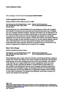
Radiographic findings Cardiomegaly, as assessed by the vertebral heart size (VHS system), is obvious in many affected dogs. The right ventricle and left ventricle are the chambers interpreted as most commonly enlarged.The left atrium appears enlarged in about half of the cases.The typical three bulges (aortic, pulmonic and auricular) on dorsoventral radiographs are only visible in a quarter of the cases (Fig. 2). Overperfusion of the pulmonary vasculature is often more prominent on the dorsoventral view than on the right lateral view. Pulmonary oedema can be present and its distribution is mainly perihilar.
Echocardiographic findings (Fig. 3) In a retrospective UK study, colour Doppler echocardiography revealed the presence of concurrent
L-R PDA
Clinical findings The clinical hallmark of a left-to-right shunting PDA is a continuous murmur, with a point of maximum intensity at the left base of the heart. The murmur can be very localised. The murmur is formed by a pressure gradient, existing in both systole and diastole, between the aorta and the pulmonary artery resulting in blood flowing through the ductus during both periods. Most PDA murmurs are loud, and therefore have an associated precordial thrill. A continuous murmur is fairly pathognomonic for a left-toright shunting PDA, however some other less common
Fig. 2: Thoracic radiographs of a dog with a PDA. The typical three bulges (aortic (black arrow), pulmonic (red arrow) and auricular (blue arrow)) on dorsoventral radiographs.
defects in 10% of cases. Analysis of the M-Mode echocardiographic findings showed that not all animals did have the typical volume overload findings of increased left ventricular diameter in diastole and systole. Left ventricular wall thicknesses were most often within normal limits. There was a wide variation in fractional shortening. The left atrium appeared enlarged in 35% of the scans. Continuous wave Doppler velocities across the aortic valve were increased in many cases and this was attributed to volume loading. Concurrent mitral regurgitation was present in 40% of the animals.The ductus was visualised in most cases and the typical continuous wave pattern was visualised easiest from the right parasternal short-axis view at the level of the pulmonary artery bifurcation.
Patent ductus arteriosus
Fig. 3: Echocardiographic changes in dogs with PDA. Above left: Colour flow Doppler of the right parasternal short-axis view showing entrance of the ductus (red) in the pulmonary artery (PA)(blue) Above right: Continuous wave Doppler signal at the level of the pulmonary artery showing continuous retrograde flow above the baseline highly suggestive of a PDA. Lower: Colour flow Doppler of the right parasternal long-axis view showing mitral regurgitation associated with a PDA. LA is left atrium, LV is left ventricle.
Treatment Closure of the patent ductus arteriosus has been established as mandatory in veterinary cardiology (at all ages). Currently accepted therapies for left-to-right shunting PDA include surgical ligation (by experienced surgeons!) and transcatheter closure (coils and other devices).
Outcome In the hands of experienced surgeons/interventionalists both techniques have extremely good outcomes with probably a lower fatality rate in the embolisation techniques compared to surgery but a higher residual shunting rate.
PULMONIC STENOSIS (PS) Epidemiology Pulmonic stenosis is common in certain breeds including
the Beagle, Samoyed, Chihuahua, English Bulldog, Miniature Schnauzer, Labrador Retriever, Mastiff, Chowchow, Newfoundland, Basset Hound, Terrier and Spaniel breeds.
Types PS can be valvular, subvalvular and supravalvular. In Bulldog breeds it has been associated with aberrant coronary arteries (Fig. 4).Valvular PS is the most common form in veterinary medicine.This form can be subdivided in a Type A (normal pulmonary artery diameter with parachute-like valve) and Type B (annular hypoplasia and a dysplastic valve).
Aberrant coronary artery
Fig. 4: One single aberrant coronary artery (arrow) associated with valvular pulmonic stenosis in a English Bulldog puppy.
Clinical findings It is characterised by a high frequency systolic ejection murmur, best heard over the left heart base.The murmur is crescendo-decrescendo, with maximum intensity in midsystole. It does not tend to radiate as widely as the murmur of aortic stenosis. There is, in most cases, a linear relationship between murmur grading and severity. The jugular veins are usually not distended or pulsating (unless there is concurrent tricuspid regurgitation), although a mildly exaggerated pulse may be noted in the lower third of the neck. In severe PS the pulses can be slightly weak.
ECG findings In mild to moderate PS the ECG is often normal. In dogs with severe right ventricular hypertrophy, evidence of right ventricular enlargement such as deep S-waves in leads I, II and III is commonly identified. Right bundle branch block might be observed.The P-waves are usually normal but if they are increased in amplitude concurrent right atrial enlargement due to tricuspid dysplasia should be suspected.
Radiographic findings Often the right ventricle is prominent.A pulmonary bulge (2 o’clock position on dorsoventral radiograph) can be
observed but there is no correlation between the size of the post-stenotic dilation and the severity of stenosis. Pulmonary vascular hypoperfusion is extremely rare.
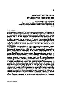
Echocardiographic findings (Fig. 5) On two-dimensional imaging the typical features of right ventricular and papillary muscle hypertrophy and a flattened septum indicating elevated right ventricular systolic pressure are often recognised. M-mode echocardiography can show the presence of paradoxical septal motion, another indicator of increased right ventricular diastolic pressure.There is often a decreased left ventricular internal diameter, secondary to decreased right ventricular cardiac output and consequently decreased left ventricular filling. The severity depends on the pressure gradient across the valve (mild 20-50 mm Hg, moderate 50-100, severe >100 mm Hg).
study in 40 dogs showed that balloon dilation valvuloplasty was successful and resulted in a sustained clinical improvement in 80% of cases showing clinical signs. However, a 7% mortality rate was present.The other types of PS can be corrected surgically but the procedures (patch graft, valvulectomy, conduits) are associated with an extremely high peri-operative mortality.
ATRIOVENTRICULAR VALVE DYSPLASIA Epidemiology Mitral dysplasia (MD) is most commonly reported in large dogs like Great Danes, German Shepherds and Rottweilers. In the UK Bull Terriers are commonly affected. Tricuspid dysplasia (TD) has a preponderance in male Labradors but Old English Sheepdogs, Great Danes, German Shepherd Dogs and Irish Setters have also been reported.
Treatment and outcome
Types
Medical treatment is palliative and aimed at reduction of
Valvular dysplasia represents a complex of thickened valve leaflets, abnormally short or long chordae tendineae, abnormal papillary muscles and abnormally low implanted atrioventricular valve.
Pulmonic stenosis
Fig. 5: Echocardiographic changes in severe pulmonic stenosis showing right ventricular papillary muscle hypertrophy (blue arrow) and a flattened septum (red arrow) indicating elevated right ventricular systolic pressure.
right ventricular hypertrophy. Beta-blockers, such as propranolol and atenolol, remain the drugs of choice and also protect against malignant ventricular arrhythmias. Correction can be surgical or interventional (balloon dilation valvuloplasty) although only cases with severe PS should be considered, since most others show no clinical signs throughout their life. Mainly valvular PS with no aberrant coronary supply will be corrected interventionally, with higher success rate for domed fused valves than with dysplastic valves. Recently a long-term
Clinical findings The murmur of mitral insufficiency is typically of mixed frequency and harsh sounding, but it may be high-pitched or musical in quality. Although this murmur is usually loudest over the mitral valve area and left atrium, it commonly radiates dorsally and to the right thorax, confounding reliable identification of tricuspid regurgitation. Intensity does not reliably indicate severity, especially when murmurs are musical. Care should be taken in broad-chested dogs (like the English Bull Terrier) where because of the conformity of the chest, important murmurs might not be audible. Doppler-flow echocardiography to exclude mitral valve dysplasia is strongly recommended if these dogs are to be used for breeding. Mitral stenosis (MS) is a severe form of mitral dysplasia and is most commonly seen in Bull Terriers.With mitral stenosis, occasionally a mid-diastolic murmur can be heard in the left apical area, but more commonly a murmur of mitral insufficiency will be detected. The intensity and duration of the systolic plateau murmur of tricuspid insufficiency tend to vary with respiration.
ECG findings
Echocardiographic findings
The ECG findings of MD are these of left atrial (widened P-wave) and ventricular enlargement (tall QRS). Supraventricular arrhythmias are more common than ventricular arrhythmias. In cases of severe atrial dilation atrial fibrillation can be observed.
Mitral dysplasia is a complex of thickened valve leaflets, abnormally short or long chordae tendineae, abnormal papillary muscles and abnormally low implanted mitral valve, all causing moderate to severe mitral valve insufficiency. Mitral stenosis is often associated with severe left atrial dilation. MS is also associated with a pathognomonic continuous wave spectral Doppler mitral inflow pattern where early diastolic filling of the left ventricle is of reduced velocity and takes longer (prolonged Edt pressure half time with tall A-wave). Tricuspid valve dysplasia includes thickened valve leaflets, abnormally short or long chordae tendineae, abnormal papillary muscles, abnormally low implanted tricuspid valves (Ebstein anomaly). Severe right atrial dilation is common.Tricuspid stenosis is uncommon and gives similar findings, but then on the right side of the heart, as MS.
In dogs with tricuspid valve dysplasia splintered QRS complexes are commonly found (Fig. 6). Supraventricular tachycardias have been associated with tricuspid dysplasia and accessory pathways in Labradors.
Radiographic findings (Fig. 7)
Tricuspid dysplasia
Treatment and outcome In mitral and tricuspid dysplasia medical therapy is palliative, and aimed at treating heart failure (ACEinhibitors and diuretics). MS and TS could be dilated by balloon catheterisation if regurgitation is not too severe.Valve replacement might be an option in young non-decompensated individuals. Fig. 6: In tricuspid valve dysplasia, a common congenital anomaly in Labradors, splintered QRS complexes can often be found on electrocardiography, as can tall P-waves.
The radiographic findings are those from respectively progressive left-sided congestive heart failure (mitral dysplasia: enlarged left atrium, pulmonary venous congestion, perihilar interstitial or more generalised alveolar pulmonary oedema) and right-sided congestive heart failure (tricuspid dysplasia: right atrial enlargement, widened caudal vena cava, hepatomegaly, ascites, pleural effusion).
A list of breed predispositions and heritability can be downloaded from www.acapulco-vet.be. © Nicole Van Israël illustrations. FURTHER READING AND REFERENCES BAUMGARTNER C., GLAUS T. M. Congenital cardiac diseases in dogs: a retrospective analysis. Schweiz Arch Tierheilkd. 2003;145(11):527-33, 535-6. BIRCHARD S. J., BONAGURA J. D., FINGLAND R. B. Results of ligation of Patent Ductus Arteriosus: 201 cases (1969-1988). J Am Vet Med Assoc 1990; 196 (12), 2011-2013. BUCHANAN J. W. Prevalence of cardiovascular disorders. In. Fox P. R., Sisson D. and Moise N. S. eds Textbook of canine and feline cardiology Philadelphia: W. B. Saunders 1999; 457-470. BONAGURA J. D., LEMHKUHL L. B. Prevalence of cardiovascular disorders. In. Fox P. R., Sisson D, and Moise N. S. eds Textbook of canine and feline cardiology Philadelphia: W. B. Saunders 1999; 471-535. BUCHANAN J. W. Patent ductus arteriosus: morphology, pathogenesis, types and treatment. J of Vet Cardiology 2001; 3 (1), 7-16. BUCHANAN J. W. Pathogenesis of single right coronary artery and pulmonic
Mitral dysplasia
Lau
stenosis in English Bulldogs. J Vet Intern Med. 2001;15 (2):101-4. Erratum in: J Vet Intern Med 2001;15(4):417.
LA
BUSSADORI C., DEMADRON E., SANTILLI R. A., BORGARELLI M. Balloon LV
LA
valvuloplasty in 30 dogs with pulmonic stenosis: effect of valve morphology and annular size on initial and 1-year outcome. J Vet Intern Med. 2001;15 (6):553-8. CHETBOUL V., TRAN D., CARLOS C., TESSIER D., POUCHELON J. L.
LV
Congenital malformations of the tricuspid valve in domestic carnivores: a retrospective study of 50 cases. Schweiz Arch Tierheilkd. 2004; 146(6):265-75. DUKES-MCEWAN J. Mitral dysplasia in Bull Terriers, Vet Annual, 1995; 35, 130-
Fig. 7: Radiographic changes in a pup with mitral dysplasia (left atrial (LA), left auricular (Lau) enlargement and rounded volumeoverloaded left ventricle (LV)).
146. HOGLUND K., FRENCH A., DUKES-MCEWAN J., HAGGSTROM J., SMITH P., CORCORAN B., KVART C. Low intensity heart murmurs in boxer dogs: inter-
observer variation and effects of stress testing. J Small Anim Pract. 2004; 45(4):178-85. JOHNSON M. S., MARTIN M., EDWARDS D., FRENCH A., HENLEY W. Pulmonic stenosis in dogs: balloon dilation improves clinical outcome. J Vet Intern Med. 2004; 18(5):656-62. KITTLESON M. D. IN KITTLESON M. D. and. KIENLE R. D. eds: Small Animal Cardiovascular Medicine. St- Louis: Mosby, 1998. KORNREICH B. G., MOISE N. S. Right AV malformations in dogs and cats: an electrocardiographic survey with emphasis on splintered QRS complexes. J Vet Intern Med. 1997 Jul-Aug;11(4):226-30. ORTON E. C., HERNDON G. D., BOON J. A., GAYNOR J. S., HACKETT T. B., MONNET E. Influence of open surgical correction on intermediate-term outcome in dogs with subvalvular aortic stenosis: 44 cases (1991-1998). J Am Vet Med Assoc. 2000 Feb 1; 216(3):364-7. SAUNDERS A. B., MILLER M. W., GORDON S. G., BAHR A. Pulmonary embolization of vascular occlusion coils in dogs with patent ductus arteriosus. J Vet Intern Med. 2004; 18(5):663-6. STAFFORD JOHNSON et al. (2004) Results of balloon valvuloplasty in 40 dogs with pulmonic stenosis. JSAP, 45,148-153. TIDHOLM A. Retrospective study of congenital heart defects in 151 dogs. J Small Anim Pract 1997; 38, 94-98. VAN ISRAËL N., FRENCH A. T., DUKES-MCEWAN J., CORCORAN B. M. Review of left to right shunting PDAs and short term outcome in 98 dogs. J Small Anim Pract 2002; 43, 395-400. VAN ISRAËL N. (2004) The echocardiographic assessment of congenital heart disease in Small Animals. British Medical Ultrasound meeting Dec 2004. Ultrasound, Nov 2004, Vol 12, N°4, p 234. VAN ISRAËL N. (2005) Congenital heart disase in small animals. Scientific Proceedings of Voorjaarsdagen Amsterdam 14-17 April; p 222-24.
CONTINUING PROFESSIONAL DEVELOPMENT SPONSORED BY P F I Z E R A N I M A L H E A LT H These multiple choice questions are based on the above text. Readers are invited to answer the questions as part of the RCVS CPD remote learning program. Answers appear on page 99. In the editorial panel’s view, the percentage scored, should reflect the appropriate proportion of the total time spent reading the article, which can then be recorded on the RCVS CPD recording form. 1. Which statement is false regarding aortic stenosis in dogs: a. The most common form is the subvalvular form. b. The lesion is always present at birth. c. The subvalvular ridge cannot always be identified on twodimensional echocardiography. d. An aortic bulge (12 o’clock position on the dorsoventral radiograph) indicates the presence of a post-stenotic dilation often associated with severe stenosis. e. Aortic stenosis commonly affects Boxers. 2. Which statement is false regarding pulmonic stenosis in dogs: a. The most common form is the valvular form. b. Bulldog breeds and Terriers are commonly affected. c. Radiographically post-stenotic dilatation is related to the severity of the stenosis. d. In Bulldog breeds it has been associated with aberrant coronary arteries. e. Balloon dilation valvuloplasty results often in a sustained clinical improvement in previously symptomatic cases. 3. Which statement is false regarding PDA in dogs: a. The hallmark of a PDA is a high grade systolic murmur at the left heart base. b. The lesion is present at birth. c. Females outnumber males by 3/1. d. The typical three bulges (aortic, pulmonic and auricular) on dorsoventral radiographs is only visible in a limited number of the cases. e. Closure of the patent ductus arteriosus has been established as mandatory in veterinary cardiology.