C H A P T E R
2 8
Common Neurologic Disorders
LEARNING OBJECTIVES
Krista M. Garner
Upon completion of this chapter, the reader will be able to: 1. Discuss the cerebral vascular circulation. 2. Outline the sensory and muscular spinal nerves. 3. Summarize the pathology of common neurologic disorders. 4. Identify evidence-based interventions for common neurologic disorders. 5. List optimal outcomes that may be achieved through evidence-based management of common neurologic disorders.
euroscience nursing has emerged as one of the fastest-growing areas of specialty practice (Hickey, 2003). Advancements in both neuroscience research and clinical practice are providing new and exciting roles for registered and advanced practice nurses. Healthcare trends have encouraged the specialty practice of neurocritical care collaborative teams based on favorable patient and financial outcome results in current literature (Suarez et al., 2004; Varelas et al., 2004). With the assumption of new roles in practice come greater responsibilities and accountability for providing the highest level of care for the neurologic patient. To achieve these goals, the neuroscience critical care nurse must have a strong neuroscience knowledge base from which to grow in complexity, as the ever-changing evidence-based research drives the practice trends to higher levels of reasoning and decision making. This chapter begins by providing a brief overview covering spinal nerve roots and cerebrovascular circulation. Understanding the complex anatomy of the cerebrovasculature is essential when caring for the critically ill neurologic patient. Complications involved with the blood supply of the brain are generally most prevalent in neurocritical care, accounting for a large percentage of poor neurologic outcomes. Cerebral circulation is the responsibility of two pairs of arteries: the two internal carotid arteries, accounting for anterior circulation, and the two vertebral arteries, accounting for posterior circulation. Each area of circulation includes distal branches as well as communicating penetrators responsible for a constant and well-networked blood supply. Table 28-1 and Table 28-2 describe the major cerebral branches and the areas they supply. The spinal cord is an elongated mass of nerve tissue from which 31 pairs of spinal nerves exit: 8 cervical, 12 thoracic, 5 lumbar, 5 sacral, and 1 coccygeal. Each spinal nerve has a dorsal root by which afferent impulses enter the cord and a ventral root by which efferent impulses leave. The dorsal roots convey sensory input from specific areas of the body known as dermatomes. The ventral roots convey motor impulses, known as myotomes, from the spinal cord to the body (Table 28-3).
N
MANAGING ELEVATED INTRACRANIAL PRESSURE Elevated intracranial pressure (ICP) is one of the major deteriorating factors in patients with intracerebral lesions (Forster & Engelhard, 2004). Because the cranium is rigid and
376
CHAPTER 28 Common Neurologic Disorders
TABLE 28-1 Major Internal Carotid Arterial Branches Artery
Area Supplied
Ophthalmic
Orbits and optic nerves
Posterior communicating (Pcom) Anterior choroidal
Connects the carotid circulation with the vertebrobasilar circulation Part of choroid plexuses of lateral ventricles; hippocampal formation; portions of globus pallidus; part of internal capsule; part of amygdaloid nucleus; part of caudate nucleus; part of putamen
Anterior cerebral (ACA)
Medial surfaces of frontal and parietal lobes; part of cingulated gyrus and “leg area” of precentral gyrus
Recurrent artery of Heubner
Special branch of ACA; penetrates the anterior perforated substances to supply part of the basal ganglia and genu of internal capsule (also called medial striate artery)
Middle cerebral (MCA)
Entire lateral surfaces of the hemisphere except for the occipital pole and the inferolateral surface of the hemisphere (supplied by posterior cerebral artery)
Lenticulostriate (from MCA)
Part of basal ganglia and internal capsule
Anterior communicating (Acom)
Connects the two ACAs
Source: Reprinted with permission from Emory University.
noncompliant, any increase in cerebral volume will, in turn, elevate cranial pressure (Figure 28-1). The major pathophysiologic problems associated with increased ICP are ischemia and herniation (Josephson, 2004). The normal range of ICP is 0 to 15 cm H2O. Elevations beyond these levels can rapidly lead to brain damage. Cerebral
perfusion pressure (CPP) is mean arterial pressure minus ICP. Global ischemic injury results from a critical reduction of CPP, and thus of cerebral blood flow (CBF). Mechanical compression and herniation of brain tissue—the second mechanism of injury—occurs with space-occupying mass lesions and compartment syndrome of brain contents (Josephson, 2004).
TABLE 28-2 Major Vertebral Arterial Branches and the Cerebral Areas They Innervate Artery
Area Supplied
Vertebral Branches Anterior spinal
Anterior two-thirds of spinal cord
Posterior spinal
Posterior one-third of spinal cord
Posterior inferior cerebellar (PICA)
Undersurface of the cerebellum, medulla, and choroid plexuses of fourth ventricle
Basilar Artery Branches Posterior cerebral (PCA) Posterior choroidals (from PCA) Medial posterior choroidal Lateral posterior choroidal
Occipital lobes, medial and inferior surfaces of the temporal lobes, midbrain, and choroid plexuses of third and lateral ventricle Tectum, choroid plexus of third ventricle, and superior and medial surfaces of the thalamus Penetrates the choroidal fissures and anastomosing with branches of the anterior choroidal arteries
Anterior inferior cerebellar artery (AICA)
Undersurface of the cerebellum and lateral surface of the pons
Superior cerebellar artery (SCA)
Upper surface of the cerebellum and midbrain
Pontine
Pons
Source: Reprinted with permission from Emory University.
Common Neurologic Disorders and Evidence-based Interventions
TABLE 28-3 Sensory Nerve Roots (Dermatome) and Motor Nerve Roots (Myotomes) and the Areas They Innervate Spinal Nerves
Dermatome (Sensory Nerve Roots)
Muscles (Motor Nerve Roots)
C-2
Back of head
Neck
C-3
Neck
Neck
C-4
Neck and upper shoulder
Neck, diaphragm
C-5
Lateral aspect of shoulder
Neck, diaphragm
C-6
Thumb; radial aspect of arm
Diaphragm, shoulder, elbow
C-7
Middle finger; middle palm; back of hand
Forward thrust of shoulder
C-8
Ring and little finger; ulnar forearm
Adduction/extension of arm, wrist
T-1, T-2
Inner aspect of the arm; shoulder blade
Control of thoracic, abdominal, and back muscles (T-1 to T-12)
T-4
Nipple line
T-7
Lower costal margin
377
saline. Both are extremely effective initial treatment modalities when bolused for the reduction of ICP.
Anesthetic and Paralytic Agents Sedation using hypnotic, narcotic, and paralytic agents is performed to reduce stress and to control ICP (Forster & Engelhard, 2004). The drugs are chosen to achieve the effects of decreased cerebral metabolic rate and reduced CBF and cerebral blood volume (CBV).
Ventilation
The respiratory alkalosis caused by induced mechanical hyperT-12, L-1 Groin region ventilation can quickly and efFlexion of hip L-2 Anterior thigh and upper fectively lower ICP by causing buttocks cerebral vasoconstriction and reExtension of leg L-3, L-4 Anterior knee and lower leg duced CBV (Josephson, 2004). Flexion of foot L-5 Dorsum of foot; great toe Hyperventilation is conducted to Perineal area and sphincters S-1, S-2, S-3 Foot, toes, medial thigh get the PCO2 at a level between S-4, S-5 Genitals area 30 and 35 mm Hg. Even though it is an effective acute interSources: Hickey, 2003; Wijdicks, 2003. vention for increased ICP, prolonged hyperventilation should be avoided due to the increased Following is a list of the most current options in the literature risk of cerebral ischemia with excessive vasoconstriction today for the treatment of increased ICP. Chapter 27 discusses (Steiner et al., 2005). ICP monitoring in more detail. Other treatment modalities for the treatment of increased ICP drawing interest in the literature include induced hypotherHead and Body Position mia (Commichau, Scarmeas, & Mayer, 2003; Shiozaki et al., 2003) and early surgical decompression via craniectomy Because cerebral venous outflow is obstructed, ICP increases (Albanese, Leone, & Alliez, 2003; Cho, Chen, & Lee, 2003). when the head is in a non-neutral position (Forster & Engelhard, 2004). Durward, Amacher, and DelMaestro (1983) COMMON NEUROLOGIC DISORDERS AND have shown that elevation of the head to 15 to 30 degrees proEVIDENCE-BASED INTERVENTIONS duces a consistent reduction of ICP. Neurologic dysfunction can be a result of a multitude of difOsmotic Agents ferential diagnoses and, when severe enough, will require intensive critical care with specific, timely, and well-organized In osmotic drug therapy, an osmotic pressure difference is innursing care. Critical care management is not always standard duced between the blood and the brain, thereby causing extracprocedure, but rather varies depending on the case and the tion of water from the cerebrum into the intravascular space patient. Thus it allows room for interpretation and interven(Josephson, 2004). Two osmotic drugs are most commonly tion based on research, experience, and advanced knowledge. used in this manner: mannitol (Osmitrol®) and hypertonic T-10
Umbilical region
378
CHAPTER 28 Common Neurologic Disorders
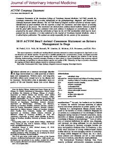
FIGURE 28-1 ICP Waveform Pulsatility—Cerebral Hyperemia Increased Blood Volume Raised ICP
Hemorrhagic Stroke
There are two types of hemorrhagic stroke: intracranial hemorrhage (ICH), which occurs with bleeding into the brain tissue, and subarachnoid hemorrhage (SAH), which occurs with bleeding into the subarachnoid space beneath the arachnoid mater of the meninges. Clinically, ICH provokes the same effect on the brain as space-occupying lesions and is often related to contusions from traumatic brain injury. If unable to be surgically evacuated, ICH is often associated with a poor outcome and increased mortality, secondary to increased ICP with potential for brain herniation. SAH is most often the result of a ruptured cerebral aneurysm. Source: Reprinted with permission from Marshall & Mayer (1997) On Call: Neurology (p. 159). Typically, the patient presents acutely, without warning, with The following are some of the most common neurologic diagwhat is described as “the worst headache” of the patient’s life. noses and current evidence-based interventions for care releAfter aneurysmal rupture, 10% of patients die suddenly, bevant to the intensive care unit (ICU) nurse. fore ever receiving medical attention. Of the patients who reach the emergency department or neuroscience ICU, 20% to 30% Cerebrovascular Complications arrive comatose and die within three months (Wijdicks, 2003). Hemorrhagic and ischemic stroke are two of the principal The primary step in the management of SAH is aneuryscomplications of the cerebral vasculature. Of those patients mal repair, either by surgically clipping the neck of the suffering from strokes in the United States each year, approxaneurysm or by occluding the sac by endovascular coiling techimately 80% to 85% experience ischemic stroke, while 15% to nique. Whether the aneurysm is repaired or not, intensive neu20% undergo hemorrhagic stroke. rologic monitoring is required due to the high prevalence of secondary complications imposed by the initial traumatic Ischemic Stroke event (Box 28-2). An ischemic stroke results from the acute interruption of SAH is graded on a scale from grade I (asymptomatic or blood flow to a volume of brain tissue supplied by an artery minimal headache) to grade V (presenting in a deep coma). (Singh, 2004). It is, therefore, a central cause of brain damage The Hunt and Hess grading scale was developed in 1958 to in neurologic patients, making prevention a primary mission classify cerebral aneurysms. Several complications are assofor the medical community. The term “time is brain” rings ciated with an SAH: rebleeding, cerebral vasospasm, and voltrue for the intensive care patient, because prolonged reducume and osmolar disturbances, such as hypernatremia. tion or absence of blood flow to a certain area of the brain Management of patients with an SAH includes blood presresults in irreversible neuronal injury. Goals for managing the sure control and aneurysm precautions: a quiet, dark room patient with acute ischemic stroke are twofold: (1) enhancewith minimal stimulation; elevation of the head of the bed to ment of CBF and (2) neuroprotection, with the aim to reduce 30 degrees; blood pressure control; and pain control for the intrinsic vulnerability of brain tissue to ischemia (Singh, headache. To prevent vasospasms, triple-H therapy is uti2004) (Box 28-1). lized: hypervolemia, hemodilution, and hypertensive ther-
Common Neurologic Disorders and Evidence-based Interventions
379
Box 28-1
Treatment Interventions for Managing the Patient with Ischemic Stroke Thrombolytic Therapy (tPA) • • • •
Supported through research Reopen occluded vessels with IV medications such as tPA Narrow time window (3–6 hours) Strict inclusion/exclusion criteria
Data from National Institute of Neurological Disorders and Stroke rt-PA Stroke Study Group, 1995.
Anticoagulant/Antiplatelet Therapy • Controversial • Prevents progression or reoccurrence of stroke using IV or low-molecular-weight heparin Data from Caplan, 2004.
Management of Increased ICP and Cerebral Edema • • • •
Mannitol, 3% saline Hyperventilation Sedation Decompressive surgery
Data from National Institute of Neurological Disorders and Stroke rt-PA Stroke Study Group, 1995; Caplan, 2004.
Blood Pressure Management • Controversial • Some trials suggest lowering BP can have adverse effects • Lowering BP usually deferred unless end-organ damage is present or MAP �130 mm Hg or SBP � 220 mm Hg Data from Ahmed, Nasman, & Wahlgren, 2000; Adams, Brott, & Cromwell, 1994.
Strict Glycemic Control • Associated with improved outcome in stroke patients • Goal is to maintain blood glucose 80–110 mg/dL with IV insulin Data from Juvela, Siironen, & Kuhmonen, 2005; Paolino & Garner, 2005.
Fever Control/Induced Hypothermia • Shown to improve cerebral ischemic injury especially in cardiac arrest patients • Neuroprotective strategy Sources: Commichau, Scarmeas, & Mayer, 2003.
apy. The first goal of therapy is to keep the patient’s central venous pressure reading at 10 to 12 mm Hg and pulmonary artery occlusive pressure at 15 to 18 mm Hg through the use of volume expanders such as colloids or crystalloids. The second goal is to maintain a patient’s hematocrit between 33% and 38% by administering blood transfusions or performing phlebotomy treatment on polycythemic patients. The third goal
with triple-H therapy is to manage hypertension by maintaining a systolic blood pressure between 110 and 160 mm Hg.
Status Epilepticus Status epilepticus (SE), or continuous seizure activity, is a medical emergency that requires rapid and vigorous treatment to prevent neuronal damage and systemic complications.
380
CHAPTER 28 Common Neurologic Disorders
Box 28-2
Secondary Complications as a Result of Aneurysmal SAH Rebleeding of Unrepaired Aneurysm • Highest percentage within the first 24 hours of the initial bleed • 30% risk in the first month Data from Ohkuma,Tsurutani, & Suzuki, 2001.
Acute Hydrocephalus • Occurs as a result of blood obstructing the reabsorption process from the subarachnoid space • Many times relieved with the placement of a ventricular drain • Dangerous elevation in ICP if CSF is not diverted via external shunting
Respiratory Failure • Mainly a product of decreased level of consciousness with an inability to protect the airway • Requires intubation through acute phase of illness
Neurogenic Cardiac Stunning • Transient ischemic injury to the myocardium • Produces inefficient cardiac function
Vasospasm • • • •
Delayed cerebral ischemia occurring in 70% of patients with aneurysmal SAH May lead to symptomatic brain ischemia or infarct in 36% of all patients Classically occurs during post initial bleed days 4–15 Treatment options include: triple-H therapy (hemodilution, hypertension, and hypervolemia), cerebral angiogram with angioplasty, and systemic or intra-arterial calcium channel blockers
Sources: Liu-Deryke & Rhoney, 2006; Sen & Albon, 2003.
Management of the patient with SE should focus on termination of seizures, prevention of seizure recurrence once status is controlled, management of precipitating causes of SE, and management of complications (Chapman, Smith, & Hirsch, 2001). Along with clinical correlation, electroencephalographic confirmation of SE should be accomplished as early as possible so that treatment strategies (Box 28-3) can quickly be implemented. Cerebral metabolic decompensation occurs after approximately 30 minutes of uncontrolled convulsive activity (Chapman et al., 2001). Four types of seizures may occur: focal motor, generalized tonic-clonic, complex partial, and nonconvulsive status. A focal motor seizure involves the face or limb and often moves from distal to proximal. A generalized tonic-clonic seizure results in a convulsion with tongue biting followed by a postictal period of altered sensorium and urinary loss. A complex partial seizure manifests with an aura that can be followed by a tonic-clonic seizure. With a nonconvulsive status seizure,
there is a loss of consciousness or altered sensorium with minimal face or limb movement.
Infections of the Nervous System A multitude of infectious etiologies affect the nervous system. The two most common that are addressed in the neuroscience ICU are bacterial meningitis and viral encephalitis. Both types of infections can be extremely toxic, imposing life-threatening complications to the acutely ill patient.
Bacterial Meningitis Bacterial meningitis is a pyogenic, or purulent, infection that involves the pia-arachnoid layers of the meninges, the subarachnoid space, and the cerebral spinal fluid (CSF) (Hickey, 2003). This type of infection primarily occurs in one of three ways: (1) bacteria gain access either via blood or through the spread of nearby infections, such as sinusitis oritis; (2) CSF is contaminated through surgical procedures or by penetration
Common Neurologic Disorders and Evidence-Based Interventions
381
Box 28-3
Emergency Medical Management for Status Epilepticus 1. Standard ABCs of Life Support • Support airway (most likely requires intubation) • Maintain blood pressure • Support circulation 2. Pharmacologic Management • Recommended initial first-line therapy is a benzodiazepine; lorazepam (Ativan®) 0.1 mg/kg at � 2 mg/min • Act as agonists at GABAa receptors and promote inhibition of neuronal firing If seizure activity persists • Second-line therapy is hydantoins, phenytoin (Dilantin®) 20 mg/kg at � 50 mg/min, fosphenytoin (Cerebyx®) 20 mg/kg at 150 mg/min • Followed by scheduled daily dosing If seizure activity persists • Midazolam (Versed®) 0.1–0.3 mg/kg followed by continuous IV infusion of 0.05–2.0 mg/kg/hr • Shown to rapidly control seizures that have not responded to traditional first- and second-line agents If seizure activity persists • Propofol (Diprivan®) initial dose 3–5 mg/kg followed by continuous IV infusion of 1–15 mg/kg/hr If seizure activity persists • Phenobarbital (Luminal®) 10–20 mg/kg at � 50 mg/min or pentobarbital (Nembutal®) 5–12 mg/kg followed by continuous infusion of 1–10 mg/kg/hr • Goal of therapy is to achieve a comatose state with a “burst suppression” pattern on EEG 3. Once there is a cessation of seizure activity for over 12 to 24 hours, a slow taper of continuous IV infusions should begin with the goal of treating complications and preventing recurrent seizure activity. Sources: Bassin, Smith, & Bleck, 2002; Chapman, Smith, & Hirsch, 2001.
of invasive catheters that reside in the brain; or (3) bacteria invade the meninges through the skull or other dural defects. Community-acquired bacterial meningitis is typically caused by such organisms as Streptococcus pneumoniae, Haemophilus ylococcus influenzae, and Neisseria meningitides, whereas hospital-acquired meningitis is usually caused by Staphylococcus aureus, Streptococcus A and B, Escherichia coli, Klebsiella, Proteus, and Pseudomonas. Both community- and hospitalacquired meningitis produce similar classic signs and symptoms, including fever, headache, nuchal rigidity, altered level of consciousness (LOC), and photophobia. The gold standard for the diagnosis of meningitis is direct CSF evaluation via lumbar puncture or aspiration of CSF from ventricular tubing. Treatment measures should be immediate, with initial interventions including general supportive treatments for complications such as septic shock, seizures, and increased ICP. Drug therapy should begin immediately, using broad-spectrum antibiotic coverage until the causative organism is identified. In most cases, intravenous antibiotics are
warranted for many weeks to ensure complete irradication of the infectious organism. Highly resistant organisms, such as methicillin-resistant staphylococcus aureus, may require direct antimicrobial contact through the intrathecal route in conjunction with intravenous therapies.
Viral Encephalitis Viral encephalitis is of sporadic or epidemic occurrence and can be caused by a multitude of atypical viruses, including herpes simplex virus, Epstein-Barr virus, and mosquito-transmitted organisms (malaria, West Nile, Eastern equine, Western equine, and St. Louis). The severity depends on the virus type. Viral encephalitis can produce rapid deterioration in a patient’s neurologic status. Its many signs and symptoms vary according to the invading organism and the area of the brain involved (Hickey, 2003). Basic symptoms, such as fever, headache, stiff neck, and change in LOC, are similar to those exhibited with bacterial meningitis. More severe symptoms progress to seizure activity, coma, and paralysis.
382
CHAPTER 28 Common Neurologic Disorders
Viral encephalitis is diagnosed by serological and CSF analysis, as well as through the patient’s clinical exam. There is no definitive treatment for viral encephalitis and, in most cases, a specific viral etiology is never identified. Supportive care measures for secondary complications focus on the usual standard of care and can last weeks or months, depending on the severity of symptoms and the rate of neurologic improvement (Box 28-4). Herpes simplex encephalitis type 1, which is associated with the common cold sore, is a particularly severe form of encephalitis with a very high morbidity and mortality rate. Intravenous acyclovir (Zovirax®), given prior to the onset of coma, is the standard of treatment.
Brain Death Brain death is defined as a total loss of brain function, meaning life has come to an end and the patient has passed away (Wijdicks, 2003). Life support is futile, but is continued briefly to make organ donation possible. Brain death in adults is frequently a consequence of severe traumatic brain injury or of compartment syndrome of the brain from a massive hemorrhage causing cerebral herniation. The task of declaring brain death, which includes a precise clinical examination, interpretation of diagnostic findings, ruling out conditions that mimic brain death, and proper execution of brain death determination practices, is extremely complicated for the neuroscience specialist. Complete destruction of the brainstem, where most autonomic reflex function is controlled, is required to confirm brain death. Box 28-5 describes the clinical criteria for determining brain death in adults.
Box 28-4
Common Supportive Care Measures for Viral Encephalitis • Steroids to control malignant cerebral edema • Mechanical ventilation for respiratory support • Anticonvulsants/EEG monitoring for seizure prevention • Continuous or frequent analgesics for pain control • Sedatives for anxiety and to decrease cerebral metabolic demand • Antipyretics/endovascular cooling to control hyperthermia
Box 28-5
Clinical Criteria for Brain Death in Adults • Coma—no eye opening to a painful stimulus • Absence of motor response to a painful stimulus • Absence of pupillary response to light— pupils midposition and dilated 4–6 mm (CNs II, III) • Absence of corneal reflexes—reflexive eyelid closure with corneal stimulus (CNs V, VII) • Absence of caloric response—reflexive eye deviation toward cold stimulus injected directly into the inner ear (CNs VIII, III, VI) • Absence of a gag and cough reflex in response to deep suctioning (CNs IX, X) • Positive apnea test—absence of a respiratory drive at a partial pressure of arterial carbon dioxide (PCO2) that is 60 mm Hg or 20 mm Hg above normal baseline values The clinical examination and apnea testing are generally performed by two separate physicians 6 hours apart although requirements vary per institution. Sources: Wijdicks, 2001; Wood, Becker, McCartney, D’Alessandro, & Coursin, 2004.
In addition to the clinical exam, confirmatory diagnostic testing may be used at the physician’s discretion, or if situations such as hypothermia, sedation, or metabolic derangements cloud the diagnosis of brain death. To document cessation of blood flow through the cerebrovascular system, bedside testing is preferred. Once brain death is confirmed, organ procurement is an option if vital functions are artificially maintained, the family requests or agrees to donation, the patient fits donation criteria, and the patient’s organs are not irreversibly damaged. The Federal Conditions of Participation of the Centers for Medicare and Medicaid Services require hospitals to notify their local organ-procurement organization in a timely manner of impending death. “Timely” means notification prior to brain death or before the withdrawal of life support. Potential donors must be ruled in or out based on exclusion criteria. Once a potential donor is identified, discussions regarding donation can be initiated with the family (Wood, Becker, McCartney,
Case Study
D’Alessandro, & Coursin, 2004). Chapter 59 provides more detail on organ donation.
PATIENT OUTCOMES
383
ucation. Knowledgeable ICU nurses are extremely vital practitioners to the team, striving to accomplish the primary goal of quality, cost-efficiency, and compassionate care.
Nurses can help ensure attainment of optimal patient outcomes such as those listed in Box 28-6 through the use of evidencebased interventions.
SUMMARY Being an ICU nurse in the care of a neurologically compromised patient is challenging and requires high levels of clinical judgment in the management of symptoms. Collaborative care teams have shown to improve morbidity and mortality in this critically ill population. The medical and nursing community can continue to improve this trend by practicing under evidence-based guidelines and moving toward greater achievements in research through data collection and continuing ed-
Box 28-6
Optimal Patient Outcomes 1. Intracranial pressure in expected range 2. Neurologic findings in expected range 3. Patient safety maintained during seizure activity 4. Absence of signs and symptoms of infection 5. Family uses effective coping strategies 6. Patient/family expresses readiness for death
CASE STUDY B.H. is a 36-year-old female who was brought into the emergency department with a history of being confused during the morning hours. Her confusion was apparent when she went to the bank and could not remember how to drive herself back home. She stopped to ask for directions and ultimately asked someone to call her husband, who came to take her to the hospital. She complained of having the “worst headache” of her life.
Physical Examination The patient had a Glasgow Coma Scale score of 14, intact cranial nerves, headache with a score of 10 on a scale of 1 to 10, neck stiffness, photophobia, and low back pain. Her blood pressure was 178/92 with heart rate of 88. The monitor displayed a normal ECG. She was afebrile.
Diagnostic Tests CT scan of the head revealed a small subarachnoid hemorrhage indicating a grade I SAH. The patient had a CT angiography and then an intra-arterial digital subtraction angiography with three-dimensional imaging. A decision was made to proceed with microsurgical treatment. Microsurgical treatment involved clipping the aneurysm using nonmagnetic titanium clips.
Postoperative Management The patient was monitored and treated for several days in the ICU by a team of nurses, physicians, and other healthcare professionals. The patient was transferred to a neuro stepdown unit for several days prior to being discharged to a rehabilitation unit.
CRITICAL THINKING QUESTIONS 1. 2. 3. 4.
What are three symptoms of cerebral aneurysm? What are the components of triple-H therapy? List five aneurysm precautions for the preoperative patient. Design a teaching plan for the family of an SAH patient.
384
CHAPTER 28 Common Neurologic Disorders
5. Use the form to write up data and a plan of care for a patient with SAH in the clinical setting. Rate the patient as a level 1, 3, or 5 on each characteristic. Identify the level of nurse characteristics needed in the care of this patient. 6. Take one patient outcome for a patient and list evidence-based interventions found in a literature review for this patient.
Using the Synergy Model to Develop a Plan of Care
Patient Characteristics
Subjective and Objective Data
Evidence-based Interventions
Outcomes
SYNERGY MODEL
Resiliency Vulnerability Stability Complexity Resource availability Participation in care Participation in decision making Predictability
Online Resources American Stroke Association: www.strokeassociation.org/presenter.jhtml?identifier=1200037 National Stroke Association: http://nfo.stroke.org/site/PageServer?pagename=HOME National Institute of Neurological Disorders and Stroke: www.ninds.nih.gov/ American Heart Association Stroke Guidelines: www.americanheart.org/presenter.jhtml?identifier=3004586 Stroke Coding Guide of the American Academy of Neurology: www.stroke-site.org/guidelines/stroke_coding.html Epilepsy information: www.epilepsy.com/ Meningitis Research Foundation: www.meningitis.org/ The Brain Aneurysm Foundation: http://www.bafound.org/info/subarachnoid.php
References
385
REFERENCES Adams, H. P., Brott, T. G., & Cromwell, R. M. (1994). Guidelines for the management of patients with acute ischemic stroke: A statement for healthcare professionals from a special writing group of the Stroke Council, American Heart Association. Stroke, 25, 1901–1914. Ahmed, N., Nasman, P., & Wahlgren, N. G. (2000). Effect of intravenous nimlodipine on blood pressure and outcome after acute stroke. Stroke, 31, 1250–1255. Albanese, J., Leone, M., & Alliez, J. R. (2003). Decompressive craniectomy for severe traumatic brain injury: Evaluation of the effects at one year. Critical Care Medicine, 31, 2535–2538. Bassin, S., Smith, T. L., & Bleck, T. P. (2002). Clinical review: Status epilepticus. Critical Care, 6(2), 137–142. Caplan, L. R. (2004). Thrombolysis 2004: The good, the bad, and the ugly. Review of Neurological Disease, 1(1), 16–26. Chapman, M. G., Smith, M., & Hirsch, N. P. (2001). Status epilepticus. Anaesthesia, 56(7), 648–659. Cho, D. Y., Chen, T. C., & Lee, H. C. (2003). Ultra-early decompressive craniectomy for malignant middle cerebral artery infarction. Surgical Neurology, 60, 227–232. Commichau, C., Scarmeas, N., & Mayer, S. (2003). Risk factors for fever in the neurologic intensive care unit. Neurology, 60(5), 837–841. Durward, Q. J., Amacher, A. L., & DelMaestro, R. F. (1983). Cerebral and cardiovascular responses to head elevation in patients with intracranial hypertension. Journal of Neurosurgery, 59, 938–944. Forster, N., & Engelhard, K. (2004). Managing elevated intracranial pressure. Current Opinion in Anaesthesiology, 17(5), 371–376. Hickey, J. V. (2003). The clinical practice of neurological and neurosurgical nursing (5th ed.). Philadelphia: Lippincott Williams & Wilkins. Josephson, L. (2004). Management of increased intracranial pressure. Dimensions of Critical Care Nursing, 23(5), 194–207. Juvela, S., Siironen, J., & Kuhmonen, J. (2005). Hyperglycemia, excess weight, and history of hypertension as risk factors for poor outcome and cerebral infarction after aneurysmal subarachnoid hemorrhage. Journal of Neurosurgery, 102(6), 998–1003. Liu-Deryke, X., & Rhoney, D. H. (2006). Cerebral vasospasm after aneurysmal subarachnoid hemorrhage: An overview of pharmacologic management. Pharmacotherapy, 26(2), 182–203.
National Institute of Neurological Disorders and Stroke rt-PA Stroke Study Group. (1995). Tissue plasminogen activator for acute ischemic stroke. New England Journal of Medicine, 333, 1581–1587. Ohkuma, H., Tsurutani, H., & Suzuki, S. (2001). Incidence and significance of early aneurysmal rebleeding before neurosurgical or neurological management. Stroke, 32(5), 1176–1180. Paolino, A. S., & Garner, K. M. (2005). Effects of hyperglycemia on neurologic outcome in stroke patients. Journal of Neuroscience Nursing, 37(3), 130–135. Sen, J., & Albon, B. A. (2003). Triple-H therapy in the management of aneurysmal subarachnoid haemorrhage. Lancet Neurolinquist, 2, 614–621. Shiozaki, T., Nakajima, Y., Taneda, M., Tasaki, O., Inoue, Y., Ikegawa, H., et al. (2003). Efficacy of moderate hypothermia in patients with severe head injury and intracranial hypertension refractory to mild hypothermia. Journal of Neurosurgery, 99(1), 47–51. Singh, V. (2004). Critical care assessment and management of acute ischemic stroke. Journal of Vascular and Interventional Radiology, 15(1, Part 2), S21–S27. Steiner, L. A., Balestreri, M., Johnston, A. J., Coles, J. P., Smielewski, P., Pickard, J. D., et al. (2005). Predicting the response of intracranial pressure to moderate hyperventilation. Acta Neurochirurgica, 147(5), 477–483. Suarez, J. I., Osama, O., Suri, M., Feen, E. S., Lynch, G., Hickman, J., et al. (2004). Length of stay and mortality in neurocritically ill patients: Impact of a specialized neurocritical care team. Critical Care Medicine, 32(11), 2311–2317. Varelas, P. N., Conti, M. M., Spanaki, M. V., Potts, E., Bradford, D., Sunstrom, C., et al. (2004). The impact of a neurointensivist-led team on a semiclosed neurosciences intensive care unit. Critical Care Medicine, 32(11), 2191–2198. Wijdicks, E. F. (2003). The clinical practice of critical care neurology (2nd ed.). New York: Oxford University Press. Wijdicks, E. F. (2001). Current concepts: The diagnosis of brain death. New England Journal of Medicine, 344(16), 1215–1221. Wood, K. E., Becker, B. N., McCartney, J. G., D’Alessandro, A. M., & Coursin, D. B. (2004). Current concepts: Care of the potential organ donor. New England Journal of Medicine, 351(26), 2730–2739.