Am. J. Trop. Med. Hyg., 79(2), 2008, pp. 159–163 Copyright © 2008 by The American Society of Tropical Medicine and Hygiene
Co-existence of Integumentary Lesions and Lung X-ray Abnormalities in Patients with Paracoccidioidomycosis (PCM) Angela Restrepo,* Angela M. Tobón, Carlos A. Agudelo, Juan E. Ochoa, David S. Rosero, Marta L. Osorio, Luz E. Cano, and Diego L. Alvarez Corporación para Investigaciones, Medellín, Colombia; Biológicas (CIB), Medellín, Colombia; Hospital La María; Facultad de Medicina, Universidad Pontificia Bolivariana, Medellín, Colombia; Clínica Universitaria Bolivariana, Medellín, Colombia; Hospital Pablo Tobón Uribe, Medellín, Colombia; Hospital Torrevieja, Alicante, Spain; Escuela de Bacteriología y Laboratorio Clínico; Facultad de Medicina, Universidad de Antioquia, Medellín, Colombia
Abstract. In paracoccidioidomycosis (PCM), the primary lung infection remains silent. In this study, attempts were done to define the primary target organ by correlating lung radiographic abnormalities with the time course of mucosal/ skin lesions concurrently exhibited at diagnosis by 63 patients in whom microscopy and/or isolation of Paracoccidioides brasiliensis from respiratory secretions had been positive. Mucosal and skin lesions were found in 65.1% and 12.7% of the patients, respectively. Odynophagia and dysphagia were present in 38.1% each. All patients had lung interstitial infiltrates, and 31.7% had also alveolar lesions; fibrosis was recorded in 46% of them. An inverse correlation was shown for fibrosis and presence of either odynophagia or dysphagia. Cluster analyzes strongly supported two sets of patients: those with mucosal damage, odynophagia/dysphagia, and alveolo-interstitial infiltrates and those with dermal lesions, dyspnea, and lung fibrosis. These groups may represent novel stages in the natural course of PCM. Domínguez Soto,12 in 1957, stressed the airborne route by reporting several patients with PCM in whom lung abnormalities had occurred previously to development of mucosal and/ or skin lesions. Additionally, Lacaz24 detected delayed hypersensitivity to paracoccidioidin in healthy Brazilian subjects and found that a number of them also had lung x-rays abnormalities and circulating anti-P. brasiliensis antibodies attesting to sub-clinical lung infection.24 Mackinnon25 delineated the most probably natural course of events in experimental animals, showing that the primary infection occurred in the lungs. Other authors have confirmed the initial lung involvement in patients and in experimental animals.8,11,26–30 Patients with the chronic PCM adult form, who are of interest in this study, exhibit progressive lung disease accompanied by mucosal, skin, lymph node, and/or adrenal lesions.1,5,9,16 However, in some patients (35%), progression occurs solely at the expense of the lungs.3–5,7,10,16,19 Furthermore, most patients (80%) with external lesions show concurrent lung involvement in the x-rays and/or image studies and also exhibit P. brasiliensis in their respiratory secretions.11,15,16,18 This study was designed to determine whether, at time of diagnosis of PCM, a correlation could be established between the type of abnormality shown by lung radiographs and the time course of the already existing mucosal and skin lesions. We aimed at elucidating the primary and the secondary target organs in this Latin American mycosis.
INTRODUCTION Paracoccidioidomycosis (PCM) is a systemic endemic mycosis restricted to tropical and subtropical areas of Latin America; its etiologic agent is the thermally dimorphic fungus Paracoccidioides brasiliensis. Brazil accounts for the largest number of cases, followed at a good distance by Colombia, Venezuela, and Ecuador; the mycosis has also been reported, albeit with lesser frequency, in the remaining South and Central American countries with the exception of Chile. PCM is diagnosed only rarely in the Caribbean Islands. The disorder afflicts mainly adult men working in agriculture.1–6 Several different clinical forms are recognized: the regressive sub-clinical infection, the progressive disease that may be either chronic (adult type) or acute/sub-acute (juvenile type), and the residual form.2,3,5,7,8 Radiographic pulmonary abnormalities are found at diagnosis in > 85% of active cases, but neither the patient’s symptoms nor the lung auscultation findings show their presence and some go unnoticed.7,9–14 Often the external lesions (mucosae, skin, lymph nodes) are the ones that prompt the patient to seek medical consultation, and it is later that lung damage is discovered.2,7,12,15,16 Pulmonary lesions consist of bilateral alveolar and interstitial infiltrates that progress to fibrosis, bringing forth incapacitating sequelae.3,7,9,13,16–19 Even at diagnosis it is common to find advanced lung lesions indicative of a disorder that has progressed over time.18–20 Modern image procedures such as computed tomography (CT), magnetic, and gallium scans have confirmed the predominance of lung lesions (95%) over those abnormalities present in other organs/systems and, furthermore, all autopsy cases have showed the fungus in their lungs.2,9,13,18 Despite the fact that P. brasiliensis’ natural habitat has not been precisely determined, it is accepted that the infection is acquired through inhalation of the fungus’ spores (conidia), disregarding older and conflicting theories on the portal of entry (traumatic implantation, transmission by a vector, or through an animal reservoir).21–23 Gonzalez Ochoa and
MATERIALS AND METHODS This is a retrospective cohort study based on the collected records of patients with PCM diagnosed and treated between TABLE 1 Lesions of the integument in 63 patients with PCM with concurrent lung x-ray abnormalities*
* Address correspondence to Angela Restrepo, Corporación para Investigaciones Biológicas, Carrera 72A 78B-141, Medellín, Colombia. E-mail:
[email protected]
Type of lesion
Number of patients
Percent of patients
Mucosae Skin Skin and mucosae Hypertrophied lymph nodes Hepato splenomegaly
41 8 14 12 2
65.1 12.7 22.2 19.1 3.2
* A patient could have presented more than one type of lesion.
159
160
RESTREPO AND OTHERS
TABLE 2 Signs and symptoms in 63 patients with PCM presenting concurrent mucosal/skin lesions and lung x-ray abnormalities Total patients with signs/symptoms*
Patients [N (%)]
Constitutional symptoms Fever Weight loss Anorexia Asthenia, adynamia Respiratory symptoms Cough Expectoration Dyspnea Hemoptysis Thoracic pain Upper respiratory signs Ronchi Rales Wheezings Upper respiratory symptoms Odinophagia/disphagia Dysphonia
48 (76.2) 23 (36.6) 41 (65.1) 8 (12.7) 23 (35.5) 53 (84.1) 46 (73.0) 45 (71.4) 35 (55.6) 14 (22.2) 5 (7.9) 20 (31.7) 7 (11.1) 5 (7.9) 13 (20.6) 35 (55.6) 24 (38.1) 18 (28.6)
ance of mucosal and skin lesions would tend to indicate the duration of the primary pulmonary lesion: short if infiltrates were present and long if fibrosis was seen. The significance of the data was determined by the 2 and Fisher exact tests. Associations among areas represented by mucosal and skin lesions, clinical aspects, and x-ray abnormalities were examined by clustering using direct linkage and Euclidian distances. The Kolmogorov Smirnov and the Spearman correlation tests were used to compare the time of initiation of the mucosal/dermal lesions with the type of lung abnormality observed in the x-rays, including fibrosis.31,32 RESULTS
* A patient could have presented more than one sign or symptom.
1978 and 2005 in the laboratory of the Medical and Experimental Mycology Unit of the Corporación para Investigaciones Biológicas (CIB), Medellín, Colombia. The procedures followed in the evaluation of the patients were in accordance with the CIB’s ethical committee and with the 1975 Helsinki Declaration, as revised in 1983. From 209 records analyzed, 63 (30.1%) were selected for further analysis because they corresponded to patients in whom x-ray lung abnormalities and mucosal and/or skin lesions had been concurrently present at the time of diagnosis and in whom P. brasiliensis had been observed in or cultured from respiratory secretions (sputum, broncho-alveolar fluids [BAL]). The remaining patients of the CIB files showed only lung pathology (residual or active), mucocutaneous lesions with no discernible lung abnormalities or concurrent laboratory-confirmed tuberculosis. Consequently, none of these patients were included in this study. From the 63 chosen patients, 62 had the adult, chronic form of the disease and 1 had the sub-acute juvenile presentation. Their corresponding lesions were examined taking into consideration their type and extension. The duration of the mucosal and skin lesions was determined by questioning the patient about the moment when they were first noticed and also by checking the corresponding information as annotated in the clinical record itself. We wanted to determine if the longer the course of these external lesions, the closer their relation to residual lung pathology (e.g., fibrous scarring), and vice versa, the shorter their duration, the earlier the pulmonary x-ray manifestations (infiltrates) would be. Thus, time of appear-
Clinical manifestations. As shown in Table 1, 41 of the 63 (65.1%) chosen patients presented mucosal lesions and 8 (12.7%) had skin lesions; in 14 cases (22%), both type of tissues were involved. Predominant lesions were those of the mucosae (55, 87.3%). Reticuloendothelial involvement was manifested by hypertrophied lymph nodes (12, 19.1%) and hepatosplenomegaly (2, 3.2%). Clinically manifested adrenal abnormalities were detected in < 3% of the patients, but no systematic studies were done to determine the functional integrity of these glands. Signs and symptoms (Table 2) were represented by constitutional manifestations registered in 48 (76.2%) cases, as well as by respiratory symptoms identified in 53 (84.1%) patients. Respiratory signs became apparent in 20 (31.7%) and odynophagia and dysphagia in 24 (38.1%) patients each, whereas dysphonia was found in 18 (28.6%) cases. According to the patients’ information, mucosal/skin lesions had first been noticed for 3 months or less in 12 (19.1%), had been present for 4–6 months in 23 (36.5%), and were 7 months or older in the remaining 28 patients (44.4%). Mean duration of such lesions was 10.3 months (range, 2–120 months). Because of initial selection, all patients exhibited lung interstitial infiltrates, and 20 (31.7%) had both interstitial and alveolar infiltrates; fibrosis was recorded in 29 (46%) of the patients. An analysis of the 63 patients (Table 3) showed that 41 (65.1%) presented radiographic abnormalities concurrently with mucosal damage, whereas a smaller proportion (8, 12.7%) of those with skin lesions had associated x-ray alterations. Patients who had both mucosal and skin lesions occupied an intermediate position (14 cases; 22.2%). Concerning fibrous lesions, they were detected in 17 patients (26.9%) who also had mucosal lesions and in 6 (9.5%) who had either skin lesions or skin and mucosal abnormalities (Table 3). Laboratory diagnosis. The diagnosis of PCM was ascertained by direct (KOH) examination in 61 (96.8%) of the
TABLE 3 Relationship between mucosal/skin lesions with lung involvement in 63 patients with PCM Lung radiographic abnormalities Lung infiltrates [N (%)]
Lung fibrosis [N (%)]
External lesions
Alveolo-interstitial
Interstitial
Plus alveolo-interstitial infiltrates
Plus interstitial infiltrates
Total [N (%)]
Mucosae Skin Mucosae/skin Total
10 (15.8) 0 2 (3.1) 12 (19)
14 (22.2) 2 (3.1) 6 (9.5) 22 (34.2)
5 (7.9) 1 (1.6) 2 (3.1) 8 (12.7)
12 (19) 5 (7.9) 4 (6.3) 21 (33.3)
41 (65.1) 8 (12.7) 14 (22.2) 63
161
PARACOCCIDIOIDOMYCOSIS: CLINICAL CORRELATES
TABLE 4 Diagnostic tests in 63 patients with PCM presenting concurrent lung and mucosal/skin lesions Results
Number of patients with positive results (%)
P. brasiliensis seen in: Sputum/BAL samples Mucosal exudates Skin exudates Total P. brasiliensis isolated in culture from: Sputum/BAL samples Mucosal exudates Skin exudates Total
55 (87.3)* 36 (57.1) 16 (25.4) 61 (96.8) 50 (79.4)* 35 (55.5) 14 (22.2) 59 (93.7)
* In all patients, the fungus was either seen or isolated in culture.
clinical samples examined. P. brasiliensis was found microscopically in 55 (87.3%) of the respiratory secretions (sputum, BAL fluids) studied; specimens from mucosal and skin exudates showed the fungus in 36 (57.1%) and 16 (25.4%) cases, respectively. The fungus was isolated in culture in 59 (93.7%) of the specimens analyzed, with 50 (79.4%) of the latter representing respiratory secretions, 35 (55.5%) representing mucosal exudates, and 14 (22.2%) representing skin samples (Table 4). Overall, in every patient, the fungus was either seen or isolated from respiratory specimens. Statistical analyses. When symptoms were correlated with radiographic involvement, a significant association was shown for dyspnea and fibrosis (P ⳱ 0.002). A significant but inverse association was also shown for fibrosis and presence of either odynophagia (P ⳱ 0.0007) or dysphagia (P ⳱ 0.004). Logistic regression analyses indicated that no association among duration of the mucosal lesions and presence of alveolo/interstitial infiltrates (P ⳱ 0.58) or fibrous alterations (P ⳱ 0.30), could be demonstrated. No correlation was established between the time of initiation of mucosal/skin lesions, and the presence of fibrous changes in the lung as tests showed a mean of 9.4 months in duration (range, 2–120 months) for the group with no fibrosis and of 10.7 months in duration (range, 2–72 months) for the group showing fibrosis (Kolmogorov Smirnov test, P ⳱ 1.0). Neither could we show
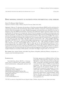
FIGURE 1.
an association (Spearman correlation) between lung x-ray abnormalities and time of initiation of either mucosal (R ⳱ 0.19, P ⳱ 0.13) or skin lesions (R ⳱ 0.32 and P ⳱ 0.1). Cluster analyses based solely on the presence of mucosal and/or skin lesions allowed us to separate the patients in two different groups: one formed by those with mucosal involvement of the oral cavity, the pharynx, the larynx, and the nose, and another made up of patients with skin involvement in various body sites, especially in the limbs and thorax (Figure 1). A different clustering designed to compare integument lesions (mucosae, skin) with both clinical manifestations and radiographic findings separated the patient population in two different groups: the first corresponding to those with mucosal lesions producing odynophagia and/or dysphagia and presence of alveolo-interstitial infiltrates in the chest radiographs. The second showed an association among skin lesions, dyspnea, and presence of fibrous lesions in the lung (Figure 2). In constructing the latter cluster, we did not take into consideration, as done when using logistic regression analysis, the time of appearance of muco-cutaneous lesions in relation to lung abnormalities. Here we analyzed only the simultaneous presence of external lesions, pulmonary x-ray alterations, and certain frequently recorded symptoms, analyses that resulted in two different groups. DISCUSSION Overall, the frequency of mucosal (87.3%) and dermal lesions 22 (34.9%) in this series agrees with previous reports with the exception of the lower rates of clinically detectable reticuloendothelial involvement.1,3–5,7,9 These data plus signs and symptoms of dissemination (fever, lymphadenopathy) corroborate the already reported systemic nature of the chronic pulmonary form of this mycosis.1,3,5,6 Logistic regression analyses in search of an association among time of initiation of the mucosal/skin lesions as related by the patients and the probable duration of the chest x-ray abnormalities, including fibrous alterations, failed. Nonetheless, it became apparent that all types of pulmonary infiltrates
Clustering by direct linkage and Euclidian distances of mucosal and skin lesions in patients with paracoccidioidomycosis.
162
RESTREPO AND OTHERS
FIGURE 2.
Clustering by direct linkage and Euclidian distances based on signs and symptoms associated with lung abnormalities.
were associated much more strongly with mucosal (65.1%) than with skin lesions (12.7%). Recent studies have shown that, regardless of its clinical form (acute/subacute juvenile, chronic adult type), PCM is a multi-organic, systemic disorder where lung lesions predominate.18 Older but also well-grounded findings such as the descriptions of the primary lymph node complex,9,33 the residual nummular lesions found in otherwise healthy subjects,9,15,34,35 and the uncommon calcified lesions detected at autopsy36 have all objectively showed the predominance of lung involvement, thus favoring the inhalation route.13,22,23 It should be remembered, however, that PCM may go unnoticed15,28,29 and that P. brasiliensis may enter into latency for decades, thus creating difficulties in tracing the actual moment of contact. Our results showed that fibrosis brings forth dyspnea (P ⳱ 0.002), and lung interstitial infiltrates associate with mucosal lesions causing odynophagia (P ⳱ 0.0007) and dysphagia (P ⳱ 0.004), whereas none of the patients with odynophagia had fibrosis, despite the fact that one half of them presented dyspnea. Accepting that fibrosis is a latecomer in the host–parasite interaction,9,13,17,18 it seems that, in patients with the chronic adult form of PCM, the initial stages of such interactions cannot be traced with certitude. Despite the fact that this study failed to show a time correlation between duration of the integument lesions and that of the lung’s radiographic abnormalities (infiltrates, fibrosis), comparisons by cluster analysis allowed us to identify two well-delimited groups of patients differing in their manifestations: those with mucosal damage, odynophagia/dysphagia, and alveolo-interstitial infiltrates and those with dermal lesions, dyspnea, and fibrous lung damage. These could well represent different stages of the mycosis’ natural course corresponding to specific syndromes. On the basis of these findings, it seems feasible to suggest that the concurrent presence of mucosal involvement, odynophagia/dysphagia, and alveolo-interstitial infiltrates may represent an early stage of the natural course of PCM. On the other hand, the coexistence of skin lesions, dyspnea, and fibrosis would attest to a later stage in the course of the mycosis.
The retrospective nature of this study and the fact that fibrosis was defined on the basis of x-rays rather than on image studies preclude a more precise definition of these findings. More studies are thus needed to confirm the importance of these data. Received December 13, 2007. Accepted for publication May 10, 2008. Acknowledgments: The authors thank the members of the Medical and Experimental Mycology Unit, Corporación para Investigaciones Biológicas for cooperation in the diagnosis of the patients. Financial support: This study was supported by the Corporación para Investigaciones Biológicas, Medellín, Colombia. Disclosure: The authors report no conflicts of interest. Authors’ addresses: Angela Restrepo, Corporación para Investigaciones Biológicas (CIB), Carrera 72A 78B-141, Medellín, Colombia, Tel: 574-441-0855, Fax: 574-441-5514, E-mail:
[email protected]. Angela M. Tobón, Corporación para Investigaciones Biológicas (CIB), and Universidad Pontificia Bolivariana y Hospital La María, Carrera 72A 78B-141, Medellín, Colombia, Tel: 574-441-0855, Fax: 574-441-5514, E-mail:
[email protected]. Carlos A. Agudelo, Universidad Pontificia Bolivariana, Clínica Universitaria Bolivariana, and Hospital Pablo Tobón Uribe, Calle 78B 72A-109, Medellín, Colombia, Tel: 574-493-6300, E-mail:
[email protected]. Juan E. Ochoa, Corporación para Investigaciones Biológicas, Carrera 72A 78B-141, Medellín, Colombia, Tel: 574-441-0855, Fax: 574-441-5514, E-mail:
[email protected]. David S. Rosero, Hospital Torrevieja, Alicante, Spain, E-mail:
[email protected]. Marta L. Osorio, Hospital La María, Calle 92CC 68A-48, Medellín, Colombia, Tel: 574-267-7511, E-mail:
[email protected]. Luz E. Cano, Corporación para Investigaciones Biológicas and Escuela de Bacteriología y Laboratorio Clínico, Universidad de Antioquia, Carrera 72A 78B-141, Medellín, Colombia, Tel: 574-441-0855, Fax: 574-4415514, E-mail:
[email protected]. Diego L. Alvarez, Facultad de Medicina, Universidad de Antioquia, Carrera 51D 62-29, Medellín, Colombia, Tel: 574-263-5411, E-mail:
[email protected]. Reprint requests: Angela Restrepo, Corporación para Investigaciones Biológicas, Carrera 72A 78B-141, Medellín, Colombia, E-mail:
[email protected].
REFERENCES 1. Blotta MH, Mamoni RL, Oliveira SJ, Nouer SA, Papaiordanou PM, Goveia A, Camargo ZP, 1999. Endemic regions of paracoccidioidomycosis in Brazil: a clinical and epidemiologic
PARACOCCIDIOIDOMYCOSIS: CLINICAL CORRELATES
2. 3.
4. 5. 6. 7. 8.
9.
10. 11.
12. 13.
14. 15. 16.
17. 18.
study of 584 cases in the southeast region. Am J Trop Med Hyg 61: 390–394. Brummer E, Castañeda E, Restrepo A, 1993. Paracoccidioidomycosis: an Update. Clin Microbiol Rev 6: 89–117. Lacaz CS, Porto E, Martin JEC, Heins-Vaccari EM, Melo NT, 2002. Paracoccidioidomicose. Lacaz CS, Porto E, Martins JEC, Heins-Vaccari EM, Melo NT, eds. Lacaz Tratado de Micologia Medica. 9th edition. San Paulo: Sarvier Publishers, 639–729. Londero AT, Melo IS, 1988. Paracoccidioidomicose. J Brazil Med 55: 96–115. Negroni R, 1993. Paracoccidioidomycosis (South American blastomycosis, Lutz mycosis). Int J Dermatol 12: 847–859. Restrepo Moreno A, 2003. Paracoccidioidomycosis. Dismukes WE, Pappas PG, Sobel J, eds. Clinical Mycology. New York: Ford University Press, 328–345. Londero AT, 1986. Paracoccidioidomicose: Patogenia, formas clinicas, manifestacões pulmonares e diagnostico. J Pneumol (Brazil) 12: 41–57. Restrepo A, Tobón AM, 2005. Paracoccidioides brasiliensis. Mandell GL, Bennett JE, Dollin R, eds. Principles and Practice of Infectious Diseases. 6th edition. Philadelphia: Elsevier, 3062–3068. Angulo Ortega A, Pollak L, 1971. Paracoccidioidomycosis. Baker RD, ed. The Pathological Anatomy of the Mycosis. Human Infections with Fungi, Actinomycetes and Algae. Berlin: Springer-Verlag, 507–560. Campos EP, Padovani CR, Cataneo AM, 1991. Paracoccidioidomycosis: radiologic and pulmonary study in 58 cases. Rev Inst Med Trop S Paulo 33: 267–276. Correa AL, Restrepo A, Franco L, Gómez I, 1991. Paracoccidioidomicosis: coexistencia de lesiones extrapulmonares y patología pulmonar silente. Descripción de 64 pacientes. Acta Med Colomb 16: 304–308. Gónzalez Ochoa A, Domínguez Soto L, 1957. Blastomicosis sudamericana. Casos mexicanos. Rev Inst Salubr Enf Trop 17: 97–104. Funari M, Kavakama J, Shikanai Yasuda M, Castro LG, Bernard G, Rocha MS, Cerri GG, Muller NL, 1999. Chronic pulmonary paracoccidioidomycosis (South American blastomycosis): high-resolution CT findings in 41 patients. AJR Am J Roentgenol 173: 59–64. Machado Filho J, Lisboa Miranda J, 1960. Considerações relativas à la blastomicose sul-americana. O Hospital (Brazil) 58: 431–449. Restrepo A, Trujillo M, Gómez I, 1989. Inapparent lung involvement in patients with the subacute juvenile type of paracoccidioidomycosis. Rev Inst Med Trop S Paulo 31: 18–22. Tobón AM, Agudelo CA, Osorio ML, Alvarez DL, Arango M, Cano LE, Restrepo A, 2003. Residual pulmonary abnormalities in adult patients with chronic paracoccidioidomycosis: Prolonged follow-up after itraconazole therapy. Clin Infect Dis 37: 898–904. Tuder RM, el Ibrahim R, Godoy CE, De Brito T, 1982. Pathology of the human pulmonary paracoccidioidomycosis. Mycopathologia 92: 179–188. Yamaga LY, Benard G, Hironaka FM, Castro LG, Funari MG, de Castro CC, Guertzenstein C, Watanable T, Buchpiguel C, Cerri GG, Shikanai Yasuda MA, 2003. The role of gallium-67 scan in defining the extent of disease in an endemic deepmycosis, paracoccidioidomycosis, a predominant multifocal disease. Eur J Nucl Med Mol Imaging 30: 888–894.
163
19. Borrero J, Restrepo A, Robledo M, 1965. Blastomicosis sudamericana de forma pulmonar pura. Antioquia Méd (Colombia) 15: 503–516. 20. Igarashi T, Kurose T, Itabashi K, Nakano I, Okamoto K, Sano A, Kimura K, Kaki H, 2004. A case of chronic pulmonary paracoccidioidomycosis. Nihon Kokyuki Gakkai Zassi 42: 629–633. 21. Borelli D, 1971. Algunos aspectos ecológicos de la paracoccidioidomicosis. Dermatol Venezolana 10: 1190–1201. 22. Gonzalez Ochoa A, 1972. Theories regarding the portal of entry of Paracoccidioides brasiliensis. Proceedings First International Symposium on Paracoccidioidomycosis Scientific Publication Nº 254. Washington DC: PAHO, 278–280. 23. Restrepo A, McEwen JG, Castañeda E, 2001. The habitat of Paracoccidiodes brasiliensis: how far from solving the riddle? Med Mycol 39: 232–241. 24. Lacaz CS, 1955-1956. South American blastomycosis. An Fact Med S Paulo 29: 1–20. 25. Mackinnon JE, 1959. Blastomicosis sudamericana experimental evolutiva obtenida por vía pulmonar. An Fac Med Montevideo 4: 355–358. 26. McEwen JG, Bedoya V, Patiño MM, Salazar ME, Restrepo A, 1987. Experimental murine paracoccidioidomycosis induced by the inhalation of conidia. J Med Vet Mycol 25: 165–175. 27. Roldán JC, Tabares AM, Gómez BL, Aristizábal BE, Cock AM, Restrepo A, 2001. The oral route in the pathogenesis of paracoccidioidomycosis: An experimental study in BALB/c mice infected with Paracoccidioides brasiliensis conidia. Mycopathologia 151: 57–62. 28. Severo LC, Geyer AT, Londero AT, Porto NS, Rizzon CFC, 1979. The primary pulmonary lymph node complex in paracoccidioidomycosis. Mycopathology 67: 115–118. 29. Wanke B, Andrade EM, Lima Neto JA, Ferreira FC, 1983. Paracoccidoidomicose pulmonar assintomática e regressiva, com posterior disseminação. Relato de um caso. Rev Soc Brasil Med Trop 16: 162–167. 30. Benard G, Kavakama J, Mendes Giannini MJS, Kono A, Duarte AJ, Shikanai Yasuda MA, 2005. Contribution to the natural history of paracoccidioidomycosis: identification of the primary pulmonary infection in the severe acute form of the disease: a case report. Clin Infect Dis 40: e1–e4. 31. Zar JH, 1999. Contingency tables. Zar JH, ed. Biostatistical Analysis. 4th edition. Upper Saddle River, NJ: Prentice Hall, 486–517. 32. Hair J, Anderson R, Tatham R, Black W, 1999. Análisis de cluster. Hosmer DW, Lemeshow S, eds. Análisis Multivariante. 5th ed. Madrid: Prentice Hall, 491–546. 33. Severo LC, Geyer AT, Londero AT, Porto NS, Rizzon CFC, 1979. The primary pulmonary lymph node complex in paracoccidioidomycosis. Mycopathology 67: 115–118. 34. Santos JWA, Michel GT, Londero AT, 1997. Paracoccidioidoma: case record and review. Mycopathologia 137: 83–85. 35. Angulo Ortega A, 1975. Lesiones numulares pulmonares de origen inflamatorio: Paracoccidioidomas. Tórax (Venezuela) 11: 25–34. 36. Angulo Ortega A, 1972. Calcifications in paracoccidioidomycosis: are they the morphological manifestation of subclinical infection? Proceedings of the First Pan American Symposium on Paracoccidioidomycosis, Pan American Health Organization, Scientific Publication No. 254, Medellín, Colombia, 25–27, 1972.