Cardiopulmonar y Imaging • Original Research Lim et al. Severity of Emphysema and Lung Cancer Location in COPD
Downloaded from www.ajronline.org by 37.44.207.11 on 01/23/17 from IP address 37.44.207.11. Copyright ARRS. For personal use only; all rights reserved
Cardiopulmonary Imaging Original Research
Relationship Between Emphysema Severity and the Location of Lung Cancer in Patients With Chronic Obstructive Lung Disease Jiseun Lim1 Kyung Min Shin2 KyungSoo Lee 3 Jae Kwang Lim 2 Hye Jung Kim 2 Seung Hyun Cho 2 Seung Ick Cha4 Lim J, Shin KM, Lee KS, et al.
Keywords: chronic obstructive pulmonary disease, emphysema, lung cancer DOI:10.2214/AJR.14.13992 Received October 14, 2014; accepted after revision March 18, 2015. 1 Department of Preventive Medicine, Eulji University School of Medicine, Daejeon, Korea. 2 Department of Radiology, Kyungpook National University Medical Center, Kyungpook National University School of Medicine, 807, Hoguk-ro, Buk-gu, Daegu 702-210, Korea. Address correspondence to K. M. Shin (
[email protected]). 3 Department of Radiology, Samsung Medical Center, Sungkyunkwan University School of Medicine, Seoul, Korea. 4 Department of Internal Medicine, Kyungpook National University School of Medicine, Daegu, Korea.
AJR 2015; 205:540–545 0361–803X/15/2053–540 © American Roentgen Ray Society
540
OBJECTIVE. New phenotypes of chronic obstructive pulmonary disease (COPD) based on emphysema severity have been recognized recently. The purpose of this study was to determine the relationship between emphysema severity (phenotype) and lung cancer location in patients with COPD. MATERIALS AND METHODS. Four hundred patients with 405 primary lung cancers confirmed pathologically between January 2010 and March 2014 were included in the study. Of these, 193 patients received a diagnosis of COPD according to the Global Initiative for Chronic Obstructive Lung Disease guidelines. We scored emphysema severity (0–4) on thinsection CT and assigned the anatomic tumor location of lung cancer as peripheral or central. RESULTS. Patients with COPD had a higher proportion of centrally located lung cancer compared with those without COPD (36.4% vs 17.4%; p < 0.001). In patients with COPD, lower emphysema grades (odds ratio [OR], 0.69; 95% CI, 0.51–0.93; p = 0.016) and reduced ratio of forced expiratory volume in 1 second (FEV1) to forced vital capacity (FVC) (OR, 0.94; 95% CI, 0.89–0.99; p = 0.024) were associated with central location. After adjusting for age, smoking, and spirometry results, the proportion of central location was approximately four times higher in patients with lower emphysema grades (0–2, < 25%) than in those with severe grades (grade 4, > 51%). CONCLUSION. Lower emphysema grades and reduced FEV1/FVC seemed to be independent predictors of central location of lung cancer in COPD. Therefore, in patients with COPD with lower grade emphysema and airway-predominant disease, additional screening tools may have to be considered for central lung cancer detection along with thin-section CT.
L
ung cancer is the leading cause of cancer-related mortality in the world. Cigarette smoking is a well-established cause of both chronic obstructive pulmonary disease (COPD) and lung cancer. Although smoking is a main contributor to the development of lung cancer, only 10–15% of long-term smokers develop lung cancer [1]. COPD is independently and closely related to the development of lung cancer [2, 3], and the risk of lung cancer in patients with COPD is twoto fivefold greater than that in smokers without COPD [4–6]. This observation suggests that lung cancer and COPD share underlying host susceptibility to smoking-induced chronic lung injury [7]. COPD is a disease characterized by progressive airflow limitation caused by a combination of abnormal inflammatory responses in the small airways and destruction of lung parenchyma (emphysema). The relative
contribution of these two mechanisms varies from person to person [8, 9]. Recently, new phenotypes of COPD based on emphysema severity determined by CT have been introduced, replacing the historical classification known as “pink puffers” and “blue bloaters” [10–12]. If patients with COPD show the same degree of airflow limitation, patients with minimal evidence of emphysema on CT could be considered to have a phenotype with predominant airway disease [10]. Therefore, we hypothesized that lung cancer location might be associated with emphysema severity in COPD—that is, central airway cancers have a tendency to occur in subjects with minimal emphysema, whereas peripheral lung parenchymal cancers tend to occur in those with severe emphysema. Previous studies have described the clinicopathologic characteristics of patients with early central lung cancer, such as heavy smoking and the predominance of squamous
AJR:205, September 2015
cell carcinoma on histologic examination [13, 14]. However, these studies have not evaluated the relationship between the anatomic location of the lung cancer (central vs peripheral) and emphysema severity in COPD nor that between central tumor location and clinical characteristics in patients with and without COPD. Because various screening methods have different abilities to detect lung cancer, depending on the central or peripheral location [15], determining central tumor location predictors has significant clinical importance. Therefore, we executed this study to test the hypothesis that emphysema severity may influence the peripheral versus central location of lung cancers in COPD. We also evaluated the association between cancer location (peripheral vs central) and demographic and clinical characteristics, including smoking and spirometry results, to identify the predictive factors for central location of lung cancer in patients with and without COPD. Materials and Methods Our institutional review board approved our retrospective study and waived informed consent (approval number KNUMC14–1053), although at our institution, written informed consent is routinely obtained from all patients for CT studies.
Patients All patients who had received a diagnosis of primary lung cancer from January 2010 through March 2014 and who had undergone pulmonary function tests before their treatment in Kyungpook National University Hospital and Kyungpook National University Medical Center were recruited. During the study period, primary lung cancer was diagnosed in 719 patients. Of these, 454 patients (63.1%) had undergone pulmonary function tests before receiving treatment. We excluded five patients with small cell lung cancer (SCLC) whose lung cancers were manifested as mediastinal or hilar lymphadenopathy without lung nodules or were beyond the reach of bronchoscopy. We also excluded 49 patients with pulmonary fibrosis who were identified to have a predilection to peripheral lung cancers [16, 17]. Of the remaining 400 patients, 193 patients with a history of smoking were categorized with COPD according to Global Initiative for Chronic Obstructive Lung Disease criteria [18]. Additionally, five patients with COPD received a diagnosis of metachronous or synchronous cancers according to current criteria [19]. We regarded multiple lung adenocarcinomas presenting as multifocal ground-glass opacity nodules in the peripheral lung parenchyma as one lesion. Fi-
nally, we included 405 lung cancers from 400 patients. Smoking history was determined by reviewing medical records. Lesions were categorized by histologic examination as non–small cell lung cancer (NSCLC) and SCLC. NSCLC was further classified as squamous cell carcinoma, adenocarcinoma, large cell carcinoma, or other types of cancers (i.e., poorly differentiated carcinoma, undifferentiated carcinoma, and pleomorphic sarcomatoid carcinoma). The seventh edition of the Union for International Cancer Control and American Joint Committee on Cancer TNM classification was used to determine the final pathologic stage [20]. SCLC was classified as limited or extensive according to standard guidelines. Spirometry was performed according to American Thoracic Society and European Respiratory Society Guideline [21] standards within 2 months of lung cancer diagnosis. Using the criteria established by the Global Initiative for Chronic Obstructive Lung Disease, subjects with a ratio of forced expiratory volume in 1 second (FEV1) to forced vital capacity (FVC) of less than 70% were classified as having COPD.
Image Interpretation The severity of emphysema was visually assessed by two independent readers according to the National Emphysema Treatment Trial [22]. In brief, the extent of emphysema was graded into five groups (grades 0–4): none (0%; grade 0), mild (1–10%; grade 1), moderate (11–25%; grade 2), severe (26–50%; grade 3), and very severe (> 50%; grade 4). If there was a disagreement between the two readers on the extent of emphysema, a third reader (another chest radiologist) determined the final score. In a different reading session, the tumor location was determined by two additional readers (a chest radiologist and a pulmonologist) who were blinded to the research hypothesis. To our knowledge, no standard definition of central and pe-
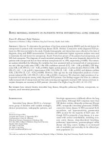
Fig. 1—Proportion of central lung cancers by emphysema severity in patients with chronic obstructive pulmonary disease. Proportion was adjusted for age (years), smoking pack-years), percentage forced expiratory volume in 1 second, and ratio of forced expiratory volume in 1 second to forced vital capacity.
Proportioon of Central Lung Cancers (%)
Downloaded from www.ajronline.org by 37.44.207.11 on 01/23/17 from IP address 37.44.207.11. Copyright ARRS. For personal use only; all rights reserved
Severity of Emphysema and Lung Cancer Location in COPD
50
ripheral locations of lung tumors has been used in previous different studies. In this study, central lung cancer was defined as cancer with an endobronchial lesion observed on bronchoscopy. On the axial and coronal reformatted CT images, we determined the approximate locations of the tumors and then categorized the locations as central or peripheral after considering the bronchoscopic findings [23]. All cases judged to be concordant central (n = 174), concordant peripheral (n = 222), or ambiguous (n = 9) were reviewed by two readers together, and a final decision was taken by consensus. Because our interest was central airway versus peripheral lung parenchyma, we used the center of geometric mass as a marker for the presumed initial starting point of the tumor.
Data Analysis Statistical analyses were performed using SPSS (version 17.0, IBM) and STATA (version 11.0, StataCorp). Data were expressed as the mean (± SD) or frequency (percentage). We compared the differences in the means of measures between the subgroups with unpaired t tests for continuous variables and with the Mann-Whitney U test for ordinal variables, such as emphysema severity. We used the chi-square or Fisher exact test to compare the differences in proportions of central location across groups; p values for trend were calculated by linear-by-linear test. We applied the multivariate analysis using the logistic regression method. We rejected the null hypotheses if p < 0.05. Adjusted proportions were calculated using STATA’s ADJROP procedure.
Results Characteristics of Patients With and Without Chronic Obstructive Pulmonary Disease Patients with COPD showed statistically significantly higher frequencies or values in terms of age (years), male sex, smoking and smoking pack-years, and emphysema
45.9
43.0
45 40
43.8
35
37.7 42.1 35.9
30
25.8
25.0
25 20
21.7
Crude Proportion
15
Adjusted Proportion
10
12.5
5 0
Grade 0
Grade 1
Grade 2
Grade 3
Grade 4
Emphysema Grade
AJR:205, September 2015 541
Downloaded from www.ajronline.org by 37.44.207.11 on 01/23/17 from IP address 37.44.207.11. Copyright ARRS. For personal use only; all rights reserved
Lim et al. morbidity and severity and statistically significantly lower FEV1 percentage and FEV1/FVC values than did patients without COPD (Table 1). With respect to lung cancer location, the proportion of central lung cancer was higher in the patients with COPD than in the non-COPD group (36.4% vs 17.4%; p < 0.001). There was also a statistically significant difference in the histologic diagnosis between patients with and without COPD (p < 0.001): squamous cell carcinoma occurred most commonly in patients with COPD (50.0%) and adenocarcinoma was most common in patients without COPD (69.1%). There was no statistically significant difference in the clinical stage of lung cancers between the two groups. Patient Characteristics and Tumor Location In patients with COPD, younger age and male sex were statistically significantly associated with central tumor location, whereas male sex, older age, smoking, and more smoking pack-years were related to central tumor location in the non-COPD group (Table 2). In both the COPD and non-COPD groups, lower percentage FEV1 and the histologic profile of squamous cell cancer or SCLC were associated with central tumor location. Regarding emphysema in patients with COPD, there was no statistically significant difference in the proportion of central lung cancers relative to the presence of emphysema; however, the negative association between emphysema severity and central tumor location approached borderline significance (p = 0.059). When we evaluated the two emphysema grades, we found that the lower emphysema group (emphysema grade 0–2, < 25%) had a statistically significantly higher proportion of central lung cancer than the higher emphysema group (emphysema grade 3–4, > 26%; p = 0.037) among patients with COPD. In contrast, both the presence and higher grade of emphysema were associated with central location in the non-COPD group. However, these differences were not statistically significant after adjusting for age, smoking pack-years, percentage FEV1, and FEV1/FVC (Table 3). After additional analyses with stepwise adjustments, we found that smoking and percentage FEV1 were the first and second, respectively, strongest confounding factors affecting the association between emphysema severity and central location shown in the univariate analysis in the non-COPD group (data not shown). In patients with COPD, emphysema severity was negatively associated with central lo-
542
TABLE 1: Clinical Characteristics of Patients With and Without Chronic Obstructive Pulmonary Disease (COPD) Characteristic Age (y)
Patients With COPD (n = 198)
Patients Without COPD (n = 207)
p
67.6 ± 7.1 (50–85)
63.6 ± 10.4 (26–88)
< 0.001
Sex, no. (%) of patients
< 0.001
Male
191 (96.5)
98 (47.3)
7 (3.5)
109 (52.7)
43.5 ± 17.4 (10–110)
16.2 ± 22.2 (0–83)
< 0.001
150 (75.8)
55 (26.6)
< 0.001
1.7 ± 1.4 (0–4)
0.5 ± 1.0 (0–4)
< 0.001a
Female Smoking pack-years Emphysema, no. (%) of patients Emphysema severity score Tumor location, no. (%) of patients
< 0.001
Central region
72 (36.4)
36 (17.4)
Periphery
126 (63.6)
171 (82.6)
Percentage FEV1
79.9 ± 18.6 (0–133)
102.7 ± 19.9 (48–181)
< 0.001
FEV1/FVC
58.8 ± 8.6 (34–92)
75.6 ± 6.4 (37–100)
< 0.001
Pulmonary function tests
%Dlco
86.8 ± 21.3 (33–145)
92.0 ± 19.8 (41–150)
0.013
%TLC
107.2 ± 19.6 (33–165)
107.5 ± 18.1 (52–192)
0.885
Histologic profile, no. (%) of patients
< 0.001
NSCLC Squamous cell carcinoma
99 (50.0)
36 (17.4)
Adenocarcinoma
54 (27.3)
143 (69.1)
6 (3.0)
8 (3.9)
16 (8.1)
11 (5.3)
23 (11.6)
9 (4.3)
Large cell carcinoma Other SCLC Pathologic stage
0.082b
NSCLC I
63 (36.2)
83 (42.6)
II
36 (20.7)
26 (13.3) 33 (16.9)
IIIA
30 (17.2)
IIIB
19 (10.9)
12 (6.2)
IV
26 (15.0)
41 (21.0) 0.815c
SCLC Limited
14 (73.7)
7 (77.8)
Extensive
5 (26.3)
2 (22.2)
Note—Except where noted otherwise, data are mean ± SD (range). FEV1 = forced expired volume in 1 second, FVC = forced vital capacity, %Dlco = percent predicted diffusing capacity of lung for carbon monoxide, %TLC = percent predicted total lung capacity, NSCLC = non–small cell lung cancer, SCLC = small cell lung cancer. ap calculated by Mann-Whitney U test. bp for trend calculated by linear-by-linear test. cp calculated by Fisher exact test.
cation (odds ratio [OR], 0.69; 95% CI, 0.51– 0.93; p = 0.016); that is, a higher emphysema grade on CT was associated with peripheral location of lung cancers even after adjusting for age (years), smoking pack-years, and spirometry results (percentage FEV1 and FEV1/FVC) (Table 3). FEV1/FVC was
also negatively associated with the central location (OR; 0.94, 95% CI, 0.89–0.99; p = 0.024), but FEV1 was not statistically significantly associated with the tumor location in patients with COPD. About 40% of patients with mild emphysema (grade