Institutionen för Odontologi Medicine Magisterexamen i Odontologi Master of Medical Science in Odontology
Clinical assessment of temporomandibular joint bone tissue destruction in rheumatoid arthritis
Magdalena Magnius
Stockholm 2007 Nr 129
Clinical assessment of temporomandibular joint bone tissue destruction in rheumatoid arthritis Sammanfattning Syfte: Att undersöka relationen mellan käkledssmärta, anteriort öppet bett och strukturella förändringar i käkleden. Material och Metoder: sextiofyra patienter, 18 män och 46 kvinnor med käkledsinvolvering av rematoid artrit (RA) inkluderades i studien. 20 friska patienter användes som referensgrupp. Vid en klinisk undersökning registrerades käkledssmärta, smärta vid palpation samt graden av anteriort öppet bett. En bilateral radiologisk undersökning (tomografi) genomfördes av käkleden där förekomst eller icke förekomst av erosiva förändringar undersöktes på sex ställen på den temporala delen och sex ställen på kondylen av varje käkled. Summan av höger och vänster käkled användes i analysen. Resultat: Anteriort öppet bett var relaterat till pågående eller tidigare pågående strukturella förändringar i käkleden genom dess relation till förekomst av radiologiska tecken på erosioner. Anteriort öppet bett var också relaterat till käkledsknäppningar och käkledssmärta. Erosioner i käkleden var endast relaterat till knäppningar. Grad 2 eller högre av anteriort öppet bett förefaller vara abnormalt. Slutsats: Den här studien tyder på att anteriort öppet bett, men inte käkledssmärta, är relaterat till strukturella förändringar i käkleden hos patienter med käkledsinvolvering av RA. Detta indikerar i sin tur att det troligen förekommer skillnader i modulering av smärta och bendestruktion. Studien stöder att undersökning av graden av anteriort öppet bett är viktigt att inkludera vid diagnostik av pågående käkledsdestruktion hos patienter med RA.
Handledare: Docent Per Alstergren, avdelningen för klinisk oral fysiologi, Institutionen för Odontologi, Karolinska Institutet, Stockholm Examinator: Professor Anders Gustafsson/Docent Tülay Yucel-Lindberg, Institutionen för Odontologi. Karolinska Institutet, Stockholm
2
Clinical assessment of temporomandibular joint bone tissue destruction in rheumatoid arthritis
Abstract Purpose: To investigate the relation between temporomandibular joint (TMJ), pain and anterior open bite versus TMJ structional changes Materials and Methods: Sixty-four patients, 18 men and 46 female, with TMJ involvement of rheumatoid arthritis (RA) were included. 20 healthy patients were included as controls. A clinical examination performed regarding TMJ pain, tenderness to palpation and the degree of anterior open bite. A bilateral radiographic examination (tomography) of the TMJ was conducted and presence or absence of erosive changes was semiquantitively recorded in six regions in the temporal part and in six regions on the condyle of each TMJ. The sum of the left and the right side was used in the analysis Results: Anterior open bite was related to ongoing or resent structural changes in the TMJ, as indicated by the radiographic signs of erosions. Anterior open bite was also related to crepitus and TMJ pain. TMJ erosions, on the other hand, were only related to crepitus. A degree of 2 or more of anterior open bite is abnormal. Conculsion: This study indicates that the degree of anterior open bite, but not TMJ pain, is related to structional changes of the TMJ in RA, in turn indicating differences in inflammatory modulation of pain and tissue destuction. It seems that assessment of the degree of anterior open bite is of value for detection and monitoring of ongoing TMJ tissue destruction in RA.
Keywords Bone resorption, Inflammation, Pain, Rheumatoid Arthritis, Temporomandibular joint, Tumor necrosis factor.
3
Introduction Numerous inflammatory diseases may affect and involve the temporomandibular joint (TMJ), one example is rheumatoid arthritis (RA). RA is a chronic systemic inflammatory disease that primary involves the musculoskeletal system but it may also involve connective tissue in other organs (heart, lung, blood vessels, eye, kidney etc.). It is likely that this condition is related to autoimmune disorder with genetic influence (Okeson 2003). RA affects individuals over a wide age range but mainly after the age of 50 and often presents a symmetrical debut in peripheral joints such as finger and toe joints. The extent of joint involvement and severity varies considerably between affected individuals and over time but debilitating pain and functional impairment due to swelling as well as cartilage and bone tissue destruction are common results of the inflammatory process (Okeson 2003). Many patients with RA sooner or later develop RA involvement of the TMJ. Pain is the most prominent symptom in patients with TMJ involvement of RA and it is also the most common reason for patients to seek help. Almost every second individual with RA report that their TMJ symtoms appeared the first year after the onset of the general disease (Uotila 1964; Tegelberg et al. 1987). As other joints affected by RA the TMJ may show pain at rest and on movement, tenderness to palpation, stiffness, impaired movement capacity as well as structural changes of the cartilage and bone tissue (Tegelberg et al. 1987). These clinical signs and symptoms have been shown to increase with the degree of general involvement in RA (Chalmers and Blair 1973). Regarding the TMJ, the disease has been reported to start in the synovial tissue of the lower compartment soon followed by the upper compartment and comprise exudation and cellular infiltration followed by pannus tissue growth. The pannus tissue is a vascular granulation tissue, covering and destroying the articular surfaces and the disk (Jagger et al. 1994).
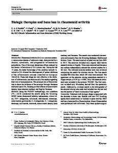
Figure 1. Severe anterior open bite in a patient with rheumatoid arthritis
A characteristic sign of severe RA in the TMJ is the progressive development of an anterior open bite due to bilateral destruction of the mandibular condyles, i.e. anterior open bite in these patients may thus be a clinical sign of bone tissue destruction within the TMJ (Jagger et al. 1994; Fig. 1). Mechanisms behind temporomandibular joint bone and tissue destruction Cytokines play important roles in the pathology of RA by mediation of acute and chronic inflammation and destruction of cartilage and bone tissue. Cytokines are extracellular peptides that via surface receptors influence nearly every cell type. Their essential role is activation and maintenance of immune system mechanisms including the inflammatory reaction. They also act as mediators of pathology in infectious, inflammatory and immune diseases (Duff 1994). Tumor necrosis factor and interleukin-1β
The very potent proinflammatory cytokines tumor necrosis factor (TNF) and interleukin- 1β (IL-1β) initiates and amplifies the inflammation in joint
5
diseases (Saxne et al. 1988). TNF and IL-1β are mainly produced by activated monocytes/macrophages, T-cells and fibroblast in the inflamed synovium and
Figure 2. Schematic overview of interleukin-1β (IL-1β) and tumor necrosis factor (TNF) mediation of bone and tissue breakdown in rheumatoid arthritis
exert cascade effects that eventually may lead to development of cartilage and bone tissue breakdown (Duff 1994). TNF and IL-1β induce several specific inflammatoric events in the synovium but also have systemic effects by stimulating the release of acute phase proteins like C-reactive protein and eliciting fever (Fig. 2). Both TNF and IL-1β are produced in large amounts during inflammation and are considered as a response to injury since they are not produced in normal tissue. TNF levels are increased in the TMJ synovial fluid of patients with RA while it is undetectable in synovial fluid from healthy individuals (Alstergren et al. 1998). Patients with chronic TMJ inflammatory disorder and anterior open bite have been found to have high levels of synovial fluid IL-1β, while
6
patients without anterior open bite had low levels of synovial fluid IL-1β (Alstergren et al. 2003). Endogenous control of the cytokine effects
There are four soluble forms of TNF and IL-1 receptors (TNFsRI, TNFsRII, IL-1sRI and IL-1sRII) that act as endogenous inhibitors to balance the effect of TNF and IL-1β. They inhibit their effects by binding to and neutralizing their activity (Alstergren et al. 2003). Indeed, insufficient endogenous control of TNF, as represented by low plasma levels of TNFsRII, seems to be a risk factor for development of structural changes in TMJ (Alstergren et al. 2006). In that study, there was no difference in plasma TNF between patients with and without anterior open bite but anterior open bite was associated with low levels of TNFsRII, i.e. a sign of insufficient endogenous TNF control (Alstergren et al. 2006). In addition, high levels of IL-1ra in synovial fluid were associated with low TMJ pain intensity and high levels of IL-1sRII in synovial fluid were associated with no or only low degree of anterior open bite. Both of these endogenous control mediators thus seem to influence the clinical expression of TMJ inflammation (Alstergren et al. 2003) Inflammatory activity
As in many chronic joint diseases a number of factors predict poor outcome. For RA patients, presence of rheumatoid factor, high C-reactive protein levels or elevated erythrocyte sedementation rate seem to predict higher inflammatory activity, systemic and/or local, as well as poorer prognosis (Voog et al. 2003). Assesement of erythrocyte sedimentation rate, C-reactive protein and thrombocyte particle count in blood is used to monitor the systemic/general inflammatory activity. The erythrocyte sedimentation rate has been found to be related to radiographic changes in late-stage RA (Tegelberg et al. 1987), to the radiographic sign of flattering (Voog et al. 2003) and to anterior open bite (Alstergren et al. 2006).
7
C-reactive protein is an acute phase protein produced in the liver and its release is modulated by TNF, IL-1β and IL-6. The serum levels of C-reactive protein increase rapidly and to a great extent during inflammation and the C-reactive protein, besides being associated with the systemic inflammatory activity, plays an important role in activating the immune system via the complement system. Elevated C-reactive protein is associated with radiographic changes in RA patients and has been suggested to be a predictor of radiographic changes in RA (Voog et al. 2003). Thrombocytes have many immunologic capabilities and contain numerous molecules with biologic activity such as histamine and serotonin and activated thrombocytes may thus directly contribute to synovial inflammation. A high thrombocyte count in synovial fluid and in blood is associated with local inflammatory activity in arthritis in patients with RA (Zeller et al. 1983). Approximately 85% of RA patients are seropositive for the rheumatoid factor, an endogenous antibody towards IgM (mainly) (Sarnat, Laskin 1991). The antigen-bound rheumatoid factor may elicit immune reactions in synovial tissue as well as activate thrombocytes in the circulation and in the synovial tissues. Indeed, high rheumatoid factor titer in blood is associated with disease activity and poor prognosis in RA patients (Sarnat, Laskin 1991). Treatment of rhematoid arthritis RA is a disease that very seldom can be cured. Today’s treatment is mainly focused on modifying the disease course with disease-modifying antirheumatic drugs (DMARDs) in combination with glucocorticoids and antiinflamatory drugs. Indeed, it seems to be important to reduce pain and joint damage in an early stage in order to maintain function in the joint for long time (Scott et al. 2000). In the last decade, the use of ”biologic drugs” that specifically targets and neutralizes pathophysiological mechanisms or mediators like TNF or IL-1β have been introduced with very good results for many patients.
8
Antiinflammatory drugs and analgesics
For symptom reduction, Non-Steroid Anti-Inflammatory Drugs (NSAIDs) have been used since decades. One of the oldest NSAID used for treatment of RA symptoms is acetylsalicylic acid but there are a number of well-established alternatives like ibuprofen and diclophenac available today. NSAIDs inhibit the prostaglandin production by inhibiting the enzyme cyklooxygenase and thereby reduce pain and inflammation and are often the first drugs to be administered to RA patients for symptom relief. NSAIDs have instant but short-lasting effects and do not affect the disease progress in RA. To reduce severe acute or chronic pains in RA analgesics with central effects like codeine and paracetamol are often used. These drugs reduce pain, but not inflammation per se, by acting on central pain mechanisms (Nitelius 1999). Glucocorticoids as prednisolon are often used systemically in RA patients to reduce the inflammation activity. Glucocorticoids have strong antiinflammatory effects on both systemic and local inflammation by inhibiting production of proinflammatory cytokines and eicosanoids while promoting production of anti-inflammatory mediators. The drug is often administrered daily in low doses to minimize side effects. One severe side effects of systemic glucocorticoid is osteoporosis (Engström-Laurent 1994). Glucocorticoids may also be used for intraarticular injections regarding the TMJ, intraarticular injection of glucocorticoid into the upper compartment in patients with chronic polyarthritides reduces TMJ resting pain, movement pain as well as TNF levels in TNJ synovial fluid (Fredriksson et al. 2005). In that study, patients with high levels of TNF in TMJ synovial fluid showed the strongest treatment effect by intraarticular glucocorticoid indicating prognostic value of high of TNF levels of the treatment effect (Fredriksson et al. 2005). Disease-modifying drugs
NSAIDs have effects on pain and inflammatory activity but can not influence the disease process itself. Treatment regimens today are therefore aiming to include drugs that can modulate the disease progress for example, to reduce the
9
destructive process. These drugs are denoted as Disease-Modifying AntiRheumatic Drugs (DMARDs). The effects of DMARDs occur slowly, often weeks or months after treatment start, but may also last some time after the administration has stopped. One often used DMARD is methotrexate, which has shown quite good results during the last two decades. Methotrexate, which also used in chemotherapy, as well as other DMARDs modulates the immune system response and is administrated to RA patients in tablet form in a low dose. Side effects of methotrexate, especially liver damage and lung disease, can be very severe and the patients are therefore monitored closely. Approximately 30% of RA patients respond well to mehotrexate treatment (van der Bijl et al. 2007). A novel type of DMARDs is called “biologics” since these drugs target specific molecular mechanisms involved in the pathophysiology. There are today several drugs available. Infliximab (Remicade™), which is a chimeric antibody towards TNF, and etanercept (Embrel™), a soluble TNF receptor, have been used successfully for more than 10 years (Elliot et al. 1994). For example, early and systemic administration of infliximab in combination with the DMARD methotrexate has been found to be very efficient in early RA patients to lower disease activity (van der Bijl et al. 2007). Today biologics are systemically administrered although the major pathology in RA is local, i.e. in the synovial tissues. Local treatment with biologics is not yet fully investigated. However, there is one case study that tried multiple intraarticular injections in the TMJ with infliximab during 15 months on a patient with psoriatic arthritis with very good results (reduction of pain, inhibition of tissue destruction) but it is not yet known if this therapy may be a future alternative for treatment of e.g. glucocorticoid-resistant patients (Alstergren et al. 2007). In the future, possible novel modalities for RA treatment may be to enhance the endogenous production and release of proteins like TNFsRI and TNFsRII to
10
Figure 3. Erosion: a local area with decreased density of the cortical joint surface (A), Sclerosis: a local area of increased density of the cortical bone (B), Subcontral speudocysts: a well-defined local area of bone rarefaction underneath an intact cortical outling of the joint surface (C), Flattering: a flat bony contour of the caput (D). Sometimes also a bony outgrowth, a osteophyte can be seen (E; adopted from Voog et al. 2004).
inhibit the strong proinflammatory TNF effects or and also to enhance endogenous production and release of IL-IRI and IL-RII that inhibit IL-1β. Temporomandibular joint bone tissue destruction The most common radiographic manifestation of RA in the TMJ is flattering of the anteriosuperior surface of the condyle and the next most common finding is erosion, which can be observed in about two thirds of the patients with RA in
11
the TMJ. Erosions are most common on the anteriosuperior condylar surface, but are also often seen on both superior and posterior surfaces of the temporal part. Osteophytes of the condyle and sclerosis of the condyle as well as the mandibular fossa are occasionally seen (Voog et al. 2004). As the destructive inflammatory process progresses the condylar outline becomes increasingly irregular and ragged and in severe manifestations with massive bone tissue loss the condyle may be more or less completely resorbed, resulting in loss of vertical support and development of the anterior open bite (Blaschke 1994; Fig. 3). Clinical assessment of temporomandibular joint bone tissue destruction
Clinical diagnosis of RA in the TMJ is not difficult in advance stages but much more so in early stages. The most common subjective symptoms of RA of the TMJ include pain both at rest and on mandibular movements, stiffness in the morning as well as difficulties in opening the mouth and chewing (Voog et al. 2004). Between 15 and 50% of patients with TMJ involvement of chronic polyarthritides develop anterior open bite, sometimes early in the disease. The degree of anterior open bite has been used as a clinical sign of TMJ bone tissue destruction in many studies and is assessed by recording of the occlusal contacts (Alstergren et al. 2006; Fig. 4). However, anterior open bite alone may not be sufficiently specific and sensitive to detect ongoing or previous TMJ bone tissue destruction since it does not seem to be a clear relation between clinical signs of TMJ arthritis, i.e. pain, and degree of anterior open bite (Alstergren et al. 2006). Development and validation of a clinical instrument that includes anterior open bite as well as other factors of importance would be of great use to detect ongoing tissue destruction in the TMJ. Impact of rheumatoid arthritis on daily life A chronic disease like RA carries important psychological and social consequences for the patient and demand significant adjustments in daily life of the affected individuals. The illness leads to impairment in musculoskeletal function, caused by pain and joint damage, and systemic complications may
12
Figure 4. The degree of anterior open bite is assessed by recording of the occlusal contacts on hard biting from the canines to the second molars.
lead to decreased health and living standards for the patients. The disease often impairs the social, work and family life and the person may become more dependent on others to manage daily life. As the disease progresses the affected patient has to reduce working time or leave a working life behind. Living with RA is also living a life with chronic pain. Adjustment to the pain and understanding of the pain and its impact are important factors in order to maintain a psychological well-being as the disease advances (Nagyova et al. 2005). In addition, the unpredictability of the disease activity has great impact on quality-of-life and also creates worries that are part of the suffering (Brown et al 1995). Pain in the TMJ caused by RA affects the quality of daily life and the capacity to perform daily tasks, mostly when the patients move the jaw but also during physical activity (Voog et al. 2003). There are also other, potentially severe, consequences for the patients that have developed an anterior open bite: chewing impairment and sometimes even speech and esthetical problems.
13
Anterior open bite is moreover very difficult for dentists to treat, especially considering the ongoing, chronic disease character of RA. Therefore, clinical detection of patients with anterior open bite due to ongoing TMJ bone destruction or at risk for such destruction would be of utmost importance in order to enable early and successful treatment as well as to minimize the potentially severe consequences for the individual patient. Aim The aim of this particular study was to investigate the relation between TMJ pain and anterior open bite versus TMJ structural changes.
14
Materials and Methods Patients
In this retrospective patient-study part, all patients examined in the Department of Clinical Oral Physiology between 1993 and 2006 with TMJ involvement of RA in whom TMJ tomography (conventional or computerized) according to had been performed and scored using the scoring system described below were included. All in all, 64 patients, 18 males and 46 females, with RA according to the diagnostic classification criteria for RA by the American College of Rheumatology (Arnett et al. 1988) were identified and included (Table 1). The patients were referred to the Department of Clinical Oral Physiology at the Institute of Odontology, Karolinska Institutet from the Department of Rheumatology, Karolinska University Hospital in Huddinge, Sweden due to suspected TMJ involvement of RA. Inclusion criteria beyond RA were recent (within 2 months) diagnosis of RA, age 18 - 80 years, TMJ pain (pain on mandibular movement, at rest or upon
Table 1. Demographic data for 64 patients with TMJ involvement of chronic polyarthritides.
PATIENTS Age Gender Duration of general joint involvement Duration of TMJ involvement Rheumatoid factor
(years) (M/F) (years) (years)
Median
IQR
n
59
20
2 1
5 5
64 18/46 61 39
(pos/neg)
28/31
Diagnoses Rheumatoid arthritis, seropositive Rheumatoid arthritis, seronegative Psoriatic arthritis HEALTHY INDIVIDUALS Age Gender
33 23 5
(years) (M/F)
29
18
20 7/13
IQR = 75th- 25th percentile, n = number of patients, M = males, F = females.
15
digital palpation) and verbal consent. Exclusion criteria were current malignancies, TMJ surgery or trauma within 2 years, recent intra-articular glucocorticoid injection in TMJ (within 1 month) and diseases other than RA causing orofacial pain. Healthy individuals
For comparison, 20 healthy individuals were included (Table 1) from the Institute of Odontology (personell) and one community dentistry clinic in Stockholm, Sweden (personell) and examined during the fall of 2006. Inclusion criteria were a statement by the individual that he or she was completely healthy. Exclusion criteria were current anti-inflammatory or analgesic medication, current malignancies, TMJ intra-articular glucocorticoid injection, surgery or trauma within 2 years and other diseases that cause orofacial pain. Clinical examination
The clinical examination of the patients was performed by one of three calibrated dentists (PA, SK or AKH) and the examination of the healthy individuals was performed by the author. The author was calibrated by PA regarding the clinical examination by verbal instruction and clinical practice/training immediately before the examination of the healthy individuals begun. A 10-cm visual analogue scale (ACO, Stockholm, Sweden; score 0 - 10) with end-point marked with "No pain" and "Worst pain ever experienced" was used to assess the current degree of overall body pain intensity as well as TMJ pain intensity at rest and on maximum mouth opening for each TMJ. A score (0 - 4) for tenderness to digital palpation of the TMJ was adopted that involved evaluation of the lateral and posterior aspect of the joint on each side. For each site, one unit was scored if the patient reported tenderness upon palpation and two units if the palpation in addition caused a palpebral reflex. The pressure pain thresholds to linearly increasing pressure over the lateral aspect of the TMJ and over glabella on the frontal bone were assessed with an electronic algometer with a 1 cm2 blunt rubber tip and a rate of increasing
16
pressure of 50 kPa/s (Somedic Sales AB, Sollentuna, Sweden). TMJ pain upon mandibular movements (maximal and voluntary mouth opening, ipsilateral and contralateral laterotrusion and protrusion) were recorded separately for each TMJ. One unit was scored for each movement causing TMJ pain on each side (score 0 - 4). The number of occluding teeth in the upper yaw as well as the degree of anterior open bite was assessed by recording of the occlusal contacts on each side upon hard biting in intercuspid position (2 x 8
m, Occlusions-Prüf-Folie,
GHM Hanel Medizinal, Nürtingen, Germany). The following scores were used on each side: 0 = occlusal contacts including the canine, 1 = no contacts anterior to the first premolar, 2 = no contacts anterior to the second premolar, 3 = no contacts anterior to the first molar, 4 = no contacts anterior to the second molar and 5 = no occlusal contact. The sum of the scores on the right and left side was used in the analysis as an estimation of the degree of anterior open bite. None of the subjects in our study was edentulous and the score thus ranged from 0 to 9. (Fig. 4) Crepitus was registrated as: no crepitus = 0, palpable crepitus = 1, audiable crepitus = 2. Radiographic examination
A radiographic examination was conducted on the 64 RA patients. Presence or absence of erosive changes was semiquantitively recorded in six regions in the temporal part and in six regions on the condyle of each TMJ using cone-beam computerized tomography images (lateral and frontal sections) assessed by an experienced radiologist. The score for each patient thus ranged from 0 to 24. Presence or absence of flattering of the condyle on the temporal part was also assessed. The score of each patient for flattering ranged from 0 to 4. Blood sampling and analysis
Venous blood samples were collected immediately before the clinical examination. For assessment of the inflammatory activity the inflammatory markers erythrocyte sedimentation rate, C-reactive protein, thrombocyte
17
particle count and rheumatoid factor were analyzed in serum. These analyses were performed according to standard procedures at the Department of Laboratory Medicine (Clinical Chemistry and Immunology), Karolinska University Hospital, Huddinge Sweden. One venous blood sample was collected into a sodium citrate tube for measurement of the erythrocyte sedimentation rate using the Westergren method by our laboratory. Statistics
Median and interquartile range (IQR) were used for descriptive analysis. The Spearman´s rank correlation (rs) test as well as the partial correlation test (rp), where one variable is normalized, were used for calculation of the significance of the correlation between two variables. A probability level less than 0.05 was considered significant.
18
Results The distributions of the clinical and radiographic variables are presented in Table 2. There was no significant difference in degree of anterior open bite between the healthy individuals and the patients (p = 0.451) although the proportion of patients with presence of anterior open bite (score 1 – 9) was higher in the healthy individuals (45%) than in the patients (24%). The 90th percentile for anterior open bite score in the healthy individuals was 2. A degree of anterior open bite of 3 or higher is therefore considered as abnormal. Eight of the patients (13%) and one healthy individual (5%) had an anterior open bite score of 3 or more. The healthy individual with anterior open bite score of 3 or more had a score of 4 and her jaw relation was considered to be due to skeletal abnormality rather than TMJ pathology by the operator. Significant correlations between anterior open bite, presence of erosions and the other variables are presented in Table 3. Briefly, anterior open bite was positively correlated to disease duration, TMJ erosions, crepitus and TMJ pain whereas TMJ erosion was positively correlated to crepitus and negatively to number of occluding teeth (Table 3). In the group of patients with erosive changes, the degree of anterior open bite was positively correlated to TMJ resting and movement pain intensity (rs = 0.45, n = 37, p = 0.005 and rs = 0.40, n = 37, p = 0.013, respectively) as well as to extension of erosive changes (rs = 0.36, n = 38, p = 0.028). In the group of patients with TMJ pain and erosive changes, the degree of anterior open bite was positively correlated to TMJ resting pain intensity (rs = 0.47, n = 21, p = 0.032) and to extension of erosive changes (rs = 0.59, n = 21, p = 0.005).
19
Table 2. Clinical, radiographic and blood variables in 64 patients with TMJ involvement of chronic polyarthritides. Median
IQR
n
%>0
0-9 mm/first h mg/L
5 24 0
4 16 18
63 58 59
100 100 68
Thrombocyte particle concentration Rheumatoid factor
10 9 /L UE
340 0
118 110
43 59
100 47
TMJ pain intensity At rest During maximum mouth opening During hard bitning
0 - 20 0 - 20 0 - 20
0 0 0
3 5 0
59 62 39
39 45 10
Painful mandibular movements
0-8
1
2
64
63
Tenderness to digital palpation Lateral, closed mouth Lateral, open mouth Posterior
0-4 0-4 0-4
0 0 0
1 0 0
45 35 31
38 23 19
Crepitus
0-4
0
1
63
43
Anterior open bite
score
0
0
62
24
Erosions
score
1
2
64
61
HEALTHY INDIVIDUALS Anterior open bite
score
0
0
20
45
PATIENTS General disease activity Number of painful joint regions Erythrocyte sedimentation rate C-reactive protein
IQR = 75th- 25th percentile, n = number of patients and %>0 = percent observations exceeding 0. Rheumatoid factor levels above 20 units were considered abnormal. UE (Swedish) = IE = international units
20
Table 3. Significant correlations between degree of anterior open bite, presence of erosions in the temporomandibular joint (TMJ) and clinical variables Anterior open bite rs Background variables General disease duration Duration of temporomandibular joint disease Clinical variables Mouth opening capacity Number of occluding teeth TMJ pain intensity at rest TMJ pain intensity during mouth opening Number of painful mandibular movements TMJ crepitus
n
p
0.42 0.45
60 0.001 37 0.046
-0.26
58 0.046
0.45 0.33 0.30 0.32
Anterior open bite
21
58 60 62 62
0.001 0.008 0.018 0.008
Erosions rs
n
p
-0.43
57 0.001
0.35
62 0.004
0.32
62 0.012
Discussion This study indicates that the clinical variable anterior open bite is partly related to ongoing or recent structural changes of the TMJ, as indicated by the radiographic sign of erosion, in patients with RA involving the TMJ. On the other hand, anterior open bite is related to both crepitus, another sign of structural cartilage changes, and TMJ pain whereas erosions are related to crepitus alone, suggesting that the mechanisms behind TMJ pain and cartilage and bone tissue destruction may differ to some extent. Anterior open bite was associated with presence and extension of erosive TMJ changes and TMJ pain at rest and on movement of the mandible. The relation between anterior open bite and crepitus supports that the anterior open bite registrered in this study is in fact due to structural changes in the TMJ. The radiographic sign of erosion is considered to be an early sign of cartilage and bone tissue destruction (Goupille et al. 1990). Such destruction may produce anterior open bite, especially if bilateral, by causing a posterior rotation of the mandible with ensuing decrease in chewing ability (Åkerman et al. 1987). However, the clinical variable anterior open bite is also influenced by factors not directly related to cartilage and bone tissue destruction. These factors include individual variation in occlusion pattern, tooth loss, prosthodontic or other restorative treatments and abnormal skeletal or dental jaw relations. The inflammatory activity in the TMJ and the progression rate may also influence the anterior open bite. For example, a slow progress may enable biological normalization of the anterior open bite by adaptation of the masticatory muscles or alveolar bone. This may be one explanation to why some patients do not have anterior open bite despite erosive signs, i.e. for anterior open bite to develop a certain rate of inflammatory activity or progression has to be present. This is corroborated by the findings that anterior open bite, but not erosions, was associated with disease duration, TMJ pain and decreased mandibular movement capacity. Nevertheless, in this study anterior open bite was related to presence and extension of erosive changes in the TMJ,
22
indicating that anterior open bite is an important variable to include in a clinical examination of patients with TMJ arthritis where cartilage and bone tissue destruction is suspected. Erosions are considered as signs of an ongoing or recent inflammatory process including cartilage and bone tissue degradation in the joint. During this inflammatory process, proteinases are released from synovial cells to degrade extracellular matrix components like collagen. A major part of the fibrous cartilage in the TMJ comprises collagen that thus may be degraded, leaving coarse articular surfaces in contrast to the very smooth articular surfaces of a normal joint. Together with structural changes of the bone tissue, these coarse surfaces may cause crepitus during articulation. In the present study, presence of crepitus was moderately associated with erosions supporting the assumption that erosion is a sign of ongoing or recent active inflammatory processes. Anterior open bite was associated with longer disease duration, both local and especially general disease, in this study. RA is a chronic and progressive disease, i.e. patients with longer duration have more total structural changes in joints (Gordon 2001), which very well may explain this relation on a group level. This relation also suggests that the general inflammatory activity eventually will influence the degree of structural changes in the TMJ in patients with RA. However, the influence of the inflammatory activity and medication over time on the development and progress of anterior open bite is unknown and out of the scope with this paper but provides a basis for a highly interesting future study. Presence and larger extension of erosions were associated with lower number of occluding teeth, which was somewhat surprising. A low number of occluding teeth may not only be explained by tooth loss, prostheses or fillings with suboptimal occlusion or abnormal skeletal/dental jaw relations but also by anterior open bite, which indeed was related to erosions. Unfortunately, the reasons for low number of occluding teeth are unknown in the present study.
23
The high intraindividual variability of the degree of anterior open bite in healthy individuals was unexpected and a degree of anterior open bite of 3 or higher seems to indicate an abnormal degree. In turn, this indicate that the use of anterior open bite as the solely parameter for clinical detection of patients with ongoing cartilage and bone tissue destruction in the TMJ has limitations, at least for small or moderate degrees. On the other hand, inclusion of this parameter into a clinical instrument where other factors are taken into account is promising and should be investigated. It would undoubtedly be of great importance to be able to early detect ongoing or progressive TMJ cartilage and bone tissue destruction in order to minimize potentially severe consequences for the patients that have developed an anterior open bite like chewing impairment and speech and esthetical problems. Anterior open bite is moreover very difficult for dentists to treat, especially considering the ongoing, chronic disease character of RA. If detection of ongoing destruction would be possible in an early stage with a clinical instrument, it would enable the possibility to early treatment with potentially better treatment effect as well as less severe consequences by the anterior open bite for the individual patient. Clinical variables could in this setting be used as indicators for a more comprehensive examination, for example radiographs of the joint or synovial fluid and blood analysis. Clinical examination focusing on specific clinical variables may enable such early detection. However, which clinical variables, alone, separate or in combination, that may predict structural changes of the TMJ is still unknown but the degree of anterior open bite seems to be important to include. In conclusion, this study indicates that the degree of anterior open bite, but not TMJ pain, is related to structural changes of the TMJ in RA, in turn indicating differences in inflammatory modulation of pain and tissue destruction. It seems that assessment of the degree of anterior open bite is of value for detection and monitoring of ongoing TMJ tissue destruction in RA.
24
Acknowledgements To my enthusiastic supervisor Associate Professor Per Alstergren. Without his great knowledge in scientific methods and support this paper would not be what it is. Dr Anna-Kari Hajati for helping me with examination and interviewing the patients and Mrs Karin Trollsås, the lab technician we all need all the time.
25
References Alstergren P, Benavente C, Kopp S. Interleukin-1beta, interleukin-1 receptor antagonist, and interleukin-1 souble receptor II in temporomandibular joint synovial fluid from patients with chronic polyarthritides J Oral Maxillofac Surg 61:1171-1178, 2003. Alstergren P, Ernberg M, Kvarnström M, Kopp S. Interleukin-1beta in synovial fluid from the arthritic temporomandibular joint and its relation to pain, mobility and anterior open bite. J Oral Maxillofac Surg 1998;56:1059-1065. Alstergren P, Kopp S. Insufficient endogenus control of tumor necrosis factoralfa contributes to temporomandibular joint pain and tissue destruction in rheumatoid arthritis. J of Rheumat 2006;33:1734-9. Brown S, Williams A. Women´s experiences of rheumatoid arthritis. Journal of adv Nursing 1995;29:695-701. Chalmers IM, Blair GS. Rheumatoid arthritis of the temporomandibular joint. A Clinical and radiological study using circular tomography Q J Med 1973 Apr;42(166):369-86. Dinarello Ca. Interleukin-1, interleukin-1 receptors and interleukin-1 receptor antagonist. Int Rev Immunol 16:457,1998. Donald D. Blaschke. Temporomandibular Joint. In: Oral Radiology Principals and Interpretation. Goaz/White. 1994. Mosby. Missouri Duff GW. Cytokines and acute phase proteins in rheumatoid arthritis. Scand J Rheumatol 1994;23 Suppl 100:9-19. Engström-Laurent A, Leden I, Nived O, Sturfelt G. Behandling In: Reumatologi 1994. Liber Utbildning. Stockholm Elliot MJ, Maini RN , Feldmann M, Kalden JR, Antoni C, Smolen JS, Leeb B, Breedveld FC, Macfarlane JD, Bijl H. Randomised double-blind comparison of chimeric monodonal antibody to tumour necrosis factor alpha (cA2) versus placebo in rheumatoid arthritis. Lancet 1994; 344: 1594-1602.
26
Fredriksson L, Alstergren P, Kopp S. Serotonergic Mechanisms Influence the Response to Glucocorticoid Treatment in TMJ Arthritis. Mediators of Inflammation 2005:4; 194-201. Gordon P, West J, Jones H, Gibson T. A 10 year old prospective follow up of patients with rheumatoid arthritis1986-96. J Rheumatol 2001;28:2409-15. Goupille P, Fouquet B, Cotty P, Goga D, Mateu J, Valat JP. The temporomandibular joint in rheumatoid arthritis. Correlations between clinical and computed tomography features. J Rheumatol 1990;17:1285-91. Jagger RG, Bates JF, Kopp S. Aetiology In: Temporomandibular Joint Dysfunction. 1994. Butterworth-Heinemann Ltd. Great Britain. Jeffery P Okeson. Threatment of temprormandibular joint disorders. In: Management of Temporormandibular Disorders and Occlusion. Okeson 2003. Mosby. Missouri. Lindström F, Ålund M. Farmakologis behandling av reumatisk sjukdom In: Reumatologi och reumakirurgi. 1996. Studentlitteratur. Lund Mishima K, Yamada T, Sugahara T. Evaluation of respiratory status ans mandibular movement after total temporomandibular joint replacement in patients with rheumatoid arthritis. Int Oral Maxillofac Surg 2003;32:275-9. Murray C. Meikle. Remodelling. In: The temporomandibular joint. A biological Basis for clinical practice. Sarnat, Larskin . W.B. Saunders Company, Philadelfia. (bild i text) Nagyova I, Stewart RE, Macejova, van Dijk JP, van den Heuvel WJ. The impact of pain on psychological well-being in rheumatoid arthritis: The mediating effects of self-esteem and adjustment to disease. Patient Educ Couns 2005;58:55-62. Nitelius E. Behandling av rematoid artrit In: Kronisk ledgångsreumatism – rheumatoid artrit 1999. Förlaget Hagman. Stockholm
27
Sarnat B, Laskin D. Pathological Aspects of Arthrites and Derangements In: The temporomandibular joint. 1991. W.B.Saunders Company. Philadelphia Saxne T, Palladino MA Jr, Heinegård D, Talal N, Wollheim FA. Detetion of tumor necrosis factor alfa but not tumor necrosis factor beta in rheumatoid arthritis synovial fluid and serum. Arthitis Rheum 1988;31:1041-5. Scot DL, Pugner K, Kaarela K, Doyle DV, Woolf A, Holmes J, Hieke K. The links between joint damage and disability in rheumatoid arthritis. Rheumatol 200;39:122-132. Tegelberg A, Kopp S, Huddenius, Forssman L. Relationship between disorders in the stomatognatic system an general joint involvement in individuals with rheumatoid arthritis. Acta Odont Scand 1987;45:391-8. Van der Bijl AB, Goekoop-Ruiterman YP, de Vries-Bouwstra JK, Ten Wolde S, Han KH, van Krugten MV, Allaart CF, Breedveld FC, Dijkmans AB. Infliximab and methotrexate as induction therapy in patients with early rheumatoid arthritis. Arthritis Rheum. 2007;7:2129-34. Voog Ü, Alstergren P, Eliasson S, Leibur E, Kallikorm R, Kopp S. Progression of radiographic changes in the temporomandibular joints of patients with rheumatoid arthritis in relation to inflammatory markers and mediators in the blood. Acta Odont Scand 2004;62:7-13. Voog Ü, Alstergren P, Eliasson S, Leibur E, Kallikorm R, Kopp S. Inflammatory mediators and radiographic changes in temporomandibular joints of patients with rheumatoid arthritis. Acta Odontol Scand 2003;61:57-64. Voog Ü, Alstergren P, Leibur E, Kallikrom R, Kopp S. Impact of temporormandibular joint pain on activities of daily living in patients with rheumatoid arthritis. Acta Odont Scand 2003;61:278-82. Uotila E. The temporomandibular joint in adult rheumatoid arthritis. A clinical and rontgenologic study Acta Odont Scand1964;22:Suppl 39:1-91.
28
Zeller J, Weissbarth E, Baruth B, Mielke H, Deicher H. Serotonin content of platelets in inflammatory rheumatic diseases. Correlation with clinical activity. Arthritis Rheum 1983;26:532-40.
29