FELLOWS CORNER
Biologic therapy in rheumatoid arthritis J. Abraham Simón* *2 nd year fellow in Rheumatology Department. Instituto Nacional de Ciencias Médicas y Nutrición Salvador Zubirán.
RESUMEN
ABSTRACT
La artritis reumatoide es una enfermedad autoinmune de causa desconocida que afecta aproximadamente al 2% de la población económicamente activa, por lo cual representa un problema de salud pública. El tratamiento con fármacos citotóxicos ha permitido mejorar la calidad de vida de estos pacientes; sin embargo, todavía existe un número importante que no responde o que tiene poca tolerancia a esta modalidad de tratamiento. El avance de la biotecnología ha permitido el surgimiento de una nueva modalidad de tratamiento conocida como terapia biológica, la cual ha creado expectativas interesantes sobre su efecto en pacientes con artritis reumatoide. El objetivo de esta publicación es revisar las diferentes modalidades de esta terapia y los resultados iniciales en pacientes con artritis reumatoide.
Rheumatoid arthritis is an autoimmune disease of unknown etiology. It represents a public health problem since it affects 2% of economically active population. Treatment with cytotoxic drugs has improved quality of life in these patients. However, a significant amount of patients do not respond or do not tolerate this treatment modality. Biotechnology advances have given rise to a new treatment approach known as biological therapy. This new approach has opened a new therapeutical perspective in these patients. The objective of this publication is to review the different aspects of biological therapy, as well as to inform the initial results of clinical trials in patients with rheumatoid arthritis.
Palabras clave clave. Artritis reumatoide. Terapia biológica.
Key words words. Rheumatoid arthritis. Biologic therapy.
INTRODUCTION Rheumatoid arthritis (RA) represents a public health problem, as it affects about 2% of the economically active population.1,2 This problem generates important expenses derived from medical attention and the loss of productivity of affected patients. Hence, the objective of RA treatment is to decrease the effects of this disease on patient functionality. The current treatment of choice is methotrexate, which, along with other recently featuring cytotoxic drugs such as cyclosporine, leflunomide and mycophenolate mofetil are contributing to that objective. Nonetheless, there is still a number of patients who do not respond to treatment. In addition, the use of these drugs increases infection risk
3 and
may even cause malignancies.4 Recent advances in the understanding of RA physiopathology and biotechnology have allowed the emergence of a new era in the treatment of this disease, known as biological therapy. 5-12 This term includes treatments based on the use of molecules, usually peptides, obtained by various biotechnological methods, which act in a more selective way on cells or soluble mediators crucial for the development of the disease.5 The appearance of this treatment has created huge expectations due to the possible beneficial effect on RA treatment, without the side effects of traditional treatment. The objective of this review is to describe the possible action targets and results of the initial studies of biological therapy in patients with RA.
452 Abraham Simón2001 J. Biological therapy of rheumatoid arthritis. Rev Invest Clin 2001; 53 (5): 452-459 La Revista de Investigación Clínica / Vol. 53, Num. 5 / September-October, / pp 452-459 Full text of this article available in internet: www.imbiomed.com.mx
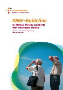
PHYSIOPATHOLOGY OF RA RA is an autoimmune disease that may potentially affect any organ or system, although the main site is the diarthrodial joint. Damage on this joint depends on 3 primary elements (all occurring in the synovial tissue) which seem to be closely linked with each other in a co-stimulation network. Such damage results in cartilage and bone destruction (Figure 1). These elements are I) inflammation, II) hyperplasia with infiltration of synovial tissue or pannus13 and III) angiogenesis.14
CD4 Citokine Th1 stimulation
Citokine Th2 blocking Cellular activation and synthesis of IL-2 and IFN-γ
IL-8 Metalloproteinases
collagenase prostaglandins
TNF-α IL-1β
Adhesion molecule activation and inflammatory cells recruitment
Phospholipases lysosimes proteinases nitrous oxide
ANGIOGENESIS, INFLAMMATION AND PANNUS
JOINT DESTRUCTION
APC: antigen-presenting cell. Mf: macrophage.
IL: Interleukin. TNF TNF: Tumor necrosis factor.
IFN: Interferon. PMN PMN: Polymorphonuclear.
Figure 1. Physiopathology of rheumatoid arthritis. The recognition of an exogenous antigen or auto-antigen seems to be the beginning of a series of events leading to joint destruction in patients with rheumatoid arthritis. This phenomenon induces the activation of CD4+Th1 lymphocytes and the subsequent release of pro-inflammatory cytokines (interleukin 2 and interferon γ), which act on fibroblasts, macrophages, polymorphonuclear and B-lymphocytes, which in turn, produce inflammation, angiogenesis and pannus through their by-products, eventually leading to joint destruction.
Damage is probably produced through antigen recognition (e.g., viruses, bacteria, and collagen) by Tlymphocytes CD4+, which together with the stimulation of different cytokines 15-17, induce differentiation in Th1 cells. On the one hand, these cells produce interleukin (IL)-2 and interferon (IFN)γ.18,19 two compounds with positive effects on T-lymphocyte and synoviocyte activation, which in turn, induces a higher production of cytokines, adherence molecules activation and chemotaxis, mainly of monocytes. On the other hand, these cells inhibit the production of antiinflammatory cytokines such as the IL-10 and the IL4 produced by Th2 cells. Thus, this phenomenon gives rise to the formation of a suitable environment for the inflammatory cells activation, even with formation of ectopic lymph tissue at synovial level.20-22 Activation of synoviocytes and the subsequent production of various pro-inflammatory cytokines, generate a vicious circle of inflammation, pannus formation, and eventual destruction.17 Type B synoviocytes (fibroblast) synthesize several molecules that may damage the joint in response to different stimuli such as the tumor necrosis factor (TNFα), the IL-1β and growth factors (FGF). Metalloprotease, prostaglandin and chemokine such as IL-8 are found amongst those molecules.23 Likewise, they stimulate the production of acute phase reactants (APR) through the synthesis of IL-6, which increases the inflammatory response.24 Moreover, these cells can induce the differentiation of B-lymphocytes in antibody-producing cells 25, which potentially participate in the destruction of the joint. Type A synoviocytes (macrophages), produce different pro-inflammatory cytokines such as IL-15 – which participates in T-lymphocyte activation and induces their differentiation to Th1-, and chemokines such as IL-8 – which stimulates polymorphonuclear chemotaxis26 and intensifies angiogenesis, respectively. However, the two cytokines with more deleterious effects on the joint are IL-1β and TNFα, both derived from macrophages. These cytokines have similar functions and present synergism in some cases. Among their main functions are fibroblast activation, autocrine activation, chemokine stimulation, and adherence molecule induction. Their most significant effect is on osteocytes. It has been observed that they may inhibit bone regeneration through osteoblasts and induce a higher re-absorption through osteoclasts.27,28 In addition to these two cells (T-lymphocytes and synoviocytes), other elements participate in the process such as growth factors,29 (FGF, TGF, GM-CSF), chemokines24 (MCP-1, MIP 1, RANTES; GRO, PF-4), acute phase reactants24 (ceruloplasmine, haptoglobu-
Abraham Simón J. Biological therapy of rheumatoid arthritis. Rev Invest Clin 2001; 53 (5): 452-459
453
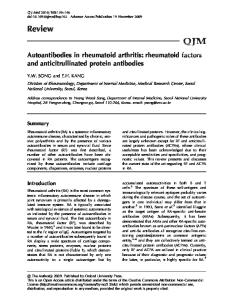
Table 1. Biological agents studied in rheumatoid arthritis Function
Target
Agent
Oral tolerization
Th2 cells
Bovine collagen Chicken collagen
Adherence molecule blockage
ICAM-1(CD54)
Murine MoAb
T lymphocyte function inhibition
CD4 CD5 CD7 CD25 CD52 TCR
Murine, chimeric and humanized MoAb Murine MoAb Murine and chimeric MoAb Chimeric MoAb Humanized MoAb (CAMPATH-1) Vaccination with TCR peptides
Anti-inflammatory cytokines
Multiple Multiple Multiple
Recombinant IL-10 Recombinant IL-4 IFN-γ
Pro-inflammatory cytokine blockage
IL-1β TNFα
IL-1 receptor antagonist TNFα soluble receptor P55-P75 and chimeric and humanized MoAb
MoAb: monoclonal antibody.
TCR: T-cells receptor.
IL: interleukin.
lin, amyloid, reactive C protein) and polymorphonuclear products26 (phospholipase A2, lysosimes, proteinases and nitrous oxide). Finally, the importance of angiogenesis lies in the fact that it provides a site for a more copious migration of inflammatory cells and permits support the metabolic requirements of a chronic inflammatory response.14 Therefore, each of the elements participating in RA physiopathology is a potentially stimulating or inhibiting site for the use of biological therapy. BIOLOGICAL THERAPY Several types of biological therapy in RA have been used (Table 1), including monoclonal antibodies (MoAb),30 soluble receptors, oral tolerization and vaccination. All these modalities have been used intended to block or stimulate various targets in this disease pathogenesis, particularly antigen, adherence molecules, T-lymphocyte, anti-inflammatory cytokines and pro-inflammatory cytokines.7 I. Antigen modulation The agent or agents causing RA are still unknown. Various candidates have been proposed to set off the disease: exogenous (e.g., Epstein Barr’s virus, cytomegalovirus, and tuberculosis) and
454
IFN: interferon.
TNF: tumor necrosis factor.
auto-antigens (e.g., type II collagen, filgastrin). 31 However, according to some researchers who talk of “oral tolerization”,32 it seems that recognition of the causal antigen is not required to inhibit the immune response directed against it. An immune response depends on various factors: I) the antigen administration route among which the digestive tract-associated lymphatic tissue seems to be the most tolerant, and II) antigen characteristics (structure and doses). It seems that the antigen, administered in small doses (low tolerance zones) through the digestive tract, is mainly recognized by Th2 lymphocytes. These cells produce anti-inflammatory cytokines —IL-4 and IL-10— regardless of the nature of the antigen. These cytokines are distributed to all the organism, which induces an anti-inflammatory response with the subsequent tolerization to related antigens.32 This basic immunological concept known as “bystander” has been used to treat RA patients orally with type II collagen. Four different reports appeared in the last years on oral tolerization with type II collagen.33-36 In this study, bovine or chicken collagen were used as antigen at different doses and at different disease stages (i.e., early phase or late state). Despite the theory supporting its use, results have not been satisfactory. An example is the multi-center doubleblind, placebo-controlled trial conducted by Barnett
Abraham Simón J. Biological therapy of rheumatoid arthritis. Rev Invest Clin 2001; 53 (5): 452-459
et al.,36 which included 274 patients with active RA. These patients received oral type II collagen (obtained from chicken cartilage) at 20, 100, 500, and 2500 µg doses every 24 hours for 24 weeks and were assessed using the Paulus’* scale and the American College of Rheumatology* (ACR) 20% scale. Results were modest as only the group which received 20 µg showed an improvement of 20% in the Paulus’ scale when compared with the placebo group. Results were not confirmed with the ACR scale nor reproduced in other studies.33-36 Several explanations could account for this failure: I) type of collagen used; although bovine and chicken collagen present great homology with that of humans, it seems that the “motif” (amino acid sequence recognized by an antibody) is substantially different, which may affect recognition and, consequently, tolerization; II) it has been difficult to define the necessary antigen dose and length of treatment to achieve stimulation of a Th2 response in the digestive tract; III) some authors suggest that for the immune response to be adequate in the digestive tract there should be an intact mucosa, which is not present in most patients who receive non-steroid anti-inflammatory agents (as happened in nearly every study), and IV) patient selection was not adequate, since most patients were late state RA and did not respond to multiple treatments. Oral tolerization has not been useful in clinical practice and research in this field has therefore stopped. II. Blockage of adherence molecules There are several families of adherence molecules that participate in RA physiopathology such as the family of CD44, selectins (L-E-P-selectin), β1 integrins proteins (VLA1,3,4,5,6), 7 integrins proteins (VCAM-1), β2 integrins proteins (LFA-1, Mac1/Mo-1, gp150), and the super-family of immunoglobulins (ICAM, PECAM). (for a more comprehensive review, see references 37 and 38). These molecules participate in recruitment and migration of inflammatory cells through the vascular endothelium. They also play a key role in the cell-cell
* The Paulus and ACR criteria are used for the assessment of clinical improvement of patients with rheumatoid arthritis. Both are comparable and include counting of painful and inflamed joints, global assessment of disease severity by the patient and the physician, and determination of an acute phase reactant such as VSG or PCR. The ACR criteria also include an impairment assessment scale (AIMS, HAQ, MACTAR etc.) For a more comprehensive review, see Arthritis Rheum 1990; 33: 477-484 and Arthritis Rheum 1995; 38: 727-735
interaction during the antigen presentation, which makes them a suitable target for biological therapy 37.38. Among these, the only one used in RA patients is ICAM-1 (CD54).39 This molecule was used as target in a Phase I-II trial, which included 10 patients with active RA of recent onset (< 6 months) who were administered intravenous R6.5 (MoAb from murine source, directed to the second extra-cellular domain of molecule ICAM-1) at 280 mg dose, fractionated in 5 days. Patients were followed-up for 340 days, and assessed using the Paulus’ scale. Despite interesting results (improvement of 60% after 8 days, sustained up to 2 months later), no conclusions were reached because of the reduced number of patients and the lack of a control group. As in the case of oral tolerization, there are no new studies that assess biological therapy whose aim is to block adherence molecules, probably due to the impact of anti-TNFα therapy (vide infra). III. Anti-T-lymphocyte therapy T-lymphocyte has a crucial participation in RA pathogenesis,15-17 which makes it a suitable target for biological therapy. Based on this, several anti-Tlymphocyte therapies have been designed directed to various surface antigens such as CD4, 40-42, CD5, 43 CD7, CD25,44 CD5245,46 and T-cells receptor.47-49 Several studies have been carried out including a large number of RA patients showing different characteristics who have undergone this therapeutic modality with bad results. An example of this was the multi-centered study carried out by Wedling and colleagues41 in 13 French universities in 1998. The study included 58 patients randomly distributed to receive BF-5 (murine MoAb directed against the second domain of antigen CD4+) at 20 mg/day doses for 10 days, or placebo. Patients were followed-up for 3 months after the application of BF-5 and assessed with the Paulus’, 20% and 50% scales. Results showed that 50% of the group receiving BF-5 did not respond to treatment, and no significant difference was found in comparison with the placebo group. In addition to the lack of efficacy, 40% of the patients presented side effects (e.g., increase in infections and lymphopenia risk). A couple of studies using anti-CD52 MoAb (CAMPATH-1H)45,46 showed some clinical benefit, although it was transient and with side effects comparable to those found in anti-CD4 MoAb. The explanation for this failure may be the nonselective depletion of T-lymphocytes. This has been demonstrated in patients treated with anti-CD4
Abraham Simón J. Biological therapy of rheumatoid arthritis. Rev Invest Clin 2001; 53 (5): 452-459
455
MoAb, in whom lymphopenia was found in peripheral blood —due to MoAb effectiveness, while synovial fluid presented persistence of auto-reactive CD4+ clones probably involved in joint damage. It is possible that the joint represents an ideal site for the presence of these cells, since in addition to be a cytokine-rich medium, it may have ectopic lymph foci. This will allows for the differentiation and proliferation in situ of T-lymphocytes, avoiding the MoAb to have any therapeutic action on these cells. 6.13.31 Nowadays, anti-T-lymphocyte therapy is interrupted. IV. Therapy with cytokines
a) Anti-inflammatory cytokines. Cytokines produced by Th2 lymphocytes (e.g. IL-4 and IL-10), in addition to reducing humoral responses, they also present anti-inflammatory properties. These inhibit macrophage activation, chemotaxis of monocytes and polymorphonuclear, different chemokines and antigen presentation. Due to these properties, the usage of recombinant anti-inflammatory cytokines was suggested for RA treatment. So far, only one study has been performed with this modality.50 This included 72 patients with active RA, who were randomly distributed to receive through daily subcutaneous injection either recombinant IL-10 or placebo for 4 weeks. Patients were evaluated with the ACR scale and serum level quantitation of TNFα, IL-1β, IL-1 receptor antagonist factor and TNF soluble receptor was performed. Despite the fact that there no significant increase was found in TNF soluble receptor and IL-1 receptor antagonist factor, as well as a decrease in circulating TNFα and IL-1β, the findings does not showed a correlation with clinical response, and only the group receiving 5 µg had a ACR response of 20%. The remaining patients, including those receiving higher doses, did not responded to treatment with recombinant IL-10. This may be produced because IL-10 effects seems to depend on co-stimulation of IL-4, because the required amount of IL-10 dose to inhibit inflammatory response is unknown. b) Anti-cytokine therapy. The “mesenchyme theory” suggests that synoviocytes are responsible for joint destruction. 6.13.31 Such destruction is due to the production of various pro-inflammatory cytokines (IL-6, IL-15, TNFα and IL-1β) and chemokines (IL-8). Among these, TNFα and IL-1β cause the most damage. 28.51 TNFα is a trimeric molecule, which requires binding with active receptors expressed in effector cells such as p-55 and
456
p75 in order to have some effect on them. On the other hand, there is a third receptor known as TNF soluble receptor (RTNFs), 28 which acts as a regulator of TNFα effects. Based on this concept, a chimeric receptor was tailored through the fusion of a P75 receptor and Fc region of immunoglobulin G 1, producing Enbrel (Etanercept). 51.54 This soluble receptor demonstrated to be useful in a multi-center double blind, placebo-controlled study, 52 which included 234 patients with active RA, with previous failure to respond to other treatments. These patients randomly received RTNFs at 10 or 25 mg doses, twice per week, for 26 weeks. They were assessed with the 20, 50, and 70% ACR scales. Although by the end of the study there were only 76% of the group on 25 mg, 68% of the group on 10 mg and 33% of the placebo group (most patients gave up treatment due to lack of response), results demonstrated a significant improvement in the two different Enbrel doses compared with placebo. The 54-31-15% of patients receiving 25 mg doses showed an ACR improvement of 20-50-75% vs. 11-5-21% in the placebo group. This was the first success of biological therapy and was reproduced in the studies conducted by Weinblatt 53 and Bathon54 who obtained similar results. Unexpectedly, these studies did not show an increase in the risk of infections or malignancies, and although 7-13% developed native anti-DNA antibodies, none developed lupus-like signs. Another modality of this therapy, also based on TNFα blockage, is the use of a chimeric MoAb (Infliximab). A multi-centered double blind, placebocontrolled study performed by Maini and collaborators 55 included 428 patients with active RA receiving 3 or 10 mg of Infliximab each 4 or 8 weeks, for 30 weeks. They were evaluated with the ACR 2050-70% scales at the end of the study. Eighty percent of the patients completed the study. Results showed a significant improvement with all Infliximab doses, using ACR 20-50 and 75% scales. Regarding infections, although percentage was very high there were no significant differences with the placebo group. Sixty percent the patients receiving Infliximab presented at least, one infectious manifestation, among which 4% were fatal. Also, 15% had severe adverse effects, one patient develop lupus-like, 13% had anti-DNA antibodies, and 3 developed neoplasia –breast carcinoma, melanoma and lymphoma. As in the case of anti-TNFα therapy, therapy against IL-1 has also demonstrated good results 56.57 . The IL-1 family includes the IL-1α, β and a third element known as IL-1 receptor antagonist
Abraham Simón J. Biological therapy of rheumatoid arthritis. Rev Invest Clin 2001; 53 (5): 452-459
(IL-1ra), which as with soluble receptors, has a regulating effect. The inhibition of the IL-1β effect in RA patients requires 100 times the amount produced by humans 23. This concept was used for IL-1ra production through recombinant engineering. A multi-center study conducted in 11 European countries 56 included 472 patients with active RA who randomly received daily 30, 75, 150 mg of IL-1ra, for 24 weeks and compared against a placebo group. These patients were evaluated with the ACR 20% scale, and with the Larsen’s and Genant’s* radiographic progression scales. Results of this study were described in two studies. The first one described clinical results56 and the second one the radiographic progression.57 Both variables showed significant differences favoring IL-1ra, when compared with the placebo group. PERSPECTIVES (GENE THERAPY) As the human genome project progresses, it is believed that gene therapy may change the course of RA. However, the onset of this disease does not depend on a single gene. Although the region of the class II DR β1 histocompatibility main complex has been considered as the main related genetic factor, this only represents about 25-40% of the risk, since other genes expressing metalloproteases, chemokines, apoptosis molecules, pro-inflammatory cytokines, anti-inflammatory cytokines and growth factors,58 may increase susceptibility to RA. Since RA is a polygenic disease, it is difficult and even risky to modify the amino acid sequence of the genes involved. Although this may be done, it could eventually induce the expression of other diseases. This therapeutic modality has been recently used to induce production of anti-inflammatory molecules in RA patients such as IL-1 soluble receptor, RTNFs, IL-1ra, IL-4, IL-10, metalloprotease inhibition factor, and cartilage anabolic factor.59.60 The production of larger quantities of these antagonist molecules in pathogenesis of RA may lead to a better regulation of the immune system. This treatment is currently under experimental phase and interesting results are expected in the next years.59 CONCLUSIONS Advances in the knowledge of RA physiopathology and in biotechnology have allowed the emergen-
ce of a new era in the RA treatment, known as biological therapy. Through this, different key elements in this disease pathogenesis have been intended to block. Among these, the only one demonstrating some clinical usefulness was the pro-inflammatory cytokine blockage, specially TNFα and IL-1β. The rest of the modalities, such as oral tolerization, anti-CD4 therapy, adherence molecule blockage and anti-inflammatory cytokines treatment, have not shown any usefulness in RA therapeutics. Although anti-TNFα and anti-IL-1β therapies have produced promising results, it is early to conclude whether or not this treatment is completely safe. It will be necessary to gather experience concerning its effectiveness and, most important, potential risks of infections, malignancies and the onset of a Lupus-like disease, as well as to define treatment duration. The possibility to selectively block different key elements of RA pathogeny may allow to regulate damage mechanisms in these patients in a better way, without the undesirable effects of non-selective drugs. ACKNOWLEDGEMENTS I wish to thank the collaboration of Luis Llorente, Ivonne Richaud, Effy Solís and my Master and Friend Jorge Alcocer-Varela, for the development of this work. GLOSARIO CD: Clusters of differentiation. CD4: Helper and inflammatory T-cells. Co-receptor for Mhc class II molecules. CD5: Unknown function, may bind to CD72. CD7: Unknown function. CD25: IL-2 receptor. CD40: Receptor for co-stimulatory signal for B cells. CD52: Unknown function . Th1: Is an alternative name for inflammatory CD4 T cells. Th2: Is an alternative name for helper CD4 T cells. IL-1: Pro-inflammatory citokine. IL-2: T-cell growth factor. IL-4: B-cell activation and anti-inflammatory citokine. IL-6: T and B-cell growth and differentiation. Acute phase protein production. IL-8: Chemotactic of mononuclear cells. IL-10: Anti-inflammatory citokine. IFNγγ : Macrophage activation. α : Tumoral nechrosis factor. Proinflammatory cytokine mainTNFα ly produced by macrophages. FGF: Fibroblast growth factor.
* These are scales that assess radiographic progression of rheumatoid arthritis and are based on presence of new erosions and/or reduction of joint spaces. For a more comprehensive review see Rheum Dis Clin North Am 1995; 21: 395-406. and Am J Med 1983; 75(Suppl. 6A): 35-47.
Abraham Simón J. Biological therapy of rheumatoid arthritis. Rev Invest Clin 2001; 53 (5): 452-459
457
TFG-β β 1: Transforming growth factor. GM-CSF: Granulocyte macrophage colony stimulating factor. MCP-1: Macrophages activation factor. MIP-1: Macrophages inflammatory protein. RANTES: T-cell modulator. Motif: Aminoacid sequence recognized by an antibody. VLA: Very late antigen. LFA: Lymphocyte function associated antigen. ICAM: Intercellular adhesion molecules. RTNFs. TNF soluble receptor. IL-1ra: IL-1 antagonist receptor. Fc: Crystallizable factor. REFERENCES 1.
2.
3.
4.
5. 6. 7. 8.
9. 10. 11.
12.
13. 14. 15.
16. 17.
18. 19.
20.
Newhall K, Ngayee J, Ramos LB, et al. Direct and indirect costs associated with the onset of seropositive rheumatoid arthritis. J Rheumatol 2000; 27: 27-5. Gabriel SE, Crowson CS, O’Fallon WM. The epidemiology of rheumatoid arthritis in Rochester, Minnesota, 1955-1985. Arthritis Rheum 1999; 42: 415-20. Hernandez B, Cardiel MH, Villa AR, Alcocer J. Development, recurrence, and severity of infections in mexican patients with rheumatoid arthritis. A nested case-control study. J Rheumatol 1998; 25; 1900-7. Beauparlant P, Papp K, Haraoui B. The incidence of cancer associated with the treatment of rheumatoid arthritis. Semin Arthritis Rheum 1999; 29: 148-58. Gracia JM. Terapias biológicas en la artritis reumatoide. Semin Fund Espan Reumatol 2000; 1:10-21 Breedveld FC. New insights in the pathogenesis of rheumatoid arthritis. J Rheumatol 1998; 53 (Suppl): 3-7. Bulpitt KJ. Biologic therapies in rheumatoid arthritis. Curr Rheumatol Rep 1999; 1: 157-63. Wallis WJ, Furst DE, Strand V, Keystone E. Biologic agents and immunotherapy in rheumatoid arthritis. Rheum Dis Clin North Am 1998; 24: 537-65. Weckmann AL, Alcocer J. Cytokine inhibitors in autoimmune disease. Semin Arthritis Rheum 1996; 26: 539-57. Cush JJ, Kavanaugh AF. Biologic interventions in rheumatoid arthritis. Rheum Dis Clin North Am 1995; 21: 797-815. Fox DA. Cytokine blockade as a new strategy to treat rheumatoid arthritis: inhibition of tumor necrosis factor. Arch Intern Med 2000; 160: 437-44. Choy EH, Kingsley GH, Panayi GS. Monoclonal antibody therapy in rheumatoid arthritis. Br J Rheumatol 1998; 37: 48490. Bresnihan B. Pathogenesis of joint damage in rheumatoid arthritis. J Rheumatol 1999; 26: 717-9. Koch AE. Review: Angiogenesis: Implications for rheumatoid arthritis. Arthritis Rheum 1998; 41: 951-62. Fox DA. The role of T cells in the immunopathogenesis of rheumatoid arthritis: New perspectives. Arthritis Rheum 1997; 40: 598-609. Yocum DE. T cells: Pathogenic cells and therapeutic targets in rheumatoid arthritis. Semin Arthritis Rheum 1999; 29: 27-35. Weyand CM, Goronzy JJ. T-cell responses in rheumatoid arthritis; systemic abnormalities-local disease. Curr Opin Rheum 1999; 11: 210-7. Breedveld FC, Verweij CL. T cells in rheumatoid arthritis. Br J Rheumatol 1997; 36: 617-9. Keystone E, Wherry J, Grint P. IL-10 as a therapeutic strategy in the treatment of rheumatoid arthritis. Rheum Dis Clin North Am 1998; 24: 629-39. Berner B, Akca D, Jung T, et al. Analysis of Th1 and Th2 cytokines expressing CD4+ and CD8+ T cells in rheumatoid ar-
458
thritis by flow cytometry. J Rheumatol 2000; 27: 1128-35. 21. Yudoh K, Matsuno H, Nakazawa F, et al. Reduced expression of the regulatory CD4+ T cell subset is related to Th1/Th2 balance and disease severity in rheumatoid arthritis. Arthritis Rheum 2000; 43: 617-27. 22. Kusaba M, Honda J, Fukuda T, Oizumi K. Analysis of type 1 and type 2 T cells in synovial fluid and peripheral blood of patients with rheumatoid arthritis. J Rheumatol 1998; 25: 146671. 23. Bresnihan B. Treatment of rheumatoid arthritis with interleukin 1 receptor antagonist. Ann Rheum Dis 1999; Nov; 58(Suppl 1): 196-8. 24. Badolato R, Oppenheim JJ. Role of cytokines, acute-phase proteins, and chemokines in the progression of rheumatoid arthritis. Semin Arthritis Rheum 1996; 26: 526-38. 25. De Clerck LS. B lymphocytes and humoral immune responses in rheumatoid arthritis. Clin Rheumatol 1995; 14(Suppl 2): 148. 26. Pillinger MH, Abramson SB. The neutrophil in rheumatoid arthritis. Rheum Dis Clin North Am 1995; 21: 691-714. 27. Moreland LW. Inhibitors of tumor necrosis factor for rheumatoid arthritis. J Rheumatol 1999; 26 (Suppl 57): 7-15. 28. Franklin CM. Clinical experience with soluble TNF p75 receptor in rheumatoid arthritis. Semin Arthritis Rheum 1999; 29: 172-81. 29. Weyand CM, Goronzy JJ. Pathogenesis of rheumatoid arthritis. Med Clin North Am 1997; 81: 29-55. 30. Köhler G, Milstein C. Continuous cultures of fused cells secreting antibody of predefines specificity. Nature 1975; 256: 495-7. 31. Kingsley G, Panayi GS. Joint destruction in rheumatoid arthritis: Biological bases. Clin Exp Rheumatol 1997; 15(Suppl 17): S3-S14. 32. Trentham DE. Oral tolerization as a treatment of rheumatoid arthritis. Rheum Dis North Am 1998; 24: 525-37. 33. McKown KM, Carbone LD, Kaplan SB, et al. Lack of efficacy of oral bovine type II collagen added to existing therapy in rheumatoid arthritis. Arthritis Rheum 1999; 42: 1204-8. 34. Sieper J, Kary S, Sorensen H, et al. Oral type II collagen treatment in early rheumatoid arthritis. A double-blind, placebocontrolled, randomized trial. Arthritis Rheum 1996; 39: 41-51. 35. Hauselmann HJ, Caravatti M, Seifert B, et al. Can collagen type II sustain a methotrexate-induced therapeutic effect in patients with long-standing rheumatoid arthritis? A double-blind, randomized trial. Br J Rheumatol 1998; 37: 1110-7. 36. Barnett ML, Kremer JM, St Clair EW, et al. Treatment of rheumatoid arthritis with oral type II collagen. Results of a multicenter, double-blind, placebo-controlled trial. Arthritis Rheum 1998; 41: 290-7. 37. Liao HX, Haynes BF. Role of adhesion molecules in the pathogenesis of rheumatoid arthritis. Rheum Dis Clin North Am 1995; 21: 715-40. 38. Mojckik CF, Shevach. Adhesion molecules. Arthritis Rheum 1997; 40: 991-1004. 39. Kavanaugh AF, Davis LS, Jain RI, et al. A phase I/II open label study of the safety and efficacy of an anti-ICAM-1 (intercellular adhesion molecule-1; CD54) monoclonal antibody in early rheumatoid arthritis. J Rheumatol 1996; 23: 1338-44. 40. Moreland LW, Pratt PW, Mayes MD, et al. Double-blind, placebo-controlled multicenter trial using chimeric monoclonal anti CD4 antibody, cM-T412, in rheumatoid arthritis patients receiving concomitant methotrexate. Arthritis Rheum 1995; 38: 1581-8. 41. Wendling D, Racadot E, Wijdenes J, et al. A randomized, double blind, placebo controlled multicenter trial of murine antiCD4 monoclonal antibody therapy in rheumatoid arthritis. J
Abraham Simón J. Biological therapy of rheumatoid arthritis. Rev Invest Clin 2001; 53 (5): 452-459
Rheumatol 1998; 25: 1457-61. 42. Van der Lubbe PA, Dijkmans BA, et al. A randomized, doubleblind, placebo-controlled study of CD4 monoclonal antibody therapy in early rheumatoid arthritis. Arthritis Rheum 1995; 38: 1097-106. 43. Olsen NJ, Brooks RH, Cush JJ, et al. A double-blind, placebocontrolled study of anti-CD5 immunoconjugate in patients with rheumatoid arthritis. The Xoma RA Investigator Group. Arthritis Rheum 1996; 39: 1102-8. 44. Breedveld FC. Monoclonal antibodies to CD4. Rheum Dis Clin North Am 1998; 24: 567-77. 45. Matteson EL, Yocum DE, St Clair EW, et al. Treatment of active refractory rheumatoid arthritis with humanized monoclonal antibody CAMPATH-1H administered by daily subcutaneous injection. Arthritis Rheum 1995; 38: 1187-93. 46. Weinblatt ME, Maddison PJ, Bulpitt KJ, et al. CAMPATH-1H, a humanized monoclonal antibody, in refractory rheumatoid arthritis. An intravenous dose-escalation study. Arthritis Rheum 1995; 38: 1589-94. 47. Moreland LW, Heck LW Jr., Koopman WJ, et al. V beta 17 T cell receptor peptide vaccination in rheumatoid arthritis: Results of phase I dose escalation study. J Rheumatol 1996; 23: 1353-62. 48. Moreland LW, Morgan EE, Adamson TC, et al. T cell receptor peptide vaccination in rheumatoid arthritis: A placebo controlled trial using a combination of Vbeta3, Vbeta14, and Vbeta17 peptides. Arthritis Rheum 1998; 41: 1919-29. 49. Bridges SL Jr., Moreland LW. T-cell receptor peptide vaccination in the treatment of rheumatoid arthritis. Rheum Dis Clin North Am 1998; 24: 641-50. 50. Maini RN, Paulus H, Breedveld PC, et al. HuIL-10 in subjects with active rheumatoid arthritis; A phase I and cytokine response study. In Arthritis and Rheumatism. Abstract Suppl. of NSC, Washington: 1997. p. 224. 51. Moreland LW. Soluble tumor necrosis factor receptor (p75) fusion protein (ENBREL) as a therapy for rheumatoid arthritis. Rheum Dis Clin North Am 1998; 24: 579-91. 52. Moreland LW, Schiff MH, Baumgartner SW, et al. Etanercept therapy in rheumatoid arthritis. A randomized, controlled trial. Ann Intern Med 1999; 130: 478-86. 53. Weinblatt ME, Kremer JM, Bankhurst AD, et al. A trial of etanercept, a recombinant tumor necrosis factor receptor: Fc fusion protein, in patients with rheumatoid arthritis receiving
methotrexate. N Engl J Med 1999; 340: 253-9. 54. Bathon JM, Martin RW, Roy M, et al. A comparison of etanercept and methotrexate in patients with early rheumatoid arthritis. N Engl J Med 2000; 343; 1586-93. 55. Maini R, St Clair EW, Breedveld F, et al. Infliximab (chimeric anti-tumour necrosis factor alpha monoclonal antibody) versus placebo in rheumatoid arthritis patients receiving concomitant methotrexate: A randomized phase III trial. ATTRACT Study Group. Lancet 1999; 354: 1932-9. 56. Bresnihan B, Alvaro-Gracia JM, Cobby M, et al. Treatment of rheumatoid arthritis with recombinant human interleukin-1 receptor antagonist. Arthritis Rheum 1998; 41: 2196-204. 57. Jiang Y, Genant HK, Watt I, et al. A multicenter, double-blind, dose-ranging, randomized, placebo-controlled study of recombinant human interleukin-1 receptor antagonist in patients with rheumatoid arthritis: Radiologic progression and correlation of Genant and Larsen scores. Arthritis Rheum 2000; 43: 1001-9. 58. Bridges SL Jr. The genetics of rheumatoid arthritis; influences on susceptibility, severity, and treatment response. Curr Rheumatol Rep 1999; 1: 164-71. 59. Chernajovsky Y. Gene transfer as therapy for rheumatoid arthritis: Why, what and how? Rheumatology 1999; 38: 804-6. 60. Evans CH, Robbins PD. Pathways to gene therapy in rheumatoid arthritis. Curr Opin Rheumatol 1996; 8: 230-4.
Abraham Simón J. Biological therapy of rheumatoid arthritis. Rev Invest Clin 2001; 53 (5): 452-459
Address for correspondence: Dr. J. Abraham Simón Departamento de Inmunología y Reumatología. Instituto Nacional de Ciencias Médicas y Nutrición Salvador Zubirán. Vasco de Quiroga No. 15, Tlalpan 14000, México, D. F. Tel: 5655-5954. Fax: 5573-2096 E-mail:
[email protected]
Receivedo: October 9, 2000. Accepted: June 28, 2001.
459