Int J Clin Exp Pathol 2011;4(7):708-712 www.ijcep.com /IJCEP1108010
Case Report Small oncocytic papillary renal cell carcinoma in diabetic glomerulosclerosis Tadashi Terada Departments of Pathology, Shizuoka City Shimizu Hospital, Shizuoka, Japan. Received August 13, 2011; accepted October 9, 2011; Epub October 16, 2011; published October 31, 2011 Abstract: Histologic and immunohistochemical features of oncocytic papillary renal cell carcinoma (RCC) have not been fully elucidated. The author herein report a case of oncocytic papillary RCC (OPRCC). A 71-year-old man with diabetes mellitus and diabetic nephropathy was found to have a small right renal tumor by CT. He had been treated with hemodialysis for chronic renal failure for 10 years. A nephrectomy was performed. Grossly, a small (1.5cm) encapsulated yellow tumor was found in the kidney. Histologically, the tumor was completely encapsulated, and consisted entirely of atypical oncocytes arranged in a diffuse papillary structure with fibrovascular cores. The oncocytes showed grade 3 atypia and pseudostratification. A few mitotic figures were seen, and psammoma bodies, foamy macrophages, and hemosiderin were scattered. Histochemically, the tumor cells were positive for colloidal iron, and negative for mucins (Alcian blue/PAS). Immunohistochemical results of the tumor were as follows: α-methylacylcoenzyme A rasemase (AMACR) +++, vimentin +++, cytokeratin (CK) 18 +++, CD10 +++, S-100 protein +, MUC1 ++, MUC2 ++, MUC5AC ++, MUC6 ++, panCK Cam5.2 +, CK7 +, CK8 +, CK14 +, CK19 +, CK20 +, p53 +, HepPar1 +, CD68 +, platelet-derived growth factor-α (PDGFRA) +, PanCK AE1/3 -, PanCK WSS -, PanCK MNF115 -, CK 35BE12 -, CK5/6 -, EMA -, desmin -, smooth muscle antigen -, α-fetoprotein -, CEA -, estrogen receptor -, progesterone receptor -, HER2 -, p63 -, and KIT -. Ki67 labeling was 6%. These results suggest that OPRCC can express colloidal iron, low molecular weight CKs, S100 protein, MUC1, MUC2, MUC5AC, MUC6, p53, PDGFRA, and HepPar1. Keywords: Oncocytic papillary renal cell carcinoma (OPRCC), immunohistochemistry
Introduction Papillary renal cell carcinoma (RCC) is characterized by diffuse papillary proliferation of RCC cells [1]. It is now classified into two subtypes, type 1 and type 2 [2]. Type 1 papillary RCC is characterized by small ovoid nuclei (grade 1) and basophilic cytoplasm, while type 2 by large oncocytic cells and large nuclei (grade 3) [2]. Type I predominate over type 2. Type 2 or its variants are also called oncocytic papillary RCC (OPRCC). Although there have been several case studies of OPRCC or similar oncocytic tumors of the kidney [1-5], its immunohistochemical features are not clear. The author herein reports a case of OPRCC, which developed in a patient with diabetic glomerulosclerosis and hemodialysis for 10 years, with an emphasis on immunohistochemical features. Case report A 56-year-old Japanese man was found to have
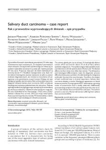
diabetes mellitus (DM), and treated by antidiabetic drugs. However, his DM deteriorated and he developed chronic renal failure due to diabetic nephropathy. At 61 years of age, the patient underwent hemodialysis. At 71 year of age, he was found to have a small tumor in the right kidney by CT. Right nephrectomy was performed. Grossly, a cortical tumor measuring 1.5 x 1.5 cm was found in the kidney (Figure 1). The tumor was encapsulated and yellowish white in color. Histologically, the tumor was completely encapsulated. The tumor cells were completely oncocytic (Figure 2A and 2B), and arranged entirely in a papillary pattern with fibrovascular cores (Figure 2A and 2B). The tumor cells have abundant oncocytic cytoplasm, and they showed stratification or pseudostratification. The nuclei was large and hyperchromatic with nucleoli (nuclear grade =3). A few mitotic figures were recognized. Psammoma bodies and hemosiderin positive for Prussian blue were scattered. Pale foamy cells were also scattered. There were no necrotic areas. Histochemically,
Oncocytic papillary renal cell carcinoma
Figure 1. Gross features of resected right kidney. The kidney shows a small tumor (arrows) of 1.5 x 1.5 x 1.5 size.
the tumor cells were stained positively with Hale’s colloidal iron (Figure 2C). Alcianblue-PAS stains revealed no mucins. An immunohistochemical study was performed
with the use of Dako’s Envision methods, as previously reported [6,7]. The immunohistochemical results were as follows: α-methylacylcoenzyme A rasemase (AMACR) +++ (Figure 3A), vimentin +++ (Figure 3B), cytokeratin (CK) 18 +++ (Figure 3C), CD10 +++, S-100 protein + (Figure 3D), MUC1 ++, MUC2 ++ (Figure 3E), MUC5AC ++ (Figure 3F), panCK Cam5.2 +, CK7 +, CK8 +, CK14 +, CK19 +, CK20 +, p53 +, HepPar1 + (Figure 3G), CD68 + (foamy cells), platelet-derived growth factor-α (PDGFRA) + (Figure 3H), PanCK AE1/3 -, PanCK WSS -, PanCK MNF115 -, CK 35BE12 -, CK5/6 -, EMA -, desmin -, smooth muscle antigen -, αfetoprotein -, CEA -, estrogen receptor -, progesterone receptor -, HER2 -, p63 -, and KIT -. Ki67 labeling was 6%. The background kidney was diabetic glomerulosclerosis. The clinical stage was pT1. The patient is now free of tumor 6 months after the nephrectomy. Discussion The present study fulfills the criteria of OPRCC. Immunoreactions of positive AMACR, vimentin,
Figure 2. Histological and histochemical findings of the tumor. A: The tumor is composed only of papillary proliferation of oncocytes with high grade atypia. HE, x100. B: High power view shows papillary oncocytic tumor with fibrovascular cores. The oncocytes have ample cytoplasm and show pseudostratification. The nuclei are large and hyperchromatic and contain nucleoli (Grade 3). Formy macrophages are seen. HE, x200. C: The tumor cells are positive for colloidal iron. Hales colloidal iron, x200.
709
Int J Clin Exp Pathol 2011;4(7):708-712
Oncocytic papillary renal cell carcinoma
Figure 3. Immunohistochemical features. The tumor cells are positive for AMACR (A), vimentin (B), CK18 (C), S100 protein (D), MUC2 (E), MUC5AC (F), HepPar1 (G), and PDGFRA (H). Immunostains, x200.
710
Int J Clin Exp Pathol 2011;4(7):708-712
Oncocytic papillary renal cell carcinoma
CD10, and CK7 are characteristics of papillary RCC [1, 4]. The positive expression of p53 suggests p53 mutations and malignant nature of the present tumor. The present tumor is different from renal oncocytoma, a benign tumor. Renal oncocytoma shows solid organoid pattern and does not usually display papillary structures [1]. In addition, the nuclear features and stratification of nuclei in the present tumor are different from renal oncocytoma. Immunohistochemically, renal oncocytoma expresses progesterone receptor and KIT [3] that was not recognized in the present tumor. The present tumor is also different from chromophobe RCC, which shows plant-like cells and KIT expression [1]. The present tumor is typical for oncocytic papillary type 2 RCC, and different from other variants of oncocytic papillary neoplasms [2, 4] In general, colloidal iron positivity is a hallmark of chromophobe RCC [1]. However, papillary RCC is infrequently weakly positive for colloidal iron [1]. The present tumor showed diffuse strong expression of colloidal iron, suggesting that OPRCC can express strong colloidal iron expression. The CK profile is not clear in OPRCC. The present OPRCC shows that lowmolecular weight CKs, but not high molecular ones, are present in the present OPRCC. S100 protein expression was once reported in OPRCC [8]. The present case also expressed S100 protein. The significance of this is unclear. Expression of MUC apomucins have been rarely reported in RCC [9, 10]. Kraus et al [9] reported that MUC1 expression in RCC was correlated with RCC progression. Leroy et al [10] reported that MUC1 and MUC3 were heterogeneously expressed in clear cell RCC. The present study showed negative reaction of Alcianblue/PAS and positive reactions of MUC1, MUC2, MUC5AC and MUC6, suggesting that nonglycosylated MUC apomucins are present in OPRCC. The expression of HepPar1 has been not examined in OPRCC. HepPar1 recognizes mitochondria-related antigen of normal and neoplastic hepatocytes. However, as is well known, HepPar1 is not specific for hepatocellular neoplasms. The present OPRCC expressed HepPar1. This may be because the oncocytic cytoplasm of OPRCC is due to abundant mitochondria [2]. PDGFRA has not been reported in OPRCC. The present case showed PDGFRA ex-
pression, although the significance is unclear. In very recent years, studies of cytogenetics in OPRCC have emerged. Park et al [5] stated that gain of chromosomes 3p, 11q and 17q, and loss of chromosome 4q was observed in RCC. Gobbo et al [8] also examined several gain and loss of chromosome 1, 2, 6, 10, and 17 in RCC. Much more cytogenetic and molecular studies of OPRCC remain to be performed. Finally, the present OPRCC is very small (1.5 cm). Most of OPRCC is large with median of 7.1 cm [2]. The small size of the present case may indicate that the present tumor is an early transformed tumor. The development of RCC in chronic renal disease and hemodialysis is much more common than in normal kidney. The estimated risk is 10-50 folds. The present OPRCC developed in chronic diabetic glomerulonephritis and hemodialysis. Conflict of Interest The author has no conflict of interest. Please address correspondence to: Tadashi Terada, MD, PhD, Department of Pathology, Shizuoka City Shimizu Hospital, Miyakami 1231 Shimizu-Ku, Shizuoka 424-8636, Japan. Tel: +81-54-336-1111, Fax: +81-54-334-1173, E-mail:
[email protected]
References [1]
[2]
[3]
[4]
[5]
711
Murphy WM, Grignon DJ, Perlman EJ. AFIP Atlas of tumor pathology series 4. Tumors of the kidney, bladder and related urinary structures. Armed Forces Institute of Pathology. Whashington DC, 2004. Hes O, Brunnelli M, Michael M, Cossu Rocca P, Hora M, Chiliosi M, Mina M, Boudova L, Menestrina F, Martignoni G. Oncocytic papillary renal cell carcinoma; a clinicopathologic, immunohistochemical, ultrastructural, and interphase cytogenetic study of 12 cases. Ann Diagn Pathol 2006; 10: 133-139. Mai KT, Kohler DM, Robertson SJ, Belanger EC, Marginean EC. Oncocytic papillary renal cell carcinoma with solid architecture; minic of renal oncocytoma. Pathol Int 2008; 58: 164168 Kunju LP, Wojno K, Wolf JS Jr, Cheng L, Shah RB, Papillary renal cell carcinoma with oncocytic cells and nonoverlapping low grade nuclei; expanding the morphologic spectrum with emphasis on clinicopathologic, immunohistochemical and molecular features. Hum Pathol 2008; 39: 96-101. Park BH, Ro JY, Park WS, Joe KJ, Kim K, Gong
Int J Clin Exp Pathol 2011;4(7):708-712
Oncocytic papillary renal cell carcinoma
[6]
[7] [8]
712
G, Cho YM. Oncocytic papillary renal cell carcinoma with inverted nuclear pattern; distinct subtype with an indolent clinical course. Pathol Int 2009; 59: 137-146. Terada, T, Kawaguchi M, Furukawa K, Sekido Y, Osamura Y. Minute mixed ductal-endocrine carcinoma of the pancreas with predominant intraductal growth. Pathol Int 52:740-746, 2002. Terada T, Kawaguchi M. Primary clear cell adenocarcinoma of the peritoneum. Tohoku J Exp Med 206:271-275, 2005. Gobbo D, Ebel JN, Delahunt B, Grignon DJ, Samaratunga H, Martognoni G, Zhang S, Wang M, Brunelli M, Cossu-Rocca P, Cheng L. Renal cell neoplasms of oncocytosis have distinct morphologic, immunohistochemical, and cytogenetic profiles. Am J Surg Pathol 2010; 34:620-6.
[9]
[10]
Kraus S, Abel PD, Nachtman C, Linsenmann HJ, Weidner W, Stamp GW, Chaudihary KS, Mitchell SE, Franke FE, Lalani el- N. MUC1 mucin and trefoil factor 1 protein expression in renal cell carcinoma; correlation with prognosis. Hum Pathol 2002; 33: 60-67. Leroy X, Copin MC, Devisme L, Buisine MP, Aubert JP, Gosselin B, Porchet N. Expression of human mucin gene in normal kidney and renal cell carcinoma. Histopathology 2002; 40: 450-457.
Int J Clin Exp Pathol 2011;4(7):708-712