ARTYKUŁY KAZUISTYCZNE Markowski J i wsp. Salivary duct carcinoma – case report
183
Salivary duct carcinoma – case report Rak z przewodów wyprowadzających ślinianek – opis przypadku Jarosław Markowski 1/, Agnieszka Piotrowska-Seweryn 1/, Anatol Wodołażski 2/, Katarzyna Kasperczyk 1/, Jarosław Paluch 1/, Piotr Wardas 1/, Michał Sokołowski 3/, Marcin Wojciechowski 3/, Wirginia Likus 4/ Katedra i Klinika Laryngologii, Wydział Lekarski w Katowicach, Śląski Uniwersytet Medyczny Katedra i Zakład Patomorfologii, Wydział Lekarski w Katowicach, Śląski Uniwersytet Medyczny 3/ Koło Naukowe przy Katedrze i Klinice Laryngologii, Wydział Lekarski w Katowicach, Śląski Uniwersytet Medyczny 4/ Katedra i Zakład Anatomii Prawidłowej, Wydział Lekarski w Katowicach, Śląski Uniwersytet Medyczny 1/ 2/
Z gruczołów ślinowych wywodzą się przynajmniej 24 różne typy nowotworów, które stanowią ok. 2% wszystkich pierwotnych raków ślinianek. Dostępne dane epidemiologiczne wskazują, że osobami najbardziej narażonymi są osoby płci męskiej powyżej 50 roku życia. Proporcja mężczyzn do kobiet dotkniętych tą chorobą wynosi 4:1. Rzadkość występowania oraz duża różnorodność histologiczna stanowi wyzwanie nie tylko w postaci trudności diagnostycznych, ale również w zrozumieniu ich patogenezy oraz poszukiwaniu nowych terapii. Dlatego też opis każdego przypadku jest tak ważny. Rak z przewodów wyprowadzających ślinianek (salivary duct carcinoma, SDC) w budowie histologicznej wykazuje podobieństwo do raka piersi. Podczas ogólnego badania laryngologicznego najczęściej jest stwierdzany guz okolicy przedusznej. Objawami towarzyszącymi mogą być: ból oraz niedowład nerwu twarzowego. 5-letni okres przeżywalności (disease-free survival, DFS) jest wciąż niekorzystny. Autorzy prezentują historię choroby 70-letniej kobiety z rakiem z przewodów wyprowadzających ślinianki przyusznej. Słowa kluczowe: nowotwór gruczołów ślinowych, rak z przewodów wyprowadzających ślinianek, gruczoł ślinowy, ślinianka, immunohistologia
The salivary glands give rise to at least 24 histologically distinct tumors which account for about 2% of all primary salivary epithelial malignancies. The epidemiological data available shows a predominance of male patients above 50 years old. The disease affects men four times more often than women. The infrequency and histological differentiation make the diagnostic process difficult and also pose a problem of understanding this malignancy and hinder the search for new therapies. Therefore, the analysis of each confirmed case is of great importance. Salivary duct carcinoma displays a similar histological appearance to ductal breast carcinoma. During a general ENT examination a growing tumor of preauricular area is usually observed. Additionally, accompanying symptoms like pain and facial paresis may occur. The 5-year disease-free survival (DFS) is still poor. The authors present a case of a 70-year-old woman suffering from salivary duct carcinoma (SDC). Keywords: parotid gland cancer, salivary duct carcinoma, salivary gland, immunohistology
© Otorynolaryngologia 2015, 14(3): 183-187
Adres do korespondencji / Address for correspondence
www.mediton.pl/orl
Dr hab. n. med. Jarosław Markowski Katedra i Klinika Laryngologii SUM Wydział Lekarski w Katowicach ul. Francuska 20-24, 40-027 Katowice
Introduction
According to World Health Organisation (WHO) annual incidence of all salivary gland tumors vary from 0.4-13.5 cases per 100,000 population. Most of them are benign. For malignant tumors, it is of 0.4 to 2.6 cases per 100,000 population. There is some variation in the frequency amongst different ethnic groups and according to place of residence [2]. For
A group of tumors of the salivary ducts by analogy to mammary duct carcinoma was first isolated in 1968 [1]. Salivary duct carcinoma is uncommon but it is characteristic for being one of the most aggressive type of salivary gland carcinomas with early distant metastases.
184
example Finnish statistical database indicates its presence as 0.2% of all human malignant neoplasm of which 4-6% are salivary duct carcinomas (SDC) [3]. In general, salivary duct carcinoma represents 9% of all malignant tumors of the salivary glands and accounts for up to 2% of all primary salivary epithelial neoplasms [4].The disease affects men four times more often than women, usually in patients over 50 years old. Parotid gland is most commonly involved [2], though several cases of SDC in submandibular gland and occasionally in minor glands have been described [5]. The etiology remains unknown. It is believed that most cases of SDC arise de novo probably from an in situ carcinoma (as a resemblance to ductal carcinoma of a breast). Rarely SDC can develop from pleomorphic adenoma [6-8]. According to McHugh so called ‘carcinoma ex-pleomorphic adenoma’ (CaExPA) is observed in around 20% of cases [8]. It has been also reported that the development of SDC of the parotid gland might be associated with IgG4-related autoimmune disease [9] and long-standing chronic obstructive sialadenitis [10]. A growing tumor is the most common initial symptom of the disease. In addition, pain and facial paresis (i.e. in case of infiltration of the facial nerve) may occur [2,4]. On examination a poorly circumscribed and often multinodular tumor is palpable. Additional diagnosis, such as ultrasound examination, might reveal scattered cysts and focal areas of necrosis. Fibrotic reaction can be also found [11]. However, CT and MR imaging act as gold standard in diagnostic procedures. From histopathological point of view SDC is characteristic for its resemblance to ductal breast cancer, within which nodules with cribriform, solid, cystic or papillary architecture can be distinguished. The usual treatment for invasive SDC covers complete surgical excision with radical neck dissection followed by adjuvant radiotherapy to the tumor bed and possibly chemotherapy [4]. It has been found that patients with HER2 subtype SDCs may benefit from targeted therapies with anti-HER2 monoclonal antibodies (Trastuzumab, Pertuzumab) or HER2 tyrosine kinase inhibitors (Lapatinib) [4]. Androgen deprivation therapy (ADT) in patients with recurrent or disseminated disease may be beneficial [12]. AR and HER2/neu receptor are expected to be used in molecular targeted therapy [13]. The clinical staging is based on TNM Staging System. A 5-year disease free survival (DFS) is still poor. According to analysis of Feinstein et al. it is
Otorynolaryngologia 2015, 14(3): 183-187
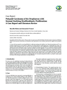
49% [14]. About 33% of patients develop local recurrence and more than 50% distant metastases at sites including lungs, bone, liver, brain and rarely skin [4,15]. Case Report The authors present a case of a 70-year-old woman suffering from salivary duct carcinoma (SDC) who presented with a progressively growing tumor-like mass on the right preauricular area. Fineneedle aspiration biopsy (FNAB) revealed tumor mixtus. On admission to the Clinical Department of Laryngology in February 2012 a hard, fixed, painless tumor of about 5 centimeters in diameter and infiltrating the external auditory meatus was palpable. Other diseases included hypertension, diabetes and myocardial insufficient disease. The patient did not report any allergies. No smoking or alcohol consumption was registered. A computed tomography (CT) revealed a tumor infiltrating left parotid gland, external ear canal (EAC) and concha cavum up to the border with head of mandibula, masseter muscle and retromandibular fossa. The diameters of the tumor were 26x26x28 centimeters (Fig. 1, Fig. 2). Several round lymph nodes of up to 8 milimiters in diameter in the region IIa and IIb on the ipsilateral side were also found. Radiological results could not differentiate between Warthin tumor, mucoepidermoid carcinoma and metastases of a squamous cell carcinoma of the EAC. The patient underwent a surgery. Due to intraoperative histopathlogical finding of a “carcinoma invasivum”, left radical parotidectomy with infiltrated facial nerve excision was performed. In the postoperative period total paresis of the left
Fig. 1. Computed tomography of head and neck. Axial projection. Hipodensive structure of 26x26x28 centimeters in the region of left parotid gland penetrating to the external ear canal (arrow). No bone infiltration has been found
Markowski J i wsp. Salivary duct carcinoma – case report
185
facial nerve could be observed. Histopathological examination of dissected tissue showed features of SDC (Fig. 3). Therefore adjuvant radiotherapy of total dose of 60 Gy to the postoperative and left submandibular region was applied. A 10-month follow up showed no local recurrences of SDC and no distant metastases. Unfortunately, no up to date information about the patient could be obtained.
Fig. 4. Patomorphological findings. H+E staining. Magnification x 100. Histologic examination reveals pleomorphic epithelioid tumour cells with a cribriform growth pattern, “Roman bridge” formation, and intraductal comedonecrosis
Discussion
Fig. 2. Computed tomography of head and neck. Coronary projection. A tumor of left parotid gland of irregular borders penetrating to the external ear canal (arrow)
Fig. 3. Histopathological examination of salivary duct carcinoma. H+E staining. Magnification x200
Salivary duct carcinoma is a rare entity. It belongs to high-grade locally aggressive malignancies with a tendency for regional and distant metastases [16]. Therefore, it seems that precise diagnosis plays a main role in proper treatment. As far as the diagnostic imaging is concerned, CT and MRI findings are nonspecific and ultrasound examination might reveal scattered cysts and focal areas of necrosis. According to Weon et al. in CT and MR images SDC is mostly seen as an ill-defined (85%) and infiltrative (60%) mass with frequent calcification (50%) and necrosis (80%) [17]. Most salivary gland tumours are brighter on T2 than T1 image, but this difference is minimal in prominently cellular tumours [2]. Although CT and MR imaging is nonspecific, comparison to histopathological examination showed that CT and MRI is useful for staging of SDC. According to Rajesh clinico-radiological tumor staging correlated well with pathologic tumor staging in 82% of the patients [17]. The assessment of the usefulness of fine-needle biopsy and its relation to pathomorphological findings also seems to be an important aspect. Some authors proved that FNAC of SDC is difficult to be interpreted due to overlapping cytomorphologic features [11]. However, FNAB remains the most important presurgical examination. In general, FNAB of parotid masses have a high accuracy with over 99% specificity and 85% sensitivity [11]. In our case, initial biopsy indicated the tumor mixtus,
Otorynolaryngologia 2015, 14(3): 183-187
186
while the postoperative histopathological findings revealed SDC. Therefore, it is our firm belief that it is crucial to take all diagnostic tests into consideration. Cytologically, recognizing the tumor as a primary tumor of the salivary gland and correct classification may be difficult. The biopsy results usually reveal cellular area, showing cribriform or pseudopapillary formations of tumor cells, often associated with necrosis. Nuclei are large and irregular with thickened membranes and prominent nucleoli. The cytoplasm is generally moderate to abundant and appears eosinophilic [18]. The most characteristic feature of salivary duct carcinoma is that it displays a similar histological appearance to ductal breast carcinoma. Histologic examination reveals pleomorphic epithelioid tumour cells with a cribriform growth pattern, “Roman bridge” formation, and intraductal comedonecrosis [2] (Fig. 4). Microscopically, large and polygonal atypical cells having abundant and finely granular cytoplasm, and prominent nucleoli, arranged in an irregular branching, cribriform, or papillary growth pattern as well as single cells formation in the background of frequent necrosis can be observed [19]. The estrogen receptor and progesterone receptor are not detected in most salivary duct carcinomas [13]. However, androgen receptors are present in over 80-90% [2,4,19] of salivary duct carcinomas. GCDFP-15 is also highly expressed in more than 90% of patients with SDC [19]. More than 20% of tumors show diffuse
and strong membranous staining for HER2/neu [13]. A cytokeratin-7 and cytokeratin-20 (CK7+/ CK20+) immunoprofile was recorded more often in salivary duct carcinomas compared to all other malignant salivary gland tumors [20]. The immunohistochemical diffenertiation of cancers that may have histological resemblance to SDC are shown in table I (Tab. I). In order to differentiate SDC metastases sialodochodysplasia may be helpful. Its presence might also support a primary salivary origin [2]. As far as the treatment is concerned, it is widely believed that surgical resection of a salivary gland is a gold standard. At the same time, no consensus on adjuvant therapy has been already established. Most authors are in favour of adjuvant radiation, but hormonotherapy seems to be an ideal solution for all HER2-positive tumors. In the described case surgical treatment with postoperative radiotherapy have been evaluated. Conclusions To sum up, salivary duct carcinoma is a rare malignancy localizing mainly in parotid gland. Fine-needle biopsy plays a great role in preoperative diagnosis, yet it does not always correspond with postoperative patomorphological findings. SDC is characteristic for its histologic resemblance to ductal breast carcinoma. Immunohistochemical examination seems to play a great role in introducing the right and most beneficial treatment after the surgical removal of the salivary gland.
Table I. Immunohistochemical differentiation of cancer that may have a similar histological findings to SDC [2,4,12] Receptors
Correlations with SDC
Additinal information
estrogen receptor α
not usually detected in SDC
ERα positivity in >25 % of tumor cells would strongly favor a breast cancer metastasis
progesterone receptor
not usually detected in SDC
seen in 2/10 acinic cell carcinomas and 3/10 mucoepidermoid carcinomas; useful for distinguishing from a breast cancer metastasis
androgen receptor (AR)
are present in over 80-90% SDC
are expected use for molecular targeted therapy
HER2 protein
positivity more than 20% of SDC tumors
are expected use for molecular targeted therapy
overexpression reported in up to 90% of cases GCDFP-15
highly expressed in more than 90%
CK-7 and CK-20
CK7 in every case; CK 20 only occasionally;
positivity in breast carcinoma
simultaneous presence is the most common of all malignant tumors of the salivary glands PSA
occasionally in SDC
S-100 protein, cytokeratins 5/7 and 14, typically negative p63, calponin, SMMHC, CD-117 and myoepthelial markers
prostate cancer at 90% does not present CK-7
Markowski J i wsp. Salivary duct carcinoma – case report
187
References 1. Kleinsasser O, Klein HJ, Hübner G. Salivary duct carcinoma. A group of salivary gland tumors analogous to mammary duct carcinoma Arch Klin Exp Ohren Nasen Kehlkopfheilkd 1968; 192(1): 100-5. 2. World Health Organization classification of tumours: pathology & genetics. Head and neck tumours. Lyon: IARCPress; 2005. 3. Luukka H, Klemi P, Leivo I, Koivunen P, Laranne J, Mäkitie A i wsp. Salivary gland cancer in Finland 199196: an evaluation of 237 cases. Acta Otolaryngolol 2005; 125(2): 207-14. 4. Simpson RH. Salivary Duct Carcinoma: New Developments – Morphological Variants Including Pure In Situ High Grade Lesions; Proposed Molecular Classification. Head Neck Pathol 2013; 7(Suppl 1): 48-58 5. van Heerden WFP, Raubenheimer EJ, Swart TJP, Boy SC. Intraoral salivary duct carcinoma: a report of 5 cases. J Oral Maxillofac Surg 2003; 61(1): 126-31. 6. Bourell LG, Chan KC, Hirsch DL. Salivary duct carcinoma ex pleomorphic adenoma of the palate: a case report. J Oral Maxillofac Surg 2015; 73(2): 370.e1-7 7. Simpson RH, Pereira EM, Ribeiro AC, Abdulkadir A, Reis-Filho JS. Polymorphous low-grade adenocarcinoma of the salivary glands with transformation to high-grade carcinoma. Histopathology 2002; 41(3): 250-9. 8. McHugh JB, Visscher DW, Barnes EL. Update on selected salivary gland neoplasms. Arch Pathol Lab Med 2009; 133(11): 1763-74. 9. Gill J, Angelo N, Yeong ML, Mclvor N. Salivary duct carcinoma arising in IgG4-related autoimmune disease of the parotid gland. Hum Pathol 2009; 40(6): 881-6. 10. Hogg RP, Ayshford C, Watkinson JC. Parotid duct carcinoma arising in bilateral chronic sialadenitis. J Laryngol Otol 1999; 113(7): 686-8. 11. Rajesh N G, Prayaga AK, Sundaram C. Salivary duct carcinoma: Correlation of morphologic features by fine needle aspiration cytology and histopathology. Indian J Pathol Microbiol 2011; 54(1): 37-41.
12. Jaspers HC, Verbist BM, Schoffelen R, Mattijssen V, Slootweg PJ, van der Graaf WT i wsp. Androgen receptorpositive salivary duct carcinoma: a disease entity with promising new treatment options. J Clin Oncol. 2011; 29(16): e473-6. 13. Nagao T, Sato E, Inoue R, Oshiro H, Takahashi RH, Nagai T i wsp. Immunohistochemical Analysis of Salivary Gland Tumors: Application for Surgical Pathology Practice. Acta Histochem Cytochem 2012; 45(5): 269–82. 14. Feinstein TM, Lai SY, Lenzner D, Gooding W, Ferris RL, Grandis JR i wsp. Prognostic factors in patients with highrisk locally advanced salivary gland cancers treated with surgery and postoperative radiotherapy. Head Neck 2011; 33(3): 318-23. 15. Cohen PR, Prieto VG, Piha-Paul SA, Kurzrock R. The „shield sign” in two men with metastatic salivary duct carcinoma to the skin: cutaneous metastases presenting as carcinoma hemorrhagiectoides. J Clin Aesthet Dermatol 2012; 5(9): 27-36. 16. Johnston ML, Huang SH, Waldron JN, Atenafu EG, Chan K, Cummings BJ i wsp. Salivary duct carcinoma: Treatment, outcomes and patterns of failure. Head Neck 2015 Apr 27. doi: 10.1002/hed.24107. 17. Weon YC, Park SW, Kim HJ, Jeong HS, Ko YH, Park IS i wsp. Salivary duct carcinomas: clinical and CT and MR imaging features in 20 patients. Neuroradiology 2012; 54(6): 631-40. 18. Mukunyadzi P. Review of fine-needle aspiration cytology of salivary gland neoplasms, with emphasis on differential diagnosis. Am J Clin Pathol 2002; 118 Suppl: S100-15. 19. Yamada S, Nabeshima A, Tabata T, Guo X, Tasaki T, Wang KY i wsp. Invasive salivary duct carcinoma ex pleomorphic adenoma of the parotid gland: a teaching case giving rise to the genuine diagnostic difficulty on an inadequate cytology specimen. Diagn Pathol 2012; 7: 61. 20. Nikitakis NG, Tosios KI, Papanikolaou VS, Rivera H, Papanicolaou SI, Ioffe OB. Immunohistochemical expression of cytokeratins 7 and 20 in malignant salivary gland tumors. Mod Pathol 2004; 17(4): 407-15.