0021-972X/04/$15.00/0 Printed in U.S.A.
The Journal of Clinical Endocrinology & Metabolism 89(10):4993– 4998 Copyright © 2004 by The Endocrine Society doi: 10.1210/jc.2004-0054
Bone Status and Fracture Prevalence in Russian Adults with Childhood-Onset Growth Hormone Deficiency R. BOUILLON, E. KOLEDOVA, O. BEZLEPKINA, J. NIJS, E. SHAVRIKHOVA, E. NAGAEVA, O. CHIKULAEVA, V. PETERKOVA, I. DEDOV, A. BAKULIN, V. OGANOV, AND A. F. ATTANASIO Katholieke Universiteit Leuven (R.B., J.N.), Laboratory of Experimental Medicine and Endocrinology, B-3000 Leuven, Belgium; Eli Lilly & Co. (E.K.), Kobe, Japan; Eli Lilly & Co. (A.F.A.), Florence, Italy; Endocrinological Research Centre (O.B., E.N., O.C., V.P., I.D.), Moscow, Russia; PSI Ltd. (E.S.), St. Petersburg, Russia; and State Research Centre (A.B., V.O.), Institute of Biomedical Problems, Russian Academy of Sciences, Space Physiology Department, Moscow, Russia The consequences of lifelong untreated childhood-onset GH deficiency (COGHD) on adult bone and especially fracture prevalence are largely unknown due to the lack of data on long-term outcome of untreated patients. Therefore, we studied adult Russian patients (n ⴝ 66; 28 females and 38 males) with idiopathic GH-untreated COGHD. Patients had isolated GH deficiency (IGHD; n ⴝ 18, age 23 ⴞ 10 yr) or multiple pituitary hormone deficiency (MPHD) with open (OMPHD; n ⴝ 27, age 23 ⴞ 5 yr) or closed growth plates (CMPHD; n ⴝ 21, age 55 ⴞ 12 yr). Bone mineral content (BMC) and bone mineral density (BMD) values were compared with 821 normal Russian controls. Fracture prevalence was ascertained from medical history and compared with similar data from 333 normal controls. Height SD score was ⴚ4.6 (range, ⴚ1.8 to ⴚ8.1). This represents 82% of the height of normal Russian adults. BMC of the lumbar spine, femoral neck, and total body of patients with IGHD was 54, 71, and 59%, respectively, of that of age- and sex-matched controls (all P < 0 0.001). A similarly decreased BMC (42– 69% of expected values) was found for all bone re-
G
H IS A major regulator of growth plate chondrocytes and bone cells. Lifelong GH deficiency (GHD) not only impairs linear growth but also decreases the threedimensional size (volume) of bone (1–3). Therefore, peak bone mass is decreased in such patients, whereas in eugonadal acromegalic patients, bone size and mass are usually increased (2– 6). In most developed societies, childhoodonset GHD (COGHD) is detected and treated early in life so that large cohorts of untreated COGHD adult patients are no longer available for studying the effects of lifelong GHD on specific tissues. Here, we report on the effects of COGHD (GHD alone or in combination with substituted other pituitary hormone deficiencies) on bone mass, bone size, and fracture prevalence in a cohort of 66 Russian patients who were virtually never treated with GH.
gions of patients with both OMPHD and CMPHD. Mean areal BMD measurements (g/cm2) varied (Z scores between ⴚ1.8 and ⴚ3.0), but the calculated true bone density (g/cm3) was normal in patients with IGHD or CMPHD and only slightly decreased (Z score, ⴚ0.8) in patients with OMPHD. Lifetime low-energy fracture prevalence was normal in patients with IGHD but substantially exceeded the expected prevalence in OMPHD (odds ratio of fracture ⴝ 3.0; 0.6 fractures per patient; P < 0.0001) or CMPHD patients (odds ratio for fracture ⴝ 7.4; 2.2 fractures per patient; P < 0.0001). In conclusion, IGHD and MPHD of childhood onset very substantially impair adult height and BMC. Although areal BMD is frankly decreased, volumetric bone density is unaffected, but nevertheless, the fracture prevalence in patients with MPHD is markedly increased. These observations demonstrate that not only volumetric density but also bone mass and shape are major determinants of bone strength. (J Clin Endocrinol Metab 89: 4993– 4998, 2004)
Patients and Methods Patients Sixty-six adult patients, all Caucasians, were studied (Table 1). They were recruited from the Moscow Endocrine Research Centre and other Moscow clinics or by dedicated search among artists (circus, theater, and musical academia). Eighteen patients (eight females and 10 males) had isolated GHD (IGHD); 27 (10 females and 17 males) had GHD with multiple pituitary hormone deficiencies (MPHD) and open growth plates (OMPHD) due to delayed sex steroid hormone replacement therapy; and 21 (10 females and 11 males) had GHD with MPHD and closed growth plates (CMPHD). The diagnostic criteria used to establish pituitary hormone deficiencies are presented in Table 2. All patients with MPHD had combined GH⫹TSH⫹LH/FSH deficiency. In addition, nine were ACTH deficient, eight were prolactin deficient, and six were ACTH and prolactin deficient. Twenty of 66 patients were working in a circus, but only three performed acrobatic activities. Oral consent to participate in this study was obtained from all subjects.
Hormonal replacement Abbreviations: BMC, Bone mineral content; BMD, bone mineral density; CMPHD, multiple pituitary hormone deficiency with closed growth plates; COGHD, childhood-onset GH deficiency; DEXA, dualenergy x-ray absorptiometry; GHD, GH deficiency; IGHD, isolated GH deficiency; MPHD, multiple pituitary hormone deficiency; OMPHD, multiple pituitary hormone deficiency with open growth plates. JCEM is published monthly by The Endocrine Society (http://www. endo-society.org), the foremost professional society serving the endocrine community.
All IGHD and 25 MPHD patients had never received any GH treatment. In the remaining 23 patients, GH therapy during childhood had been sporadic, with intermittent courses of treatment with pituitaryderived GH. Periods of exposure ranged from 2 months to a maximum of 2 yr (median, 16 months). Replacement with other hormones was made with thyroxine, testosterone propionate, or ethinyl estradiol, and prednisolone (Table 2). However, compliance with hormonal replacement therapy had not been optimal in some patients. Clinical case histories were available for all patients, and detailed
4993 Downloaded from https://academic.oup.com/jcem/article-abstract/89/10/4993/2844348 by guest on 07 June 2018
4994
J Clin Endocrinol Metab, October 2004, 89(10):4993– 4998
Bouillon et al. • Fractures and Bone Density in GHD
TABLE 1. Demographic, diagnostic, and auxological data of the patients
Age (yr) Females/males GH peak (g/liter) Height SDS Height % of controls BA/CA
IGHD (n ⫽ 18)
OMPHD (n ⫽ 27)
CMPHD (n ⫽ 21)
23.7 ⫾ 10.4 8/10 1.01 ⫾ 0.8 ⫺4.08 ⫾ 1.35b 84 ⫾ 6 0.89 ⫾ 0.14
23.1 ⫾ 5 10/17 0.55 ⫾ 0.6 ⫺4.43 ⫾ 1.66b 83 ⫾ 6 0.55 ⫾ 0.13
54.6 ⫾ 12a 10/11 0.49 ⫾ 0.52 ⫺5.27 ⫾ 1.42b 80 ⫾ 5
All patients were 18 yr or older. The large SD of their age is explained by the inclusion of a single 61-yr-old patient. Twenty of 66 patients were working in a circus, but only three patients were performing acrobatic exercises, whereas the others were not involved in physically demanding circus activities. SDS, SD score; BA/CA, bone age to chronological age ratio. a P ⬍ 0.001 vs. OMPHD and IGHD. b P ⬍ 0.001 vs. normal Russian control subjects. TABLE 2. Diagnosis of GH or other hormone deficiencies and the treatment regimens of 66 adults with COGHD Hormone deficiency
No.
Diagnosis
Treatment
GH
66
ITT with peak GH ⬍ 3 ng/ml
TSH
48
LH/FSH
48
Prolactin ACTH
5 16
fT4 ⬍ 9 pM tT4 ⬍ 65 nM M: testicular volume ⬍ 5 ml testosterone ⬍ 12 nM F: clinical ⫹ E2 ⬍ 50 pM Low plasma level Cortisol ⬍ 150 nM
None except for very short-term (m) GH/R during childhood in 23 patients; no R 12 months before study T4 (50 –150 g/d) Testosterone proprionate (125–250 mg/4 wk) Ethinyl estradiol (25 g/d) None Prednisone (2.5–5 mg/d)
ITT, Insulin tolerance test; R, replacement therapy; fT4, free T4; tT4, total T4 concentration; M, male; F, female; E2, estradiol. information on number of traumatic and nontraumatic bone fractures was obtained by detailed questionnaires and/or patient records.
Controls Normal reference values for bone mass [bone mineral content (BMC)] and bone mineral density (BMD) by dual-energy x-ray absorptiometry (DEXA) were obtained from 821 Caucasian adults (494 females and 327 males, age 18 – 80 yr) living in the Moscow area and without chronic diseases known to possibly influence bone metabolism. These data were available from the Russian Space Agency Research Program. Reference data for radiogrammetry were collected in 44 age- (⫾ 2 yr) and sexmatched healthy volunteers. To assess lifetime occurrence of fractures in the normal population, a total of 333 normal Russian adults (mean age ⫾ sd, 40 ⫾ 17 yr; range, 16 –77 yr) were given a detailed questionnaire to collect information on number, type, and cause of fractures.
Methods For height sd score calculations, the British standards (7) were used. Bone age was determined according to Greulich and Pyle (8). BMC and BMD of patients and controls were measured at the Space Physiology Department in Moscow using a Hologic QDR-1000/W instrument (Hologic, Inc., Waltham, MA) using standard company software. Quality control data, obtained by scanning regularly a Hologic spine phantom, revealed a variation coefficient of less than 0.5% and no drift in linearity of data. All acquisitions were performed by trained operators in accordance with the technical manual. The measured areal BMD data of the lumbar spine, left femoral neck, and total body were used to calculate apparent or volumetric bone density (9 –11) using the following formulae: L1–L4 ⫽ BMDL1–L4/公(areaL1–L4); femoral neck ⫽ BMC femoral neck/(area femoral neck)2; and total body ⫽ BMD total body/height. These calculated values are used whenever volumetric bone density results are mentioned in this article.
Radiogrammetry Semiautomatic measurements with a 0.01-mm readout of periosteal and endosteal width of the second metacarpal were made on the posteroanterior right-hand radiograph by a single observer (J.N.) who was blinded to the study group assignment, as described (12). The cross-
Downloaded from https://academic.oup.com/jcem/article-abstract/89/10/4993/2844348 by guest on 07 June 2018
sectional area was calculated by subtracting the square of the endosteal from the square of the periosteal diameter assuming a cylindrical model.
Statistical analysis The data were analyzed using the SAS statistical software (SAS Institute, Inc., Cary, NC). Z scores were calculated for height and BMD values using the appropriate control data. Student’s t test was used for group comparison. Group comparison for fracture risk was calculated using the Mann-Whitney U test. Data are presented as mean ⫾ sd.
Results Demographic and auxological data
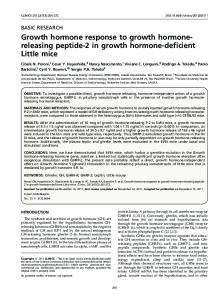
Demographic and auxological data are presented in Table 1. Gender distribution was comparable in all three groups. Although IGHD and OMPHD patients were of comparable age, CMPHD patients were significantly older (P ⬍ 0.001). Mean height sd score values in the three groups ranged from ⫺4.08 to ⫺5.27. Eight IGHD and all OMPHD patients had still open epiphyses, resulting in a ratio of bone age to chronological age of 0.89 ⫾ 0.14 in IGHD and 0.55 ⫾ 0.13 in OMGHD patients; the bone age to chronological age ratio was significantly lower in OMGHD than in IGHD patients (P ⬍ 0.02). Actual standing height values varied from 80% (CMPHD) to 84% (IGHD) of the values of age- (⫾ 2 yr) and sex-matched controls from the Russian DEXA reference population (Fig. 1). BMC and BMD
BMC was markedly decreased in all groups of patients. When compared with controls, total BMC of the lumbar spine (L1–L4) was significantly decreased in all three groups (% of normal values: OMPHD, 42 ⫾ 13%; CMPHD, 47 ⫾ 11%;
Bouillon et al. • Fractures and Bone Density in GHD
J Clin Endocrinol Metab, October 2004, 89(10):4993– 4998 4995
FIG. 1. Bone mass (BMC) and standing height in patients with COGHD compared with normal Russian adults. All values are compared with results obtained in normal controls set as 100%.
TABLE 3. Areal and calculated volumetric BMD in patients with COGHD IGHD (n ⫽ 18)
OMPHD (n ⫽ 28)
CMPHD (n ⫽ 20)
⫺2.3 ⫾ 1.1b ⫺2.2 ⫾ 1.2b ⫺1.8 ⫾ 1.2b
⫺3.6 ⫾ 0.8b ⫺3.0 ⫾ 0.9b ⫺2.8 ⫾ 0.8b
⫺2.6 ⫾ 1.0b ⫺1.9 ⫾ 1.1b ⫺1.8 ⫾ 0.7b
0.3 ⫾ 1.4 0.3 ⫾ 1.1 0.3 ⫾ 1.9
⫺1.0 ⫾ 0.9b ⫺0.6 ⫾ 0.7b ⫺0.8 ⫾ 1.4c
⫺0.4 ⫾ 1.1 0.1 ⫾ 1.9 0.6 ⫾ 1.1d
a
Areal BMD L1–L4 Femoral neck Total body Volumetric BMDa L1–L4 Femoral neck Total body
a Z score vs. Russian normal controls. Data are presented as mean ⫾ SD. b P ⬍ 0.001. c P ⬍ 0.01. d P ⬍ 0.05.
and IGHD, 54 ⫾ 17%; Fig. 1). Relative values of total BMC of the femoral neck and total body were also markedly reduced and only 47–71% of normal (Fig. 1). Areal and calculated volumetric bone densities in the three patients groups are shown in Table 3. Compared with the reference population, areal BMD was severely decreased in all patients (P ⬍ 0.001). The lowest Z score was observed in patients with OMPHD, whereas IGHD and CMPHD patients had a comparable bone deficit. On the other hand, mean values of volumetric BMD were in the normal range in these two groups and were only slightly decreased in OMPHD patients (Z score, ⫺0.6 to ⫺1.0).
Downloaded from https://academic.oup.com/jcem/article-abstract/89/10/4993/2844348 by guest on 07 June 2018
TABLE 4. Radiogrammetry of the metacarpal bones
D (mm) d (mm) Cortical area (mm2)
IGHD
CMPHD
86.2 ⫾ 9 98.9 ⫾ 11 70 ⫾ 14
91 ⫾ 10 129 ⫾ 45 68 ⫾ 14
The outer and inner diameter and cortical area (mean ⫾ SD) are compared with Russian control subjects and expressed as percentage of expected values. D, Outer diameter of second metacarpal bone; d, inner diameter of second metacarpal bone. All values are compared with values obtained in normal Russian healthy controls (set as 100%).
Radiogrammetry
Radiogrammetry was performed in IGHD and CMPHD patients only. These data are presented in Table 4. Compared with the age- and sex-matched controls, both IGHD and CMPHD patients had a reduction in the cortical area value of about 30% (IGHD, 70 ⫾ 14% and CMPHD, 68 ⫾ 14% of controls; P ⬍ 0.0005). Fracture incidence
Nontraumatic fractures were recorded in 28 OMPHD and CMPHD patients who had 70 fractures compared with 67 fractures in the 333 controls. The number of traumatic fractures (car accident and similarly severe trauma) reported in the patient and control groups was eight and 55, respectively. In IGHD patients, only traumatic fractures were recorded (7%, not significant vs. controls). By contrast, 37 and 18% of
4996
J Clin Endocrinol Metab, October 2004, 89(10):4993– 4998
Bouillon et al. • Fractures and Bone Density in GHD
the young adults with OMPHD had a history of at least one or at least two low-energy fractures, respectively (Table 5). The mean number of low-energy fractures per OMPHD patient was 3-fold higher than observed in the young control subjects (P ⬍ 0.0001). In the older group with CMPHD, the reported prevalence of at least one or at least two low-energy fractures was 62 and 33%, respectively. The mean number of low traumatic fractures per CMPHD patient was 10-fold greater than that observed in the group of older control subjects (P ⬍ 0.0001; Table 5). Most nontraumatic fractures were observed in the wrist (n ⫽ 24), hand, foot, ribs, or tibia. Few clinical vertebral fractures and no hip fractures were recorded. No radiological data were available to assess the integrity of the lumbar spine. Discussion
GH action on the growth plate is critical for longitudinal, one-dimensional growth (2, 3). However, GH also acts on periosteal bone apposition (13) and thus affects the threedimensional bone structure as well. Accordingly, GHD does not only affect skeletal height but bone size and volume as well. In addition, the reduced muscle mass and strength and increased fat mass of GHD patients may indirectly influence bone metabolism. We have evaluated the impact of GHD on bone mass, size, and mineral density and on fracture incidence in a group of adult hypopituitary GHD patients who had not been treated adequately with GH during childhood. To study patients in whom GHD had affected bone development from early childhood onwards, we selected only patients with an idiopathic cause, which can be assumed to have had its onset in early childhood. Some patients had been exposed to GH therapy, but as confirmed by the actual height values, the treatment periods had been too short to significantly affect adult outcomes. Bone mass was severely affected at all sites (⫺29 to ⫺58%), substantially more than the relative deficit in height (⫺16 to ⫺20%) would predict. Assuming that the relative height difference would also apply for the two other bone dimensions, however, the calculated relative bone volume would also be only 61% for patients with IGHD and 59 and 51% for patients with OMPHD and CMPHD, respectively. Therefore, the estimated reduction of total bone volume approximates the measured reduction in bone mass. The discrepancy with the relative height deficit is impressive (Fig. 1). We previously reported a similar type of discrep-
ancy, although to a lesser extent, in subjects with severe COGHD who had been treated with GH during childhood (6). Their height deficit was about 3–5% (⫺1 sd) of agematched controls, but their total bone mass was only 80% of age-matched controls (6). Taken together, these data suggest that, especially in GH-untreated but also in GH-treated COGHD patients, bone size and volume are significantly more affected than skeletal height. For fracture prediction, BMD rather than bone mass (BMC) has been used in nearly all clinical prospective studies (14, 15). DEXA measures bone mass and projected surface area and uses both parameters to calculate areal BMD (16). This measure, however, underestimates the volumetric density in case of reduced bone size (small height) (14). Therefore, it is not surprising that in our patients areal BMD was severely reduced (Table 3) but mean volumetric BMD was normal after closure of the growth plates. This was the case in the younger IGHD as well as the much older CMPHD patients (Table 3). This indicates that COGHD equally affects bone size and BMC but that the remaining low bone volume is adequately mineralized. These data also suggest that suboptimal hormone replacement of other pituitary hormone deficiencies, as might have been the case in some of our MPHD patients, does not cause a further deterioration of volumetric bone density beyond that observed with IGHD. These observations are in line with previous studies reporting normal volumetric BMD in GHD patients who had been adequately treated with GH during childhood (3, 17, 18). Compared with IGHD and CMPHD patients, volumetric BMD in OMPHD patients was lower, but this can be assumed to be due to sex steroid hormone deficiency and delayed substitution and the resulting considerable retardation in their pubertal bone age. Although the methodology used in the present study did not address directly the relative deficit of cortical vs. trabecular bone, the radiogrammetry results indicate that bone width was severely impaired in all of our patients (Table 4). The deficit in cortical metacarpal area (⬃70% of control) is comparable to that seen for total-body BMC, which also reflects better cortical than trabecular bone mass. Such a deficit of cortical bone could also explain why the fractures occur especially in bones with predominant cortical bone (see below). Indeed, the bending strength of bone is proportional to the fourth power of its distance to the central axis of long bones (19).
TABLE 5. Lifetime prevalence of nontraumatic fractures
One or more fractures (%) Odds ratio Confidence limits Two or more fractures (%) Odds ratio Confidence limits Mean no. of fractures per patient ⫾ P
IGHD (n ⫽ 18)
OMPHD (n ⫽ 27)
CMPHD (n ⫽ 21)
0
37 3.05 1.25–7.45 18 35 3.9 –313 0.59 ⫾ 0.9 ⬍0.0001
62 7.41 2.84 –19.36 33 12.2 3.7–39.8 2.19 ⫾ 3.7 ⬍0.0001
0
SD
0
Normal controls Young (n ⫽ 186)
Older (n ⫽ 147)
16
18
0.64
3.9
0.17 ⫾ 0.39
0.22 ⫾ 0.53
The lifetime prevalence of nontraumatic fractures in 66 Russian patients with COGHD was calculated on the basis of a detailed questionnaire and compared with similar data obtained in 186 young (25 ⫾ 5 yr) and 147 older (53 ⫾ 12 yr) normal Russian control subjects. OMPHD patients were compared with the young controls, and CMPHD patients were compared with the older controls.
Downloaded from https://academic.oup.com/jcem/article-abstract/89/10/4993/2844348 by guest on 07 June 2018
Bouillon et al. • Fractures and Bone Density in GHD
The incidence or prevalence of fractures in GHD patients has been evaluated previously in three cross-sectional studies (20 –22) and in one observational database (23). A 2- (20, 21) to 4.9-fold (23) increase in fracture risk was observed in GHD patients with substituted MPHD, and a similar trend was also found in a limited number of older IGHD patients (22). Data on adults with lifelong untreated COGHD are virtually nonexistent. In the present study, we found a low prevalence of fractures in young adults with IGHD, which is in agreement with published data in GH-treated COGHD patients (18). The discrepancy between the prevalence of fractures in young patients with IGHD and older patients with MPHD is obvious but unresolved. Unfortunately, we were unable to find sufficient, older, GH-untreated IGHD patients to assess their lifetime fracture prevalence. On the other hand, the significantly reduced bone size and volume found in our young IGHD adults is identical to that of the older MPHD patients. Moreover, previous studies have found identical bone histology and bone turnover markers in IGHD and MPHD patients (24, 25). This suggests that the quality of bone is not different in both conditions. Therefore, the low BMC values of IGHD patients probably also carry a high risk of fractures at older ages, but only specific data can validate this conclusion. Conversely, our data in both MPHD groups show that fractures are an early complication of combined GHD and MPHD in the absence of GH treatment, specifically if hormonal replacement of other hormone deficiencies has not been adequate. Compared with controls, MPHD patients had a 3- to 7-fold increased fracture prevalence (Table 5). At a mean age of 55 yr, CMPHD patients had, on average, 2.2 fractures per patient with 37 and 62% of patients having sustained at least one or at least two fractures, respectively. The sample size of the present study was too small to evaluate gender differences in fracture rates. The fracture prevalence of 16 –18% found in the Russian control subjects is very consistent with an 18% prevalence reported in a Finnish study (7217 women with a mean age of 53 yr) (26). The fracture prevalence of untreated GHD Russian adults even exceeds the lifetime fracture prevalence observed in Rochester anorexia nervosa patients (27). Nearly all fractures involved the radius or other bones (hand/foot, tibia) whereby cortical bone strength is probably involved. Because volumetric BMD is normal in CMPHD patients and only one Z score below normal in OMPHD patients, one can conclude that reduced bone mass, bone size, or bone quality are the main culprits and not volumetric density. Because no structural abnormalities but only a decreased bone turnover have been found in adult GHD patients (24), bone size and associated low bone mass seem to be the causative factors. Similarly, because gender differences in volumetric bone density are small (13, 28), differences in fracture prevalence between females and males might be more related to gender differences in the geometry of bone. Our data and conclusions are supported by findings in other clinical situations with impaired GH-IGF-I axis. Short stature, decreased BMC, and decreased areal BMD, but normal volumetric bone mineral apparent density, were also reported in patients with GH receptor deficiency (Laron patients), GHRH receptor deficiency (25, 29), and mutations of
Downloaded from https://academic.oup.com/jcem/article-abstract/89/10/4993/2844348 by guest on 07 June 2018
J Clin Endocrinol Metab, October 2004, 89(10):4993– 4998 4997
the IGF-I gene (30). In addition, normal structure of trabecular and cortical bone was described in COGHD adults (24) and in GH receptor-deficient patients (25). Finally, in GH receptor and IGF-I knockout mice (31, 32), total-body calcium and areal BMD are decreased, but volumetric BMD is normal. Total-body BMC is reduced to the same extent as body weight in the GHRH receptor-deficient (little) mouse, which displays a growth pattern very similar to that of GHD rodents (33). These human and animal data strongly indicate that COGHD, and not the other pituitary hormone deficiencies, determines bone size and mass. Because we do not have an older group of IGHD patients, we cannot extend this conclusion to fracture prevalence. Do these results have implications for non-GHD adults? Indeed, we think that the present study clearly reveals the importance of geometry and size as a major determinant, apart from density and quality, for bone strength and fracture risk. Recent long-term prospective data on postmenopausal women indicate that section modulus, a geometric parameter calculated mainly on the basis of bone width, is a substantially better predictor of fractures than BMD (34). In conclusion, bone mass is severely (⫾ 50%) reduced in GH-untreated patients with idiopathic COGHD, irrespective of the presence of other pituitary hormone deficiencies. Lifetime nontraumatic fracture prevalence is significantly increased when GHD is coupled with MPHD. Further study is needed to determine whether IGHD is associated with an increased risk of fractures. The present study demonstrates not only the critical role of GH in the development and maintenance of normal bone but also that bone mass and size, rather than volumetric density, are major determinants of bone strength. Acknowledgments We thank Eli Lilly for supporting this study and the members of the International Hypopituitary Control and Complications Study Advisory Board for scientific advice. We thank Prof. Steven Lamberts and Dr. Mark L. Hartman for their critical review of the manuscript. Received January 13, 2004. Accepted June 30, 2004. Address all correspondence and requests for reprints to: Roger Bouillon, Legendo, Gasthuisberg, Herestraat 49, B-3000 Leuven, Belgium. E-mail:
[email protected]. This work was supported by Eli Lilly & Co. This work was presented in part at the American Bone and Mineral Research Society 2001 meeting in Phoenix, Arizona.
References 1. Bouillon R, Koledova K, Bezlepkina O, Chernikhova E, Nagajeva E, Bakulin A, Nijs J, Peterkova V, Dedov I, Oganov O, Attanasio A 2001 Lifelong hypopituitarism and growth hormone deficiency decreases bone size but not volumetric density yet increases prevalence of fractures. J Bone Miner Res 16:S224 2. Ohlsson C, Bengtsson BA, Isaksson OG, Andreassen TT, Slootweg MC 1998 Growth hormone and bone. Endocr Rev 19:55–79 3. de Boer H, Blok GJ, Van der Veen EA 1995 Clinical aspects of growth hormone deficiency in adults. Endocr Rev 16:63– 86 4. Bouillon R 1991 Growth hormone and bone. Horm Res 36(Suppl 1):49 –55 5. Bolanowski M, Wielgus M, Milewicz A, Marciniak R 2000 Axial bone mineral density in patients with acromegaly. Acad Radiol 7:592–594 6. Attanasio AF, Howell S, Bates PC, Frewer P, Chipman J, Blum WF, Shalet SM 2002 Body composition, IGF-I and IGFBP-3 concentrations as outcome measures in severely GH-deficient (GHD) patients after childhood GH treat-
4998
7. 8. 9. 10. 11. 12. 13. 14. 15. 16. 17.
18. 19. 20. 21.
22.
J Clin Endocrinol Metab, October 2004, 89(10):4993– 4998
ment: a comparison with adult onset GHD patients. J Clin Endocrinol Metab 87:3368 –3372 Tanner JM, Goldstein H, Whitehouse RH 1970 Standards for children’s height at ages 2–9 years allowing for heights of parents. Arch Dis Child 45:755–762 Greulich WW, Pyle SI 1959 Radiographic atlas of skeletal development of the hand and wrist. 2nd ed. Stanford, CA: Stanford University Press; 1–256 Melton III LJ, Khosla S, Atkinson EJ, Oconnor MK, Ofallon WM, Riggs BL 2000 Cross-sectional versus longitudinal evaluation of bone loss in men and women. Osteoporos Int 11:592–599 Katzman DK, Bachrach LK, Carter DR, Marcus R 1991 Clinical and anthropometric correlates of bone mineral acquisition in healthy adolescent girls. J Clin Endocrinol Metab 73:1332–1339 Henry YM, Eastell R 2000 Ethnic and gender differences in bone mineral density and bone turnover in young adults: effect of bone size. Osteoporos Int 11:512–517 Dequeker J 1976 Quantitative radiology: radiogrammetry of cortical bone. Br J Radiol 49:912–920 Seeman E 1997 Perspective. From density to structure: growing up and growing old on the surface of bone. J Bone Miner Res 12:509 –521 Genant HK, Gluer CC, Lotz JC 1994 Gender differences in bone density, skeletal geometry, and fracture biomechanics. Radiology 190:636 – 640 Bass S, Delmas PD, Pearce G, Hendrich E, Tabensky A, Seeman E 1999 The differing tempo of growth in bone size, mass, and density in girls is regionspecific. J Clin Invest 104:795– 804 Kelly TL 1990 Bone mineral reference databases for American men and women. J Bone Miner Res 5:249 Fors H, Bjarnason R, Worent L, Albertsson-Wikland K, Bosaeust L, Bengtsson BA, Johansson G 2001 Currently used growth-promoting treatment of children results in normal bone mass and density. A prospective trial of discontinuing growth hormone treatment in adolescents. Clin Endocrinol (Oxf) 55:617– 624 Baroncelli GI, Bertelloni S, Sodini F, Saggese G 2002 Lumbar bone mineral density at final height and prevalence of fractures in treated children with GH deficiency. J Clin Endocrinol Metab 87:3624 –3631 Seeman E 2003 Periosteal bone formation. A neglected determinant of bone strength. N Engl J Med 349:320 –323 Rosen T, Wilhelmsen L, Landin-Wilhelmsen K, Lappas G, Bengtsson BA 1997 Increased fracture frequency in adult patients with hypopituitarism and GH deficiency. Eur J Endocrinol 137:240 –245 Wu¨ster C, Slenczka E, Ziegler R 1991 Erho¨ hte pra¨ valenz von osteoporose und arteriosklerose bei konventionell substituierter hypophysenvorderlappeninsuffizienz: bedarf einer zusa¨ tzlichen wachstumshormonsubstitution? Klin Wochenschr 69:769 –773 Vestergaard P, Jorgensen JO, Hagen C, Hoeck HC, Laurberg P, Rejnmark L,
Bouillon et al. • Fractures and Bone Density in GHD
23.
24.
25.
26. 27. 28. 29.
30.
31.
32. 33.
34.
Brixen K, Weeke J, Andersen M, Conceicao FL, Nielsen TL, Mosekilde L 2002 Fracture risk is increased in patients with GH deficiency or untreated prolactinomas—a case-control study. Clin Endocrinol (Oxf) 56:159 –167 Wuster C, Abs R, Bengtsson BA, Bennmarker H, Feldt-Rasmussen U, Hernberg-Stahl E, Monson JP, Westberg B, Wilton P 2001 The influence of growth hormone deficiency, growth hormone replacement therapy, and other aspects of hypopituitarism on fracture rate and bone mineral density. J Bone Miner Res 16:398 – 405 Bravenboer N, Holzmann P, de Boer H, Roos JC, Van der Veen EA, Lips P 1997 The effect of growth hormone (GH) on histomorphometric indices of bone structure and bone turnover in GH-deficient men. J Clin Endocrinol Metab 82:1818 –1822 Bachrach LK, Marcus R, Ott SM, Rosenbloom AL, Vasconez O, Martinez V, Martinez AL, Rosenfeld RG, Guevara-Aguirre J 1998 Bone mineral, histomorphometry, and body composition in adults with growth hormone receptor deficiency. J Bone Miner Res 13:415– 421 Randell KM, Honkanen RJ, Kro¨ger HSS, Saarikoski S 2002 Does hormonereplacement therapy prevent fractures in early postmenopausal women? J Bone Miner Res 17:528 –533 Lucas AR, Melton LJ, Crowson CS, O’Fallon WM 1999 Long-term fracture risk among women with anorexia nervosa: a population-based cohort study. Mayo Clin Proc 74:972–977 Seeman E 1999 The structural basis of bone fragility in men. Bone 25:143–147 Maheshwari HG, Bouillon R, Nijs J, Baumann G 2003 The impact of congenital, severe, untreated growth hormone (GH) deficiency on bone size and density in young adults: insights from genetic GH-releasing hormone receptor deficiency. J Clin Endocrinol Metab 88:2614 –2618 Woods KA, Camacho-Hubner C, Bergman RN, Barter D, Clark AJ, Savage MO 2000 Effects of insulin-like growth factor I (IGF-I) therapy on body composition and insulin resistance in IGF-I gene deletion. J Clin Endocrinol Metab 85:1407–1411 Sjogren K, Bohlooly YM, Olsson B, Coshigano K, Tornell J, Mohan S, Isaksson OG, Baumann G, Kopchick J, Ohlsson C 2000 Disproportional skeletal growth and markedly decreased bone mineral content in growth hormone receptor -/- mice. Biochem Biophys Res Commun 267:603– 608 Bikle D, Majumdar S, Laib A, Powell-Braxton L, Rosen C, Beamer W, Nauman E, Leary C, Halloran B 2001 The skeletal structure of insulin-like growth factor I-deficient mice. J Bone Miner Res 16:2320 –2329 Donahue LR, Beamer WG 1993 Growth hormone deficiency in ‘little’ mice results in aberrant body composition, reduced insulin-like growth factor-I and insulin-like growth factor-binding protein-3 (IGFBP-3), but does not affect IGFBP-2, -1 or -4. J Endocrinol 136:91–104 Ahlborg HG, Johnell O, Turner CH, Rannevik G, Karlsson MK 2003 Bone loss and bone size after menopause. N Engl J Med 349:327–334
JCEM is published monthly by The Endocrine Society (http://www.endo-society.org), the foremost professional society serving the endocrine community.
Downloaded from https://academic.oup.com/jcem/article-abstract/89/10/4993/2844348 by guest on 07 June 2018