A WORKFLOW FOR DIGITAL PHOTOGRAPHY OF FOSSIL SPECIMENS by Paul A. Selden Selden, P. A. 2014. A workflow for digital photography of fossil specimens. The Geological Curator 10 (2): 93–98. 93-97. A workflow for digital photography of fossil specimens is presented, in the hope that it will provide helpful for those wishing to set up a facility or enhance their existing setup for illustrations of geological specimens. Paul A. Selden, Department of Geology, University of Kansas, Lawrence, Kansas 66045, USA, and The Natural History Museum, Cromwell Road, London SW7 5BD, UK. Received 20th October 2014. Accepted 9th December 2014.
Introduction
Setup
Since the resolution of digital photography has increased over the years, a gradual change has been made from traditional, film photography to digital. Now that full-frame (35 mm) sensors with 24 megapixels and more have become both widely available and at reasonable cost, I have developed a workflow for getting the finest resolution out of fossil specimens, which now surpasses that obtainable by film photography. An additional impetus for this change has been the difficulty or inability to borrow specimens from some museums (e.g. in China), so that having a high-quality representation of the specimen in digital form is almost as good as having the specimen under the microscope, and frequently more convenient.
There are two setups: the microscope (Figure 1) and a macrophotography setup on a copy stand. The protocol for the microscope is given here, together with notes on the macro setup and workflow, as appropriate. In Figure 1, the labels refer to the items below: A. Eyepiece cover; remember this to keep light from entering through the eyepiece. B. Canon EOS 5D MkIII. This is a full-frame sensor DSLR. The MkII is also fine, as is any similar DSLR, but make sure that the camera allows for LiveView (the mirror flips up so that the image can be seen on the computer screen).
The protocol described here was presented at an iDigBio workshop in Michigan in 2013 (https://www.idigbio.org/wiki/index.php/Fluid_Pres erved_Invertebrate_Imaging), and has been requested by numerous people. Here, I describe the workflow in the hope that some readers might find it useful. Of course, everybody has their own needs, budgets, and existing facilities, but it is hoped that there will be some useful ideas here. The protocol has served very well with the study of fossil arthropods of various kinds and preservational types. Moreover, one photograph produced using this workflow was honoured as one (out of 14) of Nature's Images of the Year 2010 (Van Noorden 2010).
C. M42 screw adapter. Use the correct mounting adapter to enable fitting your camera body (in this case Canon) to the M42 screw thread on the phototube. Adapters are cheap from any photographic store. D. Phototube. The Leica uses a series of tubes and includes a 10× eyepiece. The whole assembly fits onto a trinocular head. This assembly will also mount on a lateral photo tube. E. I use Leica stereoscopes. Any microcope will do: when travelling, I carry the camera and phototube assembly (A-D), which will mount on any of the Leica M and DM series microscopes. The one shown is an M16; we also have M205C and DM2500M microscopes in the laboratory; the photo assembly can be transferred between them all. Make sure the iris diaphragm is wide open, any drawing 93
Figure 1. The macrophotography setup. See text for explanation.
tube is turned off, and (IMPORTANT) also cover the eyepieces and/or turn off the room lighting, otherwise you will get an image of any ceiling lighting superimposed on the photo. It took me years to discover this problem! Also, if using polarization, you do not want stray, non-polarized light entering the system.
both alcohol immersion and polarization gives the best results. For a quick snap, it is possible to just squirt ethanol over the specimen, but for longer sessions, and to reduce glare from things breaking the surface, full immersion is better.
F. Lighting: I use a variety of lighting setups: twin swan-neck (shown), and ring-illumination most commonly. For polarization on the stereoscope, I have a circular band of polarizing filter (cut by hand from a sheet) taped over the ringlight, and a photographic circular polarizing filter fits neatly between the ringlight and the objective lens. Rotating the polarizing filter to extinction provides higher definition and also cuts out any glare if immersion the specimen in alcohol. Both filters are available from photographic stores.
I. I use an Apple MacBook Pro laptop for capture and processing (more on that later). This machine has the maximum amount of RAM (16GB) and SSD drives, to manage large files in Photoshop.
H.
USB cable from camera to computer.
J. Software: any image capture software will do for capture, including that supplied by Canon (shown in the picture). However, I prefer DSLR Assistant, which is dedicated Canon-Mac capture software. It is simple to use and does a few neat things. For example, if using it on a macrophotography setup, a stack of images can be taken automatically as the software will drive the macro lens remotely. It cannot, of course, do this on the microscope.
G. The specimen shown here is dry but, normally, immersion in alcohol increases the definition of the fossil from the rock. Sometimes, 94
Workflow
cameras, but also available for Canon and Nikon DSLR. It is expensive, but if your educational institution has a contract with a software supplier, you can get it more cheaply. I batch convert all to 8bit RGB TIF files (with this setup, they are each about 60MB in size). Store the RAW files somewhere safe!
1. Turn on the lighting, connect the cable from the camera to the laptop, and turn on the camera. This usually starts up the software automatically; if not, open the application and choose LiveView: a picture of what you have under the microscope will appear on the screen.
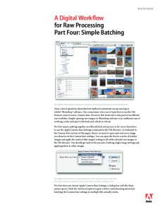
10. Now begins the process of stitching everything together in Photoshop. First, find the stacks. Photoshop has a number of ways to stack and merge. Under File > Automate, you will find Photomerge. This only works for a few files, e.g. a short panorama, or 5 or 6 well-overlapped pictures of a specimen that would not fit under the microscope completely.
2. Camera is set to P; use a low ASA (100) for lowest noise (less of a problem these days), and the software works out the exposure. However, if there is any vibration, use higher ASA (400, perhaps). Photos are taken in Camera Raw. This allows for archiving at the best quality and widest color gamut. Occasionally, I do need to go back to these raw files. 3. Name your files and folders! Choose any system you want; I use the date and specimen number (also part or counterpart, if relevant).
11. For stacking, it is usually safe to use File > Scripts > Load Files into Stack… Use the option Add Open Files to load your stack (if more files open, you can remove the unwanted ones), or else browse for the files you need. Make sure you click on the Attempt to Automatically Align Source Images option; this will align the images ready for merging. If this works (check the thumbnails), go to 13.
4. Zoom in or out for the crop you want and focus the microscope. I normally take a preliminary shot of the whole specimen together with a mm scale alongside, for future reference (see later). 5. For most fossil spiders, I take a large mosaic of high-magnification shots which are then stitched together. So, starting at one corner (e.g. top left), take a shot. NOTE: you will need some shots of bare rock, e.g. between outstretched appendages and around the edges, in order to end up with a nice, rectangular final picture for publication.
12. The fully manual method of stacking is to copy and paste all of the images you want to stack into one file (each one on its own layer). If one layer is a Background, it must be converted to a numbered layer by double-clicking on it in the Layers palette and renaming it (e.g. Layer 0). Then, select all layers in the Layers palette, and choose Edit > Auto-Align Layers… there are various options here; I just leave it at Auto and do not check the other boxes. This command will attempt to align the layers, just as in the automated method in 11.
6. For each shot, if not all is in focus, take several shots of the same area at different focus planes. Focus manually on the microscope or, if using a macro lens, this can be achieved automatically with the software. Later, these can be stacked.
13. Once the layers are aligned, they can be merged. Again, all layers you want to merge must be selected in the Layers palette (they will be already after the last step). Choose Edit > Auto-Blend Layers… The options here are: Panorama or Stack Images (choose stack for this step) and make sure you check Seamless Tones and Colors.
7. Move the specimen gently to the next shot area, remembering that about a 40% overlap is necessary for stitching successfully. If necessary, take a stack again. 8. After all shots are taken, turn off LiveView, turn off the camera, and close the software.
14. The result will be a series of layers in the Layers palette showing which parts of each layer have been used or deleted. It is worth looking through these to identify if any images are redundant. If all looks OK (the main window shows the result), then choose Layers > Merge Layers to merge them. You will probably see that the edges are a bit blurry; IMPORTANT: crop the image to exclude these artefacts.
9. Find your folder, in which will be all the shots taken. I now convert these from RAW to TIF. Photoshop can do this, but I use Capture One Pro, which is reckoned to be the best. It is a professional photography suite, similar to Aperture or Lightroom, written for the Phase One medium format digital 95
Figure 2. Photoshop screen shots showing the result of merging three shots of a specimen into a panorama. A before merge; B after merge.
96
Figure 3. Photograph of a fossil plectreurid spider from the Jurassic of China, showing how the final composite picture with high resolution can be zoomed in to examine details. A whole specimen; B zooming in to the top left, showing male palp embolus and clasper on leg I; C zooming again, showing tip of embolus (right) and clasper on leg I (left); D composite picture merging part and counterpart indicates that clasper is composite of fused macrosetae (left) and tip of embolus (right, arrowed) is revealed to have a fine, helical flagellum (from Selden & Huang 2010).
15. Having done all the z-stacks in this way (NOTE: this can also be done in Helicon Focus or other stacking software), we now need to stitch the images together into a mosaic. File > Automate > Photomerge is unlikely to work on more than a few images (see above), in which case, do it manually. Copy and paste the first image onto the second and auto-align (see 12, above). Then select all layers (remember: none can be a Background) and choose Edit > Auto-Blend Layers… as in 13. This time, select Panorama, and make sure you check Seamless Tones and Colors. The result will be a small panorama of the two images.
17. Gradually, the whole specimen can be assembled into one large mosaic. Each image starts at about 60MB, so the final image can reach 1 or 2GB in size. You will need plenty of RAM and scratch disk space to manipulate this file. 18. The final image may show distortion around the edges. To remove this, I drop in the picture I took of the whole specimen with the scale ruler. Make this layer half-transparent (Opacity 50% on the Layers palette), so both layers can be seen. Resize and rotate the new layer with the whole specimen until it is at the size of the underlying panorama. If any distortion needs correcting, the panorama layer can be distorted back to its correct proportions by selecting the layer (Select All) and then carefully using Edit > Transform > Warp to get it into shape. In practice, I
16. Continue to add each image to the new panorama, merging, and saving after each step. See Figure 2. 97
find that there is really little or no distortion and this step is unnecessary, but it is worth checking.
zoomed in to reveal the finest detail, nearly as good as having the original specimen under the microscope. Crops can then be used as illustrations of details (Figure 3B,C). Moreover, part and counterpart can be superimposed (with one reversed) and aligned to provide a view of the complete specimen. In the example given here, this allowed the discovery of a fine, helical tip to the male spider's palpal embolus (Figure 3D).
19. Before removing the additional layer, create a new layer and draw a black line along 1mm (or whatever) of the ruler to provide a scale for the main panorama. Now, the imported layer can be deleted, and what remains is a panorama with a scale on a separate layer.
Acknowledgements
20. Now, the image can be cropped to remove the ragged edges. If there are still some patches without an image (e.g. at the edge where some background is missing), then use of the Clone Stamp, Spot Healing Brushes, etc. can be used to fill in. Adjustments to colour, tone, contrast, saturation, etc. can now be applied; also some sharpening if necessary. 21.
I thank Gil Nelson (Florida State University) for encouragement.
References VAN NOORDEN, R. (ed.). 2010. Images of the year. Nature 468, 1018-1023. SELDEN, P. A. & HUANG, D-Y. 2010. The oldest haplogyne spider (Araneae: Plectreuridae), from the Middle Jurassic of China. Naturwissenschaften 97, 449-459.
A final result is shown in Figure 3.
Conclusion The final, large file can be reduced in size to produce a whole-specimen illustration (Figure 3A), or
98