Varicocele Embolization
215
12 Varicocele Embolization David Hunter and Galia T. Rosen
CONTENTS 12.1 12.2 12.3 12.4 12.5 12.6 12.7 12.8 12.9 12.10 12.11 12.12 12.13 12.14 12.15
Introduction 215 Anatomy 215 Pathophysiology 216 Diagnosis 216 Clinical Considerations 217 Alternative Therapy 217 Percutaneous Embolization 217 Catheterization Technique 218 Hot (Boiling) Contrast Sclerotherapy 220 Coil Embolization 221 Tissue Adhesives 222 Detachable Balloons 222 Complications 222 Efficacy of Treatment and Comparison to Surgery 223 Conclusion 223 References 224
12.1 Introduction Varicocele is defined as an abnormal distention of veins in the pampiniform plexus. The association between testicular atrophy and dilated scrotal veins was noticed as early as in the first century AD [1]. Ivanissevich in a 1960 article [2] remarked that the clinical association of varicocele with pain symptoms had been noted as early as 1541 by Ambrois Pare who described the varicocele as “a compact pack of vessels filled with melancholic blood”. In the same article Ivanissevich described one of the earliest extensive experiences with suprainguinal ligation of the internal spermatic vein as a curative measure [2]. In the late nineteenth century it was first shown that correction of varicocele could
D. Hunter, MD; G. T. Rosen, MD Department of Radiology, J2-447 Fairview-University Medical Center, University of Minnesota, 500 Harvard Street S.E., Minneapolis, MN 55455, USA
result in restoration of fertility [3]. Widespread acceptance of the relationship between varicocele and male factor infertility, however, came only in the 1950s based on work by Tuloch [4]. Surgical correction has always been the main therapeutic option for correction of varicocele. In 1980 Iaccarino [5] was the first to describe a percutaneous method for treatment of varicocele. The steady advancement in embolization techniques and materials has led to the development of the modern percutaneous procedure that is considered to be a safe, simple and effective alternative to surgery.
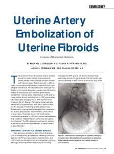
12.2 Anatomy The veins of the spermatic cord form a loose, tortuous plexus after emerging from the mediastinum of the testis. These vessels are named the pampiniform plexus. The veins in the anterior portion of the plexus coalesce to form the internal spermatic vein (ISV). The ISV passes through the inguinal canal and then ascends through the retroperitoneum alongside the spermatic artery, until it drains into either the left renal vein on the left side, or the infrarenal IVC on the right side. Additional venous drainage of the testis, which becomes important following occlusion of the ISV, includes the external pudendal, vasal and cremasteric veins (Fig. 12.1). Alternative drainage pathways of the ISV include the peri-renal, retroperitoneal and lumbar veins. Other anastomoses can exist between the ISV and other venous outflow channels in the retroperitoneum and pelvis. These anastomoses can permit reflux and varicocele formation even in the presence of a competent valve in the proximal ISV. The resulting condition is termed an aberrantly supplied varicocele. The reported rate of this phenomenon is 17%–19% of patients examined with spermatic venography. Percutaneous treatment of this type of varicocele is possible, but requires occlusion of the
D. Hunter and G. T. Rosen
216
䉴 䉴
the renal vein, particularly in cases with renal vein outflow obstruction due the compression of the vein between the aorta and superior mesenteric artery, the so-called nutcracker effect. Proponents of either theory further postulate that a bilateral effect could occur by venous crossover to the right testis [8]. There are conflicting opinions about the laterality of varicocele. Some authors feel that the condition is predominantly left-sided, with at most 30% of patients having a bilateral problem [9]. In other studies [10, 11], including one in which venograms were performed bilaterally regardless of the physical exam findings [11], bilateral ISV incompetence to an enlarged pampiniform plexus was found in 70%–80% of patients.
12.4 Diagnosis Fig. 12.1. Accessory veins. Perivesical (thin arrow), External pudendal (arrowheads), Cremasteric (white arrow), Vasal (thick arrow)
ISV at the very least above and below the segment of the vessel at the level of the venous collateral communication. Treatment of a varicocele with this type of complex anatomy has a success rate that is somewhat lower than one with classical anatomy [6].
12.3 Pathophysiology There is still debate about how, when, and to what extent varicocele affects fertility. A total of 10% of all men have a varicocele. Most are both asymptomatic and not associated with infertility. However, among infertile couples the incidence of a varicocele increases to 30% [7]. Why should the dilatation of the veins of the pampiniform plexus impair spermatogenesis? A related question in patients with a unilateral varicocele, is how unilateral venous abnormality produces bilateral testicular dysfunction? Several theories have been postulated to explain the pathophysiology of varicocele. The most popular among the theories involves the adverse effect of elevated testicular temperature on spermatogenesis [8]. Another theory is that reflux of adrenal or renal metabolites that could inhibit spermatogenesis reach the left testicle by back flow from
The clinical assessment of varicocele must start with a careful physical examination. The patient should be examined in a warm room in the standing position, and preferably after standing for 5 min. The examination should include palpation and Doppler of the scrotum during a Valsalva maneuver. The grading system as developed by Dubin and Amelar [12] is the most commonly used to classify varicoceles, and includes the following categories: x� Grade 1, varicocele palpable only during a Valsalva maneuver. x� Grade 2, varicocele palpable in the standing position. x� Grade 3, varicocele detectable by visual scrutiny alone. However, the limitations of physical examination are well documented [13], and the standard of care involves employment of additional diagnostic methods. These include thermography, color flow Doppler sonography, and venography [14–16]. Although each of these tests has reported standards that are used to make the diagnosis of varicocele, the standards are not universally accepted and the accuracy of each test has frequently been called into doubt. A varicocele that is present on an imaging study but not on physical examination is termed a subclinical varicocele. Embolization or surgical treatment of subclinical varicocele is a frequent practice in subfertile males with no other explanation for infertility. Those who advocate treating subclinical varicocele claim that even though the angiographically
Varicocele Embolization
demonstrated degree of reflux is indeed lower in subclinical cases, the improvement in semen analysis and fertility rates that is seen after embolization, appears to be about equal for the clinical and subclinical varicocele patients [17].
217
varicocele directly are becoming more common [19]. The techniques for surgical ligation have improved adequately so that percutaneous options are often not discussed with patients unless the patient has read about it, usually on the Internet, and requests the information directly. This approach clearly limits the number of percutaneous procedures performed.
12.5 Clinical Considerations Varicocele can result in pain and infertility, and either or both may be present in any patient. One important additional group of patients that has been studied in several prospective studies in Europe is adolescent boys. In most cases of adolescent varicocele, the diagnosis is made incidentally on routine physical examination. Pain and dysfunctional spermatogenesis with or without testicular atrophy is an unfortunately frequent outcome for these patients and many advocate preemptive treatment for them as well [18]. The most common semen abnormality in patients with varicocele and infertility is poor sperm motility, followed by abnormal morphology, and then depression of sperm count. The isolated finding of abnormal sperm motility has been referred to as a stress pattern. The normal World Health Organization (WHO) values [13] for the commonly evaluated parameters studied during semen analysis include the following: x� Volume: 1.5–5.0 ml x� Sperm count or density: greater than 20 million sperm/ml x� Motility: greater than 60% normal motility x� Morphology: greater than 60% normal forms x� Forward Progression (scale 1–4): 2+ x� Viscosity: no hyperviscosity x� White blood cells: 0–5 per high power field
12.7 Percutaneous Embolization The percutaneous treatment of varicocele is aimed at decreasing the engorgement of the pampiniform plexus by occluding the incompetent ISV and its collaterals. The use of percutaneous sclerotherapy as a treatment for varicocele was first described in 1980 [5]. The most commonly used embolization method in the USA, which is the use of metal coils (Fig. 12.2), was described in 1978 [20]. Ever since, additional materials and methods have been reported including modified coils such as the new Amplatz vascular plug, boiling contrast, detachable balloons,
12.6 Alternative Therapy Until the development of the percutaneous approach, surgical ligation of the ISV was the only available therapy. Several surgical procedures can be used, which differ primarily based on the level of ligation of the spermatic vein. The common sites for surgical ligation are retroperitoneal, inguinal or subinguinal. Laparoscopic methods of performing the ligation, and microsurgical operations that treat the
Fig. 12.2. After coils had been placed distally, these two coils were placed above the only large collateral. Notice how they are nested tightly inside each other. It was not considered necessary to place coils closer to the ISV origin
D. Hunter and G. T. Rosen
218
tissue adhesives, and sclerosing agents such as concentrated dextrose, sodium morrhuate, Sotradecol (sodium tetradecyl sulfate) [21], Varicocid [18], ethanolomine [10], and alcohol [22].
12.8 Catheterization Technique Most studies have reported using a femoral vein approach. The left spermatic vein requires a double curve catheter to reach the ISV origin, and the right ISV is best entered with a sidewinder type catheter. Because of the double curve required to get into either spermatic vein, the femoral technique frequently requires coaxial catheters or a catheter exchange to reach an appropriate level in the ISV. In our experience, a right trans-jugular approach to both the right and the left spermatic veins is preferable as it facilitates deep catheterization and therefore accurate delivery of the sclerosing agent or occlusion device, obviates a femoral vein puncture and the small but important risk of femoral DVT, and allows a more rapid discharge post-procedure with essentially no bleeding risk [11]. There is a small chance that manipulation through the right atrium may induce a dysrhythmia but such rhythm disturbances are almost always short-lived and resolve spontaneously. A heat re-shaped 5- or 6-F Headhunter catheter with two to six sideholes in the distal 2–3 cm (Fig. 12.3) can be used to select the left renal vein. The lordotic tertiary curve should be re-shaped into a kyphotic curve. While the catheter is being heated with a heat gun, a guidewire is kept in the lumen of the catheter to maintain catheter patency. The distal-most 3–4 mm of the tip of the catheter must be further modified to “point downstream” for selection of the right spermatic vein. Alternative catheters such as angled tip catheters (JB 1 or Vert shape), Cobra shape catheters, and variations of these shapes have all been tried but with less success. Coming from the jugular or femoral approach, the most common first maneuver is to catheterize the left renal vein. The “C” shape of the reformed headhunter catheter makes this extremely easy from the jugular approach. Entry into the left renal vein from the IJ approach occasionally requires advancing the wire far into the renal vein and then applying a counterclockwise twist or torque, thus pointing the catheter tip posteriorly. If the catheter tip passes either inferiorly or seems to be “caught” in the cen-
䉴
Fig. 12.3. The lordotic tertiary curve has been reformed into a kyphotic “C” shape (arrow). There is slight reflux into the left ISV (arrowhead)
tral left renal vein territory, a gentle hand injection of contrast can be done to check position. If the hand injection reveals that the catheter is in a lumber vein, the catheter should be pulled back and rotated slightly clockwise or anteriorly, as the lumbar vein orifice is always posterior to the left renal vein orifice. Once the catheter tip is in the mid-portion of the left renal vein, a left renal venogram is done during a forceful Valsalva maneuver to document L ISV incompetence (Fig. 12.4) and also to establish landmarks for selective L ISV catheterization. The left spermatic vein itself is also accessed using a counterclockwise rotation as the tip is pulled back, which rotates the tip of the catheter first posterior and then inferior since the curve is braced against the left renal vein origin. If the catheter tip enters the collateral from the proximal left renal vein to the adjacent paralumbar, hemiazygous system, the tip is rotated gently clockwise to point it slightly to the left and anterior where the origin of the adjacent L ISV is always located. Once the L ISV origin has been engaged, and assuming that incompetence has already been documented by the renal venogram, the catheter is advanced only 2–3 cm into the vein and the first selective diagnostic venogram is done. A forceful hand injection is performed into the L ISV using 10 cc of diluted contrast media, with the catheter tip just past the origin of the vessel. The patient is instructed once again to perform a Val-
Varicocele Embolization
Fig. 12.4. During the left renal venogram with the catheter tip nicely positioned in the mid-left renal vein, a forceful Valsalva maneuver refluxes contrast not only down the incompetent L ISV but also down the IVC
salva maneuver, while the operator looks carefully to both confirm reflux into the incompetent spermatic vein and also to define the upper L ISV anatomy. If free reflux is seen, confirming incompetence of the valve, the wire is advanced to the level of the mid to upper third of the SI joint and the catheter rotated down over the wire to the same position in approximately the mid L ISV. The catheter should be rotated and not “pushed” since that can cause buckling in the IVC or right atrium. Injections are done at approximately the mid-SI joint level to clearly define the remainder of the L ISV anatomy, confirm abnormal retrograde flow into a distended pampiniform plexus (Figs. 12.5, 12.6), and delineate any connections to other veins that could act as sources for aberrant varicocele filling or as collaterals if the L ISV is eliminated. Only after all of the diagnostic studies have been completed, can a meaningful and accurate embolization or sclerotherapy be carried out. If no reflux is seen into the L ISV on either the left renal venogram or selective L ISV origin injection, but there is clear sonographic or physical examination evidence of a varicocele, we routinely assume that the patient has retroperitoneal or pelvic bypassing collaterals and believe that embolization of the ISV is still indicated. If, however, the only abnormality is the semen analysis, and there is no physical exam or imaging evidence of reflux or varicocele,
219
Fig. 12.5. Injection into the mid L ISV demonstrates a very atypical finding of a single dominant channel of the L ISV passing down through the inguinal canal
a
b
Fig. 12.6a,b. Two patients who had recurrence of pain following surgical ligation of the L ISV. With the catheter tip in the mid L ISV just below the mid-portion of the L SI joint, injection in patient (a) demonstrates one small channel through the inguinal canal, whereas patient (b) has no patent vessels that would suggest a recurrence. Patient (a) was treated, (b) was not
D. Hunter and G. T. Rosen
220
䉴
and a competent valve is clearly seen on the L ISV origin injection, the procedure on that side can be terminated. The procedure on the right side is similar although there is no way to do a preliminary venogram to confirm incompetence. This fact alone may have led to a substantial underestimation of rightsided incompetence reflected in much of the varicocele literature. Coming from the femoral approach, the origin of the right ISV is easy to enter using a standard sidewinder catheter. From the IJ approach, the headhunter that was used for the L ISV needs to be modified so that the tip points more inferiorly (Fig. 12.7). The R ISV origin is usually located just anterior and inferior to the right renal vein. In order to find it, the catheter is placed into the right renal vein then pulled out, rotated slightly counterclockwise, pushed down below the renal vein level and manipulated up and down on the vein wall until it “catches” on the R ISV orifice. Once the tip of the catheter just barely enters the origin, the first injection is done with a gentle Valsalva to avoid dislodgement of the catheter. If the catheter tip is allowed to go too deep into the vein, the valve at the origin, which is frequently the only valve, can be bypassed, and an incorrect diagnosis of incompetence made. Once incompetence has been documented, the catheter is advanced over a floppy wire to the mid to upper third of the R ISV (Fig. 12.8), the anatomy of the remainder of the vein is clarified, and embolization or sclerotherapy performed.
Fig. 12.7. Tertiary (thin arrow), Secondary (grey arrow), Primary (thick arrow) curves are all still kyphotic, but the distal tip is bent to point downward (arrowhead)
a
b
Fig. 12.8a,b. Injection above (a) and at (b) the mid R ISV, demonstrates the typical parallel collaterals and multiple channels at the level of the inguinal ligament that make complete treatment with coil placement or surgical ligation challenging
12.9 Hot (Boiling) Contrast Sclerotherapy The use of heat to occlude veins is a well-documented and tested technique. Various heat sources have been used including boiling liquids, lasers, and radiofrequency electrodes. One distinct advantage of heat is that it induces spasm and wall damage leading to occlusion with relatively minimal thrombosis. Embolization and non-heated liquid sclerotherapy techniques result in a large amount of thrombus, which, along with any mechanical blockade causes secondary venous obliteration. The same contrast that is used during the diagnostic part of the procedure can be heated in a metal container over a heating plate for the sclerotherapy. This makes boiling contrast quick, inexpensive, universally available, non-toxic, and easily visible and therefore controllable. The advantage of a boiling liquid over other heat sources and over all embolization techniques, is that it can flow into and obliterate any potential collaterals. While the patient is receiving extra sedation and the contrast is coming to a boil, the multisideholes catheter is allowed to drain so that the ISV is as empty as possible. When boiling temperature is reached, which is signified by the liquid dem-
Varicocele Embolization
onstrating the bubbling action of a “rolling boil”, the operator fills, as best as possible, a 10 cc plastic syringe with the boiling contrast, eliminates extra air, and injects the remaining 6–9 cc directly into the catheter under careful fluoroscopic control. No stopcocks or connectors are used to minimize any heat loss. A senior member of the team concurrently compresses the ISV where it crosses over the superior pubic ramus (Fig. 12.9) to prevent any flow of boiling material into the pampiniform plexus. Any sclerosant or boiling liquid that is allowed to enter the veins of the pampiniform plexus will cause a very painful scrotal swelling and potentially testicular atrophy. Injections are done without moving the catheter since the spread of the boiling contrast in volumes of 6–9 cc is usually adequate to cover the entire extent of the ISV as well as any collaterals. In most cases, three sequential injections of 6–9 cc of boiling contrast are sufficient to ensure a complete obliteration of the injected vein unless it is exceptionally large. Overall, the angiographic or imaging success rate of the boiling contrast and other liquid sclerosing agents is considered very high with a reported recurrence rate of 2%–19% [23].
12.10 Coil Embolization The most common percutaneous technique in current use is that of coil embolization. Its primary and significant advantage over sclerotherapy techniques especially boiling contrast is that it is relatively painless requiring far less sedation. Gianturco coils (Cook), ranging in size from 5 mm to 8 mm are the most common sizes used [24], although some authors report using smaller or larger coils as needed based on the size of the vein. Coil sizing, stacking, and placement are clearly of paramount importance for technical success and avoiding complications particularly coil embolization to the heart and lungs. The size of the coil should be 10%–20% larger than the diameter of the ISV at the level of deployment. Sizing the coil too small can result in pulmonary embolization, which is aesthetically and emotionally unpleasant, but due to the size of the coils is usually without any clinical consequences. Coil removal from the pulmonary artery can usually be done with minimal problems using a snare or grasper [25]. Usually more than one coil is deployed at any given site since a single coil
221
a
b
Fig. 12.9a,b. Compression on the ramus (a) occludes retrograde flow whereas compression slightly off the ramus (b) or tilted does not
has a higher recanalization and failure rate than two or more “stacked” or “nested” coils. With the development of special tapered shape coils that are tapered in one or two directions, the need for multiple coils is no longer clear although it has not been rigorously tested. The Amplatz Vascular Plug (AGA Medical, Minneapolis, MN) is a coil type device that is longer and more completely occlusive than standard coils, is recapturable until a good “final position” has been confirmed, and will likely find an important role in the treatment of varicocele. Coils must be deployed proximal and distal to any major collaterals. If there are parallel veins, often both must be separately occluded. The minimal distance of the coils from the orifice of the ISV is somewhat debatable. Most European literature cites a distance of 6 cm from the origin as safe and effective, while others claim that embolization up to the level of the origin is preferable to avoid “dead space” in which clot could accumulate and potentially embolize. Some authors [26] favor the combination of coils and sodium tetradecyl sulfate (Thromboject 3%; Omega, Montreal, QC, Canada). The coils are delivered relatively distally, at the level of the inguinal canal, not only to occlude the vein at that level but also to prevent reflux into the pampiniform plexus. The sclerosant is delivered proximal to the coils to occlude all the side branches that could become collaterals. If necessary, coils can also be deposited in the proximal portion of the ISV to prevent pulmonary emboli. One coil that was never used in the
D. Hunter and G. T. Rosen
222
USA deserves special mention. This coil was made from tungsten, which obviously would markedly improve visibility. Unfortunately, one feature of the tungsten coil is that it was biodegradable leading to increased tungsten levels in the blood [27–29]. Though no adverse effects of tungsten in humans were ever described, the observation was concerning enough that their usage for this application has been discontinued.
12.11 Tissue Adhesives The most commonly used liquid tissue adhesives are cyanoacrylates. Their low viscosity makes their delivery easy through small coaxial microcatheters. However, their rapid polymerization when in contact with blood, can make precise and safe occlusion challenging. Usually they are mixed with an oil-based contrast media, such as Ethiodol (Savage Laboratories, Melville, NY). The contrast serves to both opacify the cyanoacrylate and slows the polymerization time [30]. Another tissue adhesive that has been reported is enbucrylate (Histoacryl; B. Braun, Tuttlingen, Germany). It can be used when multiple collaterals are seen that are too small to be selectively catheterized [26].
12.12 Detachable Balloons The use of detachable balloons to occlude the ISV was first described in 1981 [31]. Due to a lack of FDA approval for some of the devices and manufacturer related issues, the balloons have not always been readily available in the USA. From a technical standpoint, balloons were similar to coils in that they needed to be deployed above and below collateral connections. However, since the balloon occluded the vein completely, much like the Amplatz Vascular Plug, only one was necessary at any given site. As with coils, it was common for users of balloons to perform some type of sclerotherapy on the vein segments between balloons to decrease the development of collaterals. Balloons suffered from two major drawbacks that severely limited their more widespread acceptance. The first was cost. The other was that the size of the catheter required to deliver
a large balloon of 7–10 mm diameter was 7 or 8 F. In addition, the introducer catheter had to be placed to the level of the desired occlusion, a feat that was often technically very challenging especially from the femoral approach.
12.13 Complications A unique, and fortunately rare, but significant risk of the hot contrast or other liquid sclerotherapy, is distal reflux of the sclerosing agent. Inadvertent injection of a sclerosing agent into the pampiniform plexus can result in painful scrotal swelling, phlebitis, and testicular damage with depression of spermatogenesis and even irreversible testicular atrophy [11]. To prevent that a preliminary run with contrast is carried out to verify that the occlusion of the inflow into the pampiniform plexus is indeed complete and efficient. A specially designed device, which is essentially a gently curved piece of plastic with padding on the edge that will press against the skin, was developed specifically to address this and was shown to be highly effective [32]. The device must be held exactly at a 90’ angle to the bone. Fluoroscopy is used to verify its end-on position and to ensure that all contrast stays above the compressor during the injection. A different kind of complication is proximal reflux of the sclerosing agent, which can result in renal vein thrombosis. Other complications are extravasation and dissection. The ISV is a very thin-walled structure that can easily be torn by overly aggressive catheter manipulations or the use of glidewires. Gentle maneuvers and the use of a carefully controlled floppy wire will almost always prevent ISV damage. On occasion, however, a small vein may go into spasm around the wire and catheter resulting in vein avulsion or disruption during attempts to extract it. Hot contrast or sclerosants should definitely be prevented from reaching the retroperitoneum since they can cause ureteral or muscle injury and severe pain. Contrast extravasation at the time of catheter placement should prompt a change of the embolization method to one using coils or other mechanical devices. Another consideration with the use of sclerosing agents is the pain that is associated with their injection. However it is usually of very short duration, most commonly 10–15 s, although rare instances
Varicocele Embolization
lasting up to several minutes in poorly sedated patients have been seen. The pain can be both alleviated and “forgotten” with the use of agents such as Fentanyl and Versed.
12.14 Efficacy of Treatment and Comparison to Surgery Success of the embolization or sclerotherapy treatment can be defined as a technical success, which is the immediate angiographic closure of the ISV. It can also be defined as a long-term technical success based on the finding of persistent closure on the delayed or follow-up angiographic or other imaging study. Of particular note, the thermal damage caused by boiling contrast may result in some immediate spasm and stasis of contrast, but the vein generally remains patent acutely. Success can also be defined as clinical success, that is, partial or complete resolution of the clinical signs and symptoms associated with varicocele. In particular, the most meaningful definition of success in couples with an infertility issue is the successful achievement of a pregnancy. The two parameters that are usually evaluated in the assessment of the clinical efficacy of the treatment are semen analysis changes and the pregnancy rate. There is great controversy in the literature about the effectiveness of varicocele embolization. According to one recent Cochrane analysis, the existing prospective randomized trials that satisfied their criteria for inclusion showed improvement in semen parameters and symptoms, but no difference in pregnancy rate compared to no treatment [33]. However, some of the individual studies in that analysis did show a very significant improvement in pregnancy rate of the treated group [34]. When retrospective data or non-randomized control groups are included in the analysis, most authors do find a significant difference in pregnancy rates between treatment and no treatment for varicocele. The historical rates that seem to have the most widespread acceptance suggest that the pregnancy rate for couples in whom there is documented male infertility as the sole cause of infertility is approximately 30% if the male receives either surgical or percutaneous treatment of the varicocele. This compares to a pregnancy rate of 16% for couples with the same history who undergo no therapy [35]. Thus, many clinicians and infertile couples are still interested in treatment for varicocele.
223
When the percutaneous procedure is compared to surgical ligation, varicocele embolization has been shown to be an equally effective means of treatment and is associated with less post procedure discomfort and more rapid return to normal activities [24, 36–38]. The overall reported technical success rate as cited by the JVIR quality improvement guidelines are 83%– 96% with a clinical or imaging detected recurrence rate at 6 weeks of 7%–16% [23]. All of the analyses of procedural and clinical efficacy are obviously influenced and potentially severely confounded by several poorly controlled variables. One problem is that the need for treatment of both sides has never been clarified and it is clear that different authors have had markedly different opinions and angiographic findings on the subject. Another problem is that imaging follow-up is infrequent and impossible to quantify in a meaningful fashion. Therefore, the impact of aggressive and extensive treatment such as with coils plus sclerosants, boiling contrast or sclerosants is difficult to compare with simple coil or ligation therapy. In addition, the inclusion criteria, particularly with respect to semen analysis variables are poorly controlled. As an example, patients who present with a very low sperm count, below 2 million/ml, seem to have a lower response rate to treatment and yet are usually lumped with more favorable patients in most analyses. One place where embolization or sclerotherapy appears to have established a definite niche is in the treatment of recurrence or failure following surgical ligation. In these cases, repeat surgery can be done with a different technique, but fearing another failure, most surgeons will refer the patient for percutaneous diagnosis and treatment. A total of 31 patients out of 40 with recurrence after surgery in one study [16], and 33 out of 39 patients in another [39] had successful diagnoses and treatment. The ISV venogram allows a precise anatomical definition of the cause and location of the veins responsible for the recurrent varicocele and the use of steel-coil embolization in both studies provided an effective means of treatment with improvement in semen quality and pregnancy rates [16, 39].
12.15 Conclusion Even though some controversy exists about the justification for any type of treatment of varicocele with
224
wide variation in results between different studies, we feel that current recommendations should advocate percutaneous embolization of varicocele as a safe, effective and potentially first-line treatment. Its overall efficacy is comparable to that of surgical ligation with a shorter recovery period and less pain, permitting it to be conducted as an outpatient procedure. Our preferred embolization method is using boiling contrast. Other embolization techniques, such as coils, vascular plugs, other sclerosing agents, and detachable balloons are acceptable, and clearly less painful although more complex alternatives. The primary and important risk of any sclerosant type procedure is reflux into the pampiniform plexus, which might lead to orchitis and even testicular atrophy. The best way to avoid this complication is to use a compression device, rather than relying on manual compression.
Cookbook:
Catheter: x� IJ approach #1: Heat reformed Headhunter 1 with extra sideholes x� IJ approach #2: Heat reformed JB 1 with extra sideholes Femoral approach, renal double curve guider left and Sidewinder 1 right Microcatheter: None needed from the IJ approach Renegade from the femoral approach Embolic agent: Boiling contrast Amplatz vascular occluder, or coils, preferably with a tornado or other complex shape, and always at least two at each point of embolization
References 001. Page H (1989) Estimation of the prevalence and incidence of infertility in a population: a pilot study. Fertil Steril 51:571–577 002. Ivanissevich O (1960) Left varicocele due to reflux; experience with 4,470 operative cases in forty-two years. J int coll surg 34:742–755
D. Hunter and G. T. Rosen 003. Barwell R (1885) One hundred cases of varicocele treated by subcutaneous wire loop. Lancet 1:978 004. Tulloch WS (1952) A consideration of sterility factors in the light of subsequent pregnancies. IISubfertility in male. Trans Edinb Obst Soc 59:29–34 005. Iaccarino V (1980) A nonsurgical treatment of varicocele: trans-catheter sclerotherapy of gonadal veins. Ann Radiol 23:369–370 006. Marsman JW (1995) The aberrantly fed varicocele: frequency, venographic appearance, and results of transcatheter embolization. AJR 164: 649–657 007. Schlessinger MH, Wilets IF, Nagler HM (1994) Treatment outcome after varicocelectomy. Urol Clin North Am 21: 517–529 008. Goldstein M, Eid JF (1989) Elevation of intratesticular and scrotal skin surface temperature in men with varicocele. J Urol. 142: 743–745 009. Bigot JM, Le Blanche AF, Carette MF, Gagey N, Bazot M, Boudghene FP (1997) Anastomoses between the spermatic and visceral veins: a retrospective study of 500 consecutive patients. Abdom Imaging. 22:226–232 010. Gat Y, Bachar GN, Zukerman Z, Belenky A, Gornish M (2004) Varicocele: a bilateral disease. Fertil Steril. 81:424– 429 011. Hunter DW, King NJ 3rd, Aeppli DM, Yedlicka JW Jr, Castaneda-Zuniga WR, Hulbert JC, Kaye K, Amplatz K (1991) Spermatic vein occlusion with hot contrast material: angiographic results. J Vasc Interv Radiol 2:507–515 012. Dubin L, Amelar RD (1970) Varicocele size and results of varicolectomy in selected subfertile men with varicocele. Fertil Steril 21:606–609 013. World Health Organization (1985) Comparison among different methods for the diagnosis of varicocele. Fertil Steril 43:575–582 014. Chiou RK, Anderson JC, Wobig RK, Rosinsky DE, Matamoros A Jr, Chen WS, Taylor RJ (1997) Color Doppler ultrasound criteria to diagnose varicoceles: correlation of a new scoring system with physical examination. Urology 50:953–956 015. Hamm B, Fobbe F, Sorensen R, Felsenberg D (1986) Varicoceles: combined sonography and thermography in diagnosis and posttherapeutic evaluation. Radiology 160:419–424 016. Morag B, Rubinstein ZJ, Madgar I, Lunnenfeld B (1985) The role of spermatic venography after surgical high ligation of the left spermatic veins: diagnosis and percutaneous occlusion. Urol Radiol 7:32–34 017. Marsman JW (1985) Clinical versus subclinical varicocele: venographic findings and improvement of fertility after embolization. Radiology 155:635–638 018. Braedel HU, Steffens J, Ziegler M, Polsky MS (1990) Outpatient sclerotherapy of idiopathic left-sided varicocele in children and adults. Br J Urol 65: 536–540 019. Donovan JF, Winfield HN (1992) Laparoscopic varix ligation. J Urol 147:77–81 020. Lima SS, Castro MP, Costa OF (1978) A new method for the treatment of varicocele. Andrologia 10:103–106 021. Trombetta C, Salisci E, Deriu M, Paoni A, Sanna M, Ganau A, Belgrano E (1993) Echo-flowmetric control 6 years after percutaneous treatment of varicocele. Arch Ital Urol Androl 65:363–367 022. Usuki N, Nakamura K, Takashima S, Takada K, Kaminoh T, Tsubakimoto M, Matsuoka T, Nakatsuka H, Oda J,
Varicocele Embolization Minakuchi K, et al. (1994) Embolization of varicocele with ethanol. Nippon Igaku Hoshasen Gakkai Zasshi 54:870– 875 (Japanese) 023. Drooz AT, Lewis CA, Allen TE, Citron SJ, Cole PE, Freeman NJ, Husted JW, Malloy PC, Martin LG, Van Moore A, Neithamer CD, Roberts AC, Sacks D, Sanchez O, Venbrux AC, Bakal CW (2003) Society of Interventional Radiology Standards of Practice Committee. Quality improvement guidelines for percutaneous transcatheter embolization. J Vasc Interv Radiol 14(9 Pt 2): S237–242 024. Shlansky-Goldberg RD, VanArsdalen KN, Rutter CM, Soulen MC, Haskal ZJ, Baum RA, Redd DC, Cope C, Pentecost MJ (1997) Percutaneous varicocele embolization versus surgical ligation for the treatment of infertility: changes in seminal parameters and pregnancy outcomes. J Vasc Interv Radiol 8:759–767 025. Chomyn JJ, Craven WM, Groves BM, Durham JD (1991) Percutaneous removal of a Gianturco coil from the pulmonary artery with use of flexible intravascular forceps. J Vasc Interv Radiol 2: 105–106 026. Tay KH, Martin ML, Lisl Mayer A, Machan LS (2002) Selective spermatic venography and varicocele embolization in men with circumaortic left renal veins. J Vasc Interv Radiol 13:739–742 027. Barrett J, Wells I, Riordan R, Roobottom C (2000) Endovascular embolization of varicoceles: resorption of tungsten coils in the spermatic vein. Cardiovasc Intervent Radiol 23:457–459. 028. Kampmann C, Brzezinska R, Abidini M et al. (2002) Biodegradation of tungsten embolisation coils used in children. Pediatr Radiol 32:839–843 029. Wells IP (2003) Biodegradation of tungsten embolisation coils used in children. Pediatr Radiol 33:288 030. Pollak JS, White RI (2001) The use of cyanoacrylate adhe-
225 sives in peripheral embolization. J Vasc Interv Radiol 12:907–913 031. White RI Jr, Kaufman SL, Barth KH, Kadir S, Smyth JW, Walsh PC (1981) Occlusion of varicoceles with detachable balloons. Radiology 139:327–334 032. Hunter DW, Bildsoe MC, Amplatz K (1989) Aid for safer sclerotherapy of the internal spermatic vein. Radiology 173:282 033. Evers J, Collins J (2004) Surgery or embolisation for varicocele in subfertile men. Cochrane Database Syst Rev 3: CD000479 034. Madgar I, Weissenberg R, Lunenfeld B, Karasik A, Goldwasser B (1995) Controlled trial of high spermatic vein ligation for varicocele in infertile men. Fertil Steril 63:120–124 035. Skoog SJ, Roberts KP, Goldstein M, Pryor JL (1997) The adolescent varicocele: what’s new with an old problem in young patients? Pediatrics 100:112–122 036. Dewire DM, Thomas AJ, Falk RM, Geisinger MA, Lammert GK (1994) Clinical outcome and cost comparison of percutaneous embolization and surgical ligation of varicocele. J Androl 15[suppl]:38–42 037. Nieschlag E, Behre HM, Schlingheider A, Nashan D, Pohl J, Fischedick AR (1993) Surgical ligation vs. angiographic embolization of the vena spermatica: a prospective randomized study for the treatment of varicocele related infertility. Andrologia 25:233–237 038. Parsch EM, Schill WB, Erlinger C, Tauber R, Pfeifer KJ (1990) Semen parameters and conception rates after surgical treatment and sclerotherapy of varicocele. Andrologia 22:275–278 039. Punekar SV, Prem AR, Ridhorkar VR, Deshmukh HL, Kelkar AR (1996) Post-surgical recurrent varicocele: efficacy of internal spermatic venography and steel-coil embolization. Br J Urol 77:124–128