[Chinese Journal of Cancer 27:10, 358-362; October 2008]; ©2008 Sun Yat-Sen University Cancer Center
Clinical Research Paper
Amenorrhea after uterine fibroid embolization A report of six cases Wen-Bo Guo,* Jian-Yong Yang, Wen Chen and Wen-Quan Zhuang Department of Interventional Radiology; The First Affiliated Hospital; Sun Yat-sen University; Guangzhou, Guangdong P.R. China
Key words: uterine neoplasm, embolization, amenorrhea, complication
Background and Objective: Uterine fibroid embolization is an effective treatment alternative for uterine fibroids. However, amenorrhea after uterine fibroid embolization occurs in some patients. This study was to investigate the causes of amenorrhea after uterine fibroid embolization. Methods: Bilateral transcatheter uterine artery embolization was performed in 487 patients with uterine fibroids. Pingyangmycin (6–16 mg) dispersed in lipiodol (8–15 mL) was used as embolic agent in 104 patients; polyvinyl alcohol (80–150 mg) was used in 158 patients; absorbable gelatin sponge (1–2 g) was used in 225 patients. All patients had been followed up for 1 year to observe amenorrhea occurrence. Results: Uterine fibroid embolization was effective in 483 (98.97%) patients, but failed in five (1.03%) patients. Amenorrhea after embolization was found in six (1.23%) patients. Of the six patients, three were in lipiodol plus pingyangmycin group with lipiodol deposited in the ovarian region, one was in polyvinyl alcohol group and the other two were in absorbable gelatin group. Except for one patient in absorbable gelatin group, the rest five patients had estradiol (E2) decreasing and follicle stimulating hormone (FSH) rising due to ovarian function failure. In absorbable gelatin group, multiple ultrasonography examinations revealed that the endometrium was only 3 mm thick in one amenorrhea patient who had normal levels of E2 and FSH, and hysteroscope examination confirmed endometrial atrophy in this patient. Conclusions: Routine embolic agents have the chance in inducing amenorrhea after uterine fibroid embolization. The occurrence rate of amenorrhea after uterine fibroid embolization is about 1.23%. Ovarian function failure and endometrial atrophy are the most related factors. Uterine fibroid embolization has been gradually accepted as a safe and effective treatment for uterine leiomyoma since it was first *Correspondence to: Wen-Bo Guo; Department of Interventional Radiology; The First Affiliated Hospital; Sun Yat-sen University; Guangzhou, Guangdong 510080 P.R. China; Tel.: 86.20.87755766.8722; Fax: 86.20.87330903; Email: atrickguo@ tom.com Submitted: 02/16/08; Revised: 04/29/08; Accepted: 07/23/08 This paper was translated into English from its original publication in Chinese. Translated by: Wei Liu on 07/21/08. The original Chinese version of this paper is published in: Ai Zheng (Chinese Journal of Cancer), 27(10); http://www.cjcsysu.cn/cn/article.asp?id=14694 Previously published online as a Chinese Journal of Cancer E-publication: ttp://www.landesbioscience.com/journals/cjc/article/7978 358
reported by Ravina et al.1 in 1995. However, because some uterine leiomyoma patients would develop amenorrhea with unknown reason after uterine fibroid embolization, uterine leiomyoma tends to occur in women of a younger age and women now tend to have children at a relatively older age, the clinical application of uterine fibroid embolization for uterine leiomyoma is affected to a certain extent, especially for young patients.2-4 In order to promote the development of uterine fibroid embolization techniques, we analyzed the clinical data of 487 uterine leiomyoma patients who developed amenorrhea after uterine fibroid embolization and explored underlying causes.
Data and Methods Clinical data. Regular follow-up was performed in 487 uterine leiomyoma patients who underwent uterine fibroid embolization at the First Affiliated Hospital of Sun Yat-sen University from January 2000 to March 2007. All diagnoses were confirmed by gynecological examination and gynecologic ultrasound examination. The patients were 24–48 years old with a median age of 35 years. Of all patients, 453 showed excessive menstruation or prolonged menses, 28 showed oppressive symptoms such as sychnuria and constipation, and six showed no symptoms; 439 had multiple uterine leiomyomas and 48 had single uterine leiomyoma. The volumes of uterine leiomyomas were 47.09–546.84 cm3 with an average of 120.78 cm3. Therapeutic methods. All patients were subjected to continuous epidural analgesia till the third day after embolotherapy. After anesthesia, bilateral uterine artery catheterization was performed via the right femoral artery approach. A mixture of embolic agents and contrast agents was infused via the catheter (a 5-F Yashiro catheter; Terumo, Japan). When contrast agent retention was observed in the uterine artery, gelatin sponge particles were used to reinforce the embolization of the main trunk of the uterine artery. Angiography was performed again to confirm whether the embolization was satisfactory. Three different embolic agents were used. Of the 487 patients, 104 (24–48 years old with a median age of 34 years) used lipiodol emulsion (Gubert, France) and pingyangmycin (Tianjin, China) as embolic agents at doses of 8–15 mL (average, 10 mL) and 6–16 mg (average, 8 mg), respectively; 158 (26–45 years old with a median age of 35 years) used polyvinyl alcohol particles (300–700 μm in diameter; COOK, USA) at a dose of 80–150mg (average, 100 mg); 225 (25–43 years old with an average of 36.4 years) used gelatin
Chinese Journal of Cancer
2008; Vol. 27 Issue 10
Amenorrhea after uterine fibroid embolization: a report of six cases
Table 1
Clinical data of six patients with amenorrhea after embolization
sponge particles (900–1000 μm in diameter; Lianyungang, Jiangsu Province) at a dose of 1–2 g (average, 1.5 g). Follow-up. All patients were reexamined at one, three, six and 12 months after embolotherapy. After then, the patients were reexamined every six months. The main examinations included gynecological color ultrasound as well as the observation of changes in clinical symptoms and menstrual status. Considering that the amenorrhea present at two years after embolotherapy might be caused by complex factors, the observation endpoint was 12 months after operation. Criteria for evaluating amenorrhea. According to the definition in gynecology,5 amenorrhea was concluded if the duration of menstrual cessation was more than six months or three times longer than the duration of past menstrual cycle. Calculating the volume of the uterus or uterine leiomyoma. According to reference 6, the volume of the uterus or uterine leiomyoma = 0.5233 × the longest longitudinal diameter of the uterus or uterine leiomyoma × the longest transverse diameter × the longest anterior-posterior diameter. The diameter (unit: cm) was measured by ultrasound. Calculating the volume of multiple uterine leiomyomas. The volume of multiple uterine leiomyomas was calculated by adding the volumes of the three largest leiomyomas of those that could be detected using ultrasound examination. Calculating volume decrease of the uterus or uterine leiomyoma. The percentage of volume decrease = (the volume before embolization - the volume after embolization)/the volume before embolization × 100%. Diagnosis criteria of ovarian amenorrhea. According to reference 5, ovarian amenorrhea was diagnosed when the concentration of follicle-stimulating hormone (FSH) increased to ≥ 30 mL/L and that of estradiol (E2) decreased to ≤ 30 pg/L.
Results Embolization and its clinical efficacy. All patients underwent successful bilateral uterine fibroid embolization and were followed up for one year. During follow-up, 448 of the 453 patients who showed excessive menstruation or prolonged menses recovered after embolization. Clinical symptoms were alleviated in 448 (91.99%) patients. All the 28 patients who showed sychnuria and constipation recovered. Six asymptomatic patients showed 50–64% reduction in the volume of uterine leiomyomas one year after embolization when compared with that before embolization by color ultrasound examination, and no blood flow within uterine leiomyomas was www.landesbioscience.com
Table 2
Changes of follicle-stimulating hormone (FSH) and estradiol (E2) in six amenorrhea patients before and after embolization
observed. The total effective rate was 98.97% (482/487) while the embolization failure rate was 1.03% (5/487). Five patients who had excessive menstruation or prolonged menses showed no symptom alleviation after treatment. Among these five patients, four were treated with traditional Chinese medicine to control symptoms, and the remaining one underwent myomectomy one year after embolotherapy. The incidence of amenorrhea. Six (1.23%) patients developed amenorrhea after embolization. Of the six patients, three used lipiodol emulsion and pingyangmycin, one used polyvinyl alcohol particles, and two used gelatin sponge particles. Except one patient who used gelatin sponge particles, the remaining five did not resume menstruation any longer after embolization. Clinical data of patients developed amenorrhea. The clinical data of six patients who developed amenorrhea were shown in Table 1. Except for one patient (case 6) with single uterine leiomyoma, the remaining five had multiple uterine leiomyomas. All six patients showed excessive menstruation or prolonged menses before embolization. Except for one patient (case 6), the remaining five developed menopausal symptoms, such as varying degrees of vexation, emotional volatility, sleep disorders, hectic fever, vaginal dryness, and so on, at 3–4 months after embolization. The serum levels of FSH and E2 at six months after embolization were shown in Table 2. The
Chinese Journal of Cancer
359
Amenorrhea after uterine fibroid embolization: a report of six cases
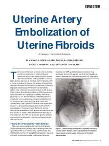
changes in FSH and E2 levels in five patients (cases 1–5) suggested ovarian amenorrhea, that is, ovarian function failure. Uterine artery angiography showed bilateral uterine artery thickening and lengthening, rich blood supply to leiomyomas and arterial network of leiomyomas in these six patients (Fig. 1). Embolic agents were injected via the catheter and the embolization met the requirement. In three patients who used lipiodol emulsion and pingyangmycin, angiography showed ovarian branches in the root of the uterine artery and contrast agents in the ovary, embolic agents were deposited in bilateral ovaries after embolization (Fig. 2). After embolization, case 6 showed hypomenorrhea, her menstrual period only lasted 2–3 days and Figure 1. Angiography of uterine fibroid embolization. (A) The arterial bed of uterine fibroids gradually decreased, and she developed amenorrhea with abundant blood supply can be seen; (B) The arterial bed of uterine fibroids was not visuat four months later. The levels of FSH and E2 alized and the contrast media was detained in main trunk of the uterine artery. Embolization showed no significant changes after embolization. was completed. After embolization, multiple ultrasound examinations showed that endometrial thickness was only 3 mm and did not provide blood supply to the ovary. Therefore, embolotherapy often change during menstrual cycle. Hysteroscopy showed atrophy and leads to disruption of ovarian blood supply via the ovarian branches attenuation of endometrium. These results proved that the amenor- of the uterine artery. Embolic agents may even enter ovarian vascular network through the ovarian branches of the uterine artery and rhea was a result of endometrial atrophy. block ovarian vascular network, thereby inducing ovarian ischemia, Discussion ovarian function failure, and eventually amenorrhea. Through examEffect of uterine fibroid embolization for uterine leiomyoma on ining ovarian artery by color Doppler ultrasound before and after 9 ovarian function. Although uterine fibroid embolization for uterine embolization, Ryu et al. found that if ovarian arterial perfusion wass leiomyoma has a high success rate, its impact on ovarian function decreased while resistance index and pulsation index were increased, has always attracted extensive attention. Because uterine leiomyoma non-target embolization of the ovarian artery was indicated. In addi10 tends to occur in women of a younger age and women now tend to tion, Payne et al. observed the pathologic changes in embolized have children at a relatively older age, the effect of this therapeutic specimens and found that embolic agents were present in the terminal method on the ovarian function of young patients who want to get branches of the ovarian artery. Thus, they speculated that embolic pregnant has attracted more attention from researchers not only in agents might enter into the ovary via the uterine artery and ovarian obstetrics and gynecology7 but also in interventional radiology.8 artery collaterals, block blood supply to the ovary and lead to ovarian Amenorrhea is not only the most serious side effect of uterine fibroid ischemia, thereby inducing ovarian function decline or failure. These embolization for uterine leiomyomas but also the greatest threat studies prove that one reason underlying the development of ovarian to patients of childbearing age. Therefore, it is essential to further amenorrhea is non-target embolization of the ovarian artery which is cuased by embolization-induced disruption of blood supply via the analyze the reasons underlying the development of amenorrhea. Among the six patients with amenorrhea in our study, five showed ovarian branches of the uterine artery and entering of embolic agents ovarian amenorrhea that was characterized by an increase in FSH into the ovary via the ovarian branches. In the three amenorrhea patients who used lipiodol emulsion level and a decrease in E2 level. These five patients used different embolic agents, suggesting that uterine fibroid embolization may and pingyangmycin in this study, uterine artery angiography in the induce a decline in ovarian function no matter which embolic mate- process of uterine fibroid embolization showed the ovarian branches of the uterine artery and the ovary, embolic agents were deposited rial is used. The results of previous studies3 suggested that the effect of embo- in the ovary after embolization. These results also confirm the lotherapy on the ovary is mainly related with the age of patients: above-mentioned speculation, that is, embolic agents can enter into patients over the age of 45 may develop upward shift of baseline FSH ovarian vascular network via the ovarian branches of the uterine artery level and might prone to amenorrhea, while young patients won't and disrupt ovarian blood supply. Since lipiodol emulsion belongs to have obvious change in ovarian function. However, recent studies liquefied embolic material and is even able to enter capillaries, it has found that the effect of embolotherapy on the ovary may be more the largest effect on the ovarian function and can result in ovarian related to the impairment of ovarian blood supply. The principle of ischemia, ovarian function failure, and eventually amenorrhea. In the remaining two amenorrhea patients, although no embolic transcatheter uterine fibroid embolization for uterine leiomyomas is to embolize the uterine vascular bed by injecting embolic agents agents were found to be deposited in the ovary, ovarian amenorrhea into the uterine artery till the blood flow in the main trunk of the still developed after embolization. Some studies have found that uterine artery is blocked. Anatomically, the ascending branch of the the ovarian branches of the uterine artery provide the main blood uterine artery sends branches to the ovary near the uterine fundus to supply to the ovary in about 6.6% of leiomyoma patients, which 360 www.landesbioscience.com
Chinese Journal of Cancer
2008; Vol. 27 Issue360 10
Amenorrhea after uterine fibroid embolization: a report of six cases
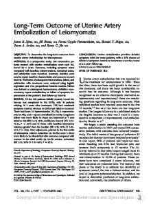
Figure 2. Angiography of the patient with amenorrhea after uterine fibroid embolization. (A) Right uterine arteriography shows that the right ovarian branch and ovarian area are visualized and stained (the arrow); (B) After right uterine artery embolization, lipiodol entered into the right ovarian area and deposited (the arrow); (C) Right uterine arteriography shows that the right ovarian branch and ovarian area are visualized and stained (the arrow); (D) After right uterine artery embolization, lipiodol entered into the right ovarian area and deposited (the arrow).
is regarded as a high risk factor for the development of amenorrhea after embolization.11 Therefore, we speculate that, in these two patients, the ovarian branches of the uterine artery are the main arteries to supply blood to the ovary. Although the diameter of polyvinyl alcohol particles and gelatin sponge particles is relatively large (300–700 μm and 900–1000 μm, respectively), the block of the main trunk of the ovarian branches would cause ovarian ischemia and function failure, and lead to amenorrhea. Effect of uterine fibroid embolization on the endometrium. The amenorrhea presented in case 6 was different from those presented in other cases, which was resulted from endometrial atrophy. We www.landesbioscience.com
found that FSH and E2 levels in this patient, who used gelatin sponge particles as embolic agent, showed no changes after embolization. Although the patient showed no menopausal symptoms, the amount of menstrual bleeding gradually declined and amenorrhea eventually developed. Moreover, her endometrium was attenuated and lost response to sex hormones, and endometrial thickness did not change with menstrual cycle change. Hysteroscopic examination proved endometrial atrophy. Therefore, embolization-induced endometrial atrophy is also an important contributing factor for amenorrhea. Currently, few reports on this contributing factor are seen. Tropeano et al.12 reported a 44-year-old uterine leiomyoma
Chinese Journal of Cancer
361
Amenorrhea after uterine fibroid embolization: a report of six cases
patient who developed continuous amenorrhea after uterine fibroid embolization: although sex hormone levels and ovarian imaging in this patient showed no changes after embolization, hysteroscopic examination showed attenuation of the endometrium, and endometrial biopsy revealed endometrial atrophy. Their findings are similar to those observed in case 6 in our study. We speculate that it may be associated with the sensitivity of the germinal epithelium of the endometrium to ischemia, which can lead to endometrial dysplasia and thus induce amenorrhea after embolization. However, its exact mechanism is unclear yet. At present, no satisfactory methods are available to prevent such complications. The main measures currently taken include using of absorbable embolic materials and monitoring of ovarian blood supply by angiography. It is particularly critical to control the degree of embolization. To sum up, amenorrhea may occur after uterine fibroid embolization for uterine leiomyomas no matter which kind of embolic agent is used, though the incidence is low. The occurrence of amenorrhea may be associated with ovarian dysfunction and endometrial atrophy. References [1] Ravina JH, Herbreteau D, Cirau-Vigneron N, et al. Arterial embolisation to treat uterine myomata [J]. Lancet, 1995,346(8976):671-672. [2] Guo WB, Yang JY, Chen W, et al.The non-target bi-ovarian branches occlusion in fibroids embolization on resumption of menses and ovarian function [J]. Chin J Radiol, 2005,39(9):934-937. [in Chinese] [3] Chrisman HB, Saker MB, Ryu RK, et al.The impact of uterine fibroid embolization on resumption of menses and ovarian function [J]. J Vasc Intervent Radiol, 2000,11(6):699703. [4] Bradley EA, Reidy JF, Forman RG, et al. Transcatheter uterine artery embolization to treat large uterine fibroids [J]. Br J Obstet Gynaecol, 1998,105(2):235-240. [5] Zhang XY. Practical obstetrics and gynecology [M]. Second edition. Beijing: People hygiene press, 2003:817-818. [in Chinese] [6] Orsini LF, Salardi S, Pilu G, et al. Pelvic organs in premenarcheal girls: real-time ultrasonography [J]. Radiology, 1984,153(1):113-116. [7] Shi YF, Li JQ. Be careful of choosing uterine fibroids embolization [J]. Modern Practical Med, 2004,16(8):449-501. [in Chinese] [8] Chen XM, Luo PF, Beware of safety of uterine fibroids embolization [J]. Chin J Radiol, 2004,38(8):790-793. [in Chinese] [9] Ryu RK, Chrisman HB, Omary RA, et al. The vascular impact of uterine artery embolization: prospective sonographic assessment of ovarian arterial circulation [J]. J Vasc Interv Radiol, 2001,12(9):1071-1074. [10] Payne JF, Robboy SJ, Haney AF. Embolic microspheres within ovarian arterial vasculature after uterine artery embolization [J]. Obstet Gynecol, 2002,100(5 pt 1):883-886. [11] Razavi MK, Wolanske KA, Hwang GL, et al. Angiographic classification of ovarian arteryto-uterine artery anastomoses: initial observations in uterine fibroid embolization [J]. Radiology, 2002,224(3):707-712. [12] Tropeano G, Litwicka K, Di Stasi C, et al. Permanent amenorrhea associated with endometrial atrophy after uterine artery embolization for symptomatic uterine fibroids [J]. Fertil Steril, 2003,79(1):132-135.
362
Chinese Journal of Cancer
2008; Vol. 27 Issue 10