What You Should Know About Pathologic Myopia By David J. Browning MD, PhD Myopia, or nearsightedness, is common, occurring in approximately one third of all adults. A small fraction of myopic people has pathologic myopia, in which the tissues of the eyes are stretched and damaged to various degrees. Approximately 1% of all people have this much more serious form of myopia. In this pamphlet we will be focusing on this small group of severely affected people. A normal eye is approximately 24 mm (about an inch) long and is spherical. A pathologically myopic eye is longer, and the eye looks more like a watermelon than a sphere. The retina, which is the lining of nerve tissue coating the back of the eye, and the choroid, the nourishing layer of blood vessels just below the retina, are both stretched and thinned to cover the larger surface area of the eye wall. This causes a number of problems, including: 1. Cracks in the barrier layer separating the retina from the choroid. These are called lacquer cracks and can allow small abnormal blood vessels to grow under the retina where they can bleed and form scars. 2. The center of the back of the eye, where the finest vision is generated, can stretch even more than usual, producing a nipple-like protrusion of the eye wall called a staphyloma. This extra stretched area can cause a hole in the central retina, or macula, called a macular hole. 3. Zones of cells die and leave bald spots in the back of the eye. Since there are no functioning photoreceptors in these areas the patient sees blank spots in the field of vision of the eye. Unfortunately, these spots are often in the center or close to it and can cause severe visual handicaps. 4. The internal limiting membrane, the retinal vessels, and the vitreous gel can all prevent the retina from adhering properly to the back wall of the eye, especially if a posterior protrusion of the eye wall is present. The tractional forces can cause the macular layers to split, called myopic foveoschisis, and can cause a submacular collection of fluid.1 Sometimes the macula develops a hole, which can be the basis of a particularly difficult type of retinal detachment to repair.2 All these changes reduce vision. 5. Thin spots in the side retina are called lattice degeneration. These can tear as the patient ages and lead to a detached retina. Symptoms of this include flashes of light, floaters in the visual field, or loss of peripheral vision, progressing from hours to days or weeks. 6. The outflow channel to the eye, called the trabecular meshwork, can be affected by the stretching, making it harder for the fluids inside the eye to exit. This raises the pressure inside the eye and damages the optic nerve that connects the eye to the brain, a condition called glaucoma.
Besides these maladies associated with eye wall stretching, patients with pathologic myopia also develop premature cataracts, or cloudy lenses. Although cataract surgery can correct this problem, this surgery increases the chance of retinal tears and detachment. In brief, patients with pathologic myopia have a host of eye maladies and require lifelong ophthalmologic monitoring and care.
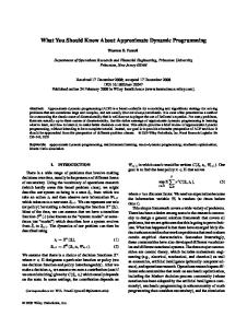
What Causes Pathologic Myopia? Pathologic myopia is primarily a genetic condition. The type of inheritance is frequently not simple, but in general an affected patient can pass it on to their children, both male and female. There is some evidence that extensive childhood close work, such as reading, can exacerbate simple myopia. This may be due to the effects of prolonged accommodation, which is the focusing of the eye’s lens by the ciliary muscle. There is evidence that reduced time spent outdoors may be another risk factor.3 Normal Retina No evidence exists to suggest that this environmental component is a true cause of pathologic myopia. Stronger evidence exists that it is caused by genetics. Clinical studies using atropine drops to reduce the effects of accommodation and help prevent simple myopia have been inconclusive and have no relevance that we know of to pathologic myopia.
Retina with Pathologic Myopia
Can Anything Be Done to Treat Pathologic Myopia? Research studies have been done to try to prevent eye wall stretching, for example by suturing sturdy bolstering tissue to the back of the eye. However, nothing has been conclusively shown to prevent the progression of the disease. For the present, our efforts are directed toward promptly treating the secondary consequences of pathologic myopia.
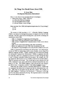
What Should I Watch For? There is no symptom of glaucoma. This is why you must have your eyes checked periodically by the ophthalmologist for the duration of your life. During your visit, your eye pressure is checked and the appearance of the optic nerve is assessed. Sometimes a visual field test is conducted to check the function of the optic nerve. The best test to check for abnormal blood vessels in the eyes is the Amsler Grid test. This test involves having the patient look at a piece of graph paper using one eye at a time. If the lines on the paper are distorted or spots on the grid are blurry, then the patient needs to see their ophthalmologist promptly. Sometimes pictures of the retina after injection of dye, called a fluorescein angiogram, may be needed to check for abnormal blood vessels. Amsler Grid To check for retinal tears or detachments, watch for symptoms such as new floaters, new or different flashes of light, particularly at night when the lights are turned out, and look for loss of peripheral vision. Check each eye individually by covering one eye at a time. Look at a clock or another object on the wall. Make sure that the eye being tested has vision in all four corners of the wall without moving the eye from the object of fixation. If a part of your visual field is missing, come in promptly to be examined.
What Kind of Treatment is Available? If patients develop blood vessel growth under the retina, we can reduce the damage using injections into the eye of a drug called bevacizumab (Avastin is the brand name). The white part of the eye is numbed with a series of Q-tips soaked in lidocaine and a tiny needle is used to inject the drug. Usually 1-3 injections over 2-4 months are needed to stop the blood vessel growth and thereafter regular monitoring is continued. Whereas this form of treatment produces outcomes superior to doing nothing, the visual acuity is usually not restored to the baseline level before the abnormal vessel grew. Optical coherence tomography scans are frequently used to monitor how treatment is working. Occasionally pictures are taken after injection fluorescein dye into a vein of the patient’s arm. These pictures (called a fluorescein angiogram) help to define location, size, and activity of the abnormal blood vessels.4,5 If the patient develops a retinal tear, then an argon laser can be used to weld around the retinal tear. This can prevent the fluids inside the eye from flowing through the retinal tear and causing a retinal detachment. This procedure is done in the office and requires no needles or stitches. It is uncomfortable, but not terribly so, and usually no pain medicine is needed. If patients advance beyond a retinal tear to develop retinal detachment before seeing the ophthalmologist, an operation to repair the detachment is required. This takes place as an outpatient at the hospital or outpatient surgery center. The procedure usually takes approximately 90 minutes. Intravenous sedatives keep the patient calm during surgery, and
numbing injections given around the eye prevent pain. Patients are usually out of work for approximately a week after surgery. Myopic foveoschisis can be helped by an operation called vitrectomy in which the tractional forces on the macula are relieved by removing the vitreous gel and the internal limiting membrane of the macula.1 A gas bubble may be used and the patient may be asked to maintain a face-down position for a few days to cause the split retinal layers and subretinal fluid to go away.6 If a pathologically myopic patient develops glaucoma, drops are often prescribed to keep the eye pressure appropriately low. Tests called Visual Fields are given from time to time to check for any progression of damage and help the doctor gauge if more or different drops are needed. A glaucoma specialist is often involved in this part of the patient’s care.
How Does Laser Vision Correction Apply to Pathologic Myopia? Laser vision correction is the use of an excimer laser, with or without a precise incision of the cornea, to change the focal point of the eye and attempt to remove the need for glasses. Laser vision correction does nothing about the tissue damage and stretching effects that underpin the problems of pathologic myopia. It can be used in people with pathologic myopia, but is generally avoided when extreme levels of refractive error exist. Most people with pathologic myopia have refractive errors greater than –6.0 diopters. Most patients with refractive errors of –10.0 diopters or greater would not be considered candidates for laser vision correction. All patients with pathologic myopia who are being considered for laser vision correction should have a thorough examination of the retina by an ophthalmologist familiar with detecting the various problems of pathologic myopia before having the procedure.
Final Comments Pathologic myopia is the 7th most common cause of legal blindness in the United States. It is particularly devastating because it often affects patients in the prime of their working lives, leading to significant disability and loss of productivity and income. Patients with pathologic myopia need regular ophthalmologic examination, and should avoid eye rubbing, which can cause hemorrhages in the thin, cracked tissues at the back of the eye. These patients should also be sure to wear protective eyewear when engaging in sports and work where eye trauma is possible. Patients have an obligation to familiarize themselves with the warning signs discussed in this pamphlet. If flashes, floaters, loss of side vision, or distorted straight lines develop, prompt examination by the ophthalmologist gives the patient the best opportunity to minimize damage in the eye. If you have questions after reading this pamphlet, please submit them using the “asking a question” link on the home page of this website. If you wish to read about these matters in greater detail, an excellent resource is Pubmed on the Internet site of the National Library of Medicine at http://www.ncbi.nlm.nih.gov/entrez/query.fcgi. Updated 1-21-2014 References
1. Taniuchi S, Hirakata A, Itoh Y, Hirota K, Inoue M. Vitrectomy with or without internal limiting membrane peeling for each stage of myopic traction maculopathy. Retina 2013;33:2025. 2. Ortisi E, Avitabile T, Bonfiglio V. Surgical management of retinal detachment because of macular hole in highly myopic eyes. Retina 2012;32:1704-1718. 3. Dharani R, Lee C-F, Theng ZX, et al. Comparison of measurements of time outdoors and light levels as risk factors for myopia in young Singapore children. Eye 2012;26:911-918. 4. Introini U, Casalino G, Querques G, et al. Spectral-domain OCT in anti-VEGF treatment of myopic choroidal neovascularization. Eye 2012;26:976-982. 5. Lai TYY, Luk FOJ, Lee GKY, and Lam DSC. Long-term outcome of intravitreal antivascular endothelial growth factor therapy with bevacizumab or ranibizumab as primary treatment for subfoveal myopic choridal neovascularization. Eye 2012;26:1004-1011. 6. Kim KS, Lee SB, Lee WK. Vitrectomy and internal limiting membrane peeling with and without gas tamponade for myopic foveoschisis. Am J Ophthalmol 2012;153:320-326.