Volume 3. Issue 2. Pages 113-302. 2008 ISSN 1934-578X (printed); ISSN 1555-9475 (online) www.naturalproduct.us
NPC
Natural Product Communications
EDITOR-IN-CHIEF DR. PAWAN K AGRAWAL Natural Product Inc. 7963, Anderson Park Lane, Westerville, Ohio 43081, USA
[email protected] EDITORS PROFESSOR GERALD BLUNDEN The School of Pharmacy & Biomedical Sciences, University of Portsmouth, Portsmouth, PO1 2DT U.K.
[email protected] PROFESSOR ALESSANDRA BRACA Dipartimento di Chimica Bioorganicae Biofarmacia, Universita di Pisa, via Bonanno 33, 56126 Pisa, Italy
[email protected] PROFESSOR DEAN GUO State Key Laboratory of Natural and Biomimetic Drugs, School of Pharmaceutical Sciences, Peking University, Beijing 100083, China
[email protected] PROFESSOR J. ALBERTO MARCO Departamento de Quimica Organica, Universidade de Valencia, E-46100 Burjassot, Valencia, Spain
[email protected] PROFESSOR YOSHIHIRO MIMAKI School of Pharmacy, Tokyo University of Pharmacy and Life Sciences, Horinouchi 1432-1, Hachioji, Tokyo 192-0392, Japan
[email protected] PROFESSOR STEPHEN G. PYNE Department of Chemistry University of Wollongong Wollongong, New South Wales, 2522, Australia
[email protected] PROFESSOR MANFRED G. REINECKE Department of Chemistry, Texas Christian University, Forts Worth, TX 76129, USA
[email protected] PROFESSOR WILLIAM N. SETZER Department of Chemistry The University of Alabama in Huntsville Huntsville, AL 35809, USA
[email protected] PROFESSOR YASUHIRO TEZUKA Institute of Natural Medicine Institute of Natural Medicine, University of Toyama, 2630-Sugitani, Toyama 930-0194, Japan
[email protected]
ADVISORY BOARD Prof. Viqar Uddin Ahmad Karachi, Pakistan Prof. Øyvind M. Andersen Bergen, Norway Prof. Giovanni Appendino Novara, Italy Prof. Yoshinori Asakawa Tokushima, Japan Prof. Maurizio Bruno Palermo, Italy Prof. Carlos Cerda-Garcia-Rojas Mexico city, Mexico Prof. Josep Coll Barcelona, Spain Prof. Geoffrey Cordell Chicago, IL, USA Prof. Samuel Danishefsky New York, NY, USA Dr. Biswanath Das Hyderabad, India Prof. A.A. Leslie Gunatilaka Tucson, AZ, USA Prof. Stephen Hanessian Montreal, Canada Prof. Michael Heinrich London, UK Prof. Kurt Hostettmann Lausanne, Switzerland Prof. Martin A. Iglesias Arteaga Mexico, D. F, Mexico Prof. Jerzy Jaroszewski Copenhagen, Denmark Prof. Teodoro Kaufman Rosario, Argentina Prof. Norbert De Kimpe Gent, Belgium Prof. Hartmut Laatsch Gottingen, Germany Prof. Marie Lacaille-Dubois Dijon, France Prof. Shoei-Sheng Lee Taipei, Taiwan
Prof. Francisco Macias Cadiz, Spain Prof. Anita Marsaioli Campinas, Brazil Prof. Imre Mathe Szeged, Hungary Prof. Joseph Michael Johannesburg, South Africa Prof. Ermino Murano Trieste, Italy Prof. Virinder Parmar Delhi, India Prof. Luc Pieters Antwerp, Belgium Prof. Om Prakash Manhattan, KS, USA Prof. Peter Proksch Düsseldorf, Germany Prof. William Reynolds Toronto, Canada Prof. Raffaele Riccio Salerno, Italy Prof. Ricardo Riguera Santiago de Compostela, Spain Prof. Satyajit Sarker Coleraine, UK Prof. Monique Simmonds Richmond, UK Prof. Valentin Stonik Vladivostok, Russia Prof. Hermann Stuppner Innsbruck, Austria Prof. Apichart Suksamrarn Bangkock, Thailand Prof. Hiromitsu Takayama Chiba, Japan Prof. Karen Valant-Vetschera Vienna, Austria Prof. Peter G. Waterman Lismore, Australia Prof. Paul Wender Stanford, USA
INFORMATION FOR AUTHORS Full details of how to submit a manuscript for publication in Natural Product Communications are given in Information for Authors on our Web site http://www.naturalproduct.us. Authors may reproduce/republish portions of their published contribution without seeking permission from NPC, provided that any such republication is accompanied by an acknowledgment (original citation)-Reproduced by permission of Natural Product Communications. Any unauthorized reproduction, transmission or storage may result in either civil or criminal liability. The publication of each of the articles contained herein is protected by copyright. Except as allowed under national “fair use” laws, copying is not permitted by any means or for any purpose, such as for distribution to any third party (whether by sale, loan, gift, or otherwise); as agent (express or implied) of any third party; for purposes of advertising or promotion; or to create collective or derivative works. Such permission requests, or other inquiries, should be addressed to the Natural Product Inc. (NPI). A photocopy license is available from the NPI for institutional subscribers that need to make multiple copies of single articles for internal study or research purposes. To Subscribe: Natural Product Communications is a journal published monthly. 2007 subscription price: US$1,395 (Print, ISSN# 1934-578X); US$1,095 (Web edition, ISSN# 1555-9475); US$1,795 (Print + single site online). Orders should be addressed to Subscription Department, Natural Product Communications, Natural Product Inc., 7963 Anderson Park Lane, Westerville, Ohio 43081, USA. Subscriptions are renewed on an annual basis. Claims for nonreceipt of issues will be honored if made within three months of publication of the issue. All issues are dispatched by airmail throughout the world, excluding the USA and Canada.
NPC
2008 Vol. 3 No. 2 199 - 203
Natural Product Communications
Phenolic Constituents of Platanus orientalis L. Leaves Taha S. El-Alfya, Hamida M.A. El-Goharya, Nadia M. Sokkara, Amani A. Sleemb and Dalia A. Al-Mahdya a
Department of Pharmacognosy, Faculty of Pharmacy, Cairo University, Egypt
b
Department of Pharmacology, National Research Center, Giza, Egypt
[email protected] Received: September 10th, 2007; Accepted: November 24th, 2007
A new flavonoid glycoside, 5,7,4´-trihydroxy-3,6-dimethoxyflavone-3´-O-β-D-xylopyranoside (axillarin-3´-O-β-Dxylopyranoside), was isolated from the ethyl acetate fraction of the ethanolic extract of Platanus orientalis L. leaves, along with nine known flavonoid aglycones and glycosides, a phenolic acid and a phenolic acid derivative. Their structures were established on the basis of detailed spectral analysis. The total ethanolic, aqueous and ethyl acetate extracts of the leaves were tested for antihepatotoxic, antioxidant and cytotoxic activities. The extracts were found to be effective in protection against CCl4 liver injury by significantly lowering alanine aminotransferase (ALT), aspartate aminotransferase (AST) and alkaline phosphatase (ALP) levels. The extracts also significantly restored depleted levels of glutathione, indicating a strong antioxidant activity, while they showed variable cytotoxic activity. Keywords: Platanus orientalis L., antihepatotoxic, antioxidant, antitumor, flavonoids.
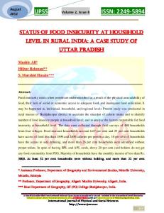
Many phytochemicals, especially phenolic constituents, have shown promise for the protection and/or treatment of hepatotoxicity cases in which oxidative stress plays a key role [1-3]. Platanus orientalis L., known as oriental plane (family Platanaceae), is a deciduous tree native to south-west Asia, south-east Europe and the Mediterranean regions [4,5]. The leaves were used in folk medicine in opthalmia, the bark was boiled in vinegar and given for diarrhea, dysentery and toothache [6,7], while the buds were used as a urinary tract antiseptic and antimicrobial [8]. Only one report has been found dealing with the flavonoidal content of the buds and their cytotoxic activity [9]. Nothing could be traced concerning either the phytochemical content or biological activities of the leaves. This work reports the isolation of a new flavonoid glycoside, 5,7,4´-trihydroxy-3,6dimethoxyflavone-3´-O-β-xylopyranoside (axillarin3´-O-β-xylopyranoside) (1), in addition to nine known flavonoid aglycones and glycosides (2-10), a phenolic acid (11), and a phenolic acid
derivative (12). Also, the antihepatotoxic, antioxidant and cytotoxic activities of the total ethanolic extract 5` 6` HO
7
8
9
O
2
H3CO
5 OH
4
H
H
3 10
OH
3`
1` 2`
6
4`
HO O
1``
OH
5`` O
OCH3
3``
2`` H
H
H
OH
4`` H
O
1
(TEE), total aqueous extract (TAE), and the flavonoid-rich ethyl acetate fraction (EAF) were evaluated and reported. Compound 1 was obtained as a yellow powder. The molecular formula was calculated as C22H22O12 from the ESI-MS [M-H]- molecular ion peak appearing at m/z 477. A peak observed at m/z 345 after ionization indicated the loss of a pentose sugar. The NMR spectra suggested a flavonoid skeleton for the compound. The 1H NMR spectrum showed signals of ring B (2 doublets at δ 6.99, d, J = 8.4 Hz and δ 7.83, d, J = 2.1 Hz) indicating ortho and meta couplings of protons 5´ and 2´ with H-6´,
200 Natural Product Communications Vol. 3 (2) 2008
respectively. This was confirmed by a doublet of doublets assigned to H-6´ (δ 7.65, dd, J = 8.4, 2.1 Hz). The downfield shift of H-2´ (0.7 ppm) indicates the presence of a substituent at the 3´position [10,11]. In addition, ring A protons were represented by a singlet at δ 6.52, assigned to H-8. The absence of other signals characteristic of ring A suggested substitution at the 3 and 6 positions, with the two methoxyl groups displayed at δ 3.80 and 3.74, respectively. The anomeric proton of the sugar unit appeared as a doublet at δ 4.79. This upfield shift, together with its coupling constant (J = 6.9 Hz), confirmed the sugar attachment to be at the 3´ position having a β- configuration [10,11]. All the 13 C NMR signals were assigned and the sugar moiety was identified as β-xylopyranoside by comparison with reported data [12]. The HMBC experiment showed correlations between δ 4.79 (H-1´´) and 144.9 (C-3´), between δ 3.74 (CH3-6) and 131.2 (C-6) and between δ 3.80 (CH3-3) and 137.3 (C-3). Thus, compound 1 was identified as 5,7,4´-trihydroxy-3,6-dimethoxyflavone-3´-O-βD-xylopyranoside. Compounds 2-12 were identified as kaempferol (2), quercetin (3), kaempferol-3-O-α-L-(2´´,3´´-di-E-pcoumaroylrhamnopyranoside) (platanoside) (4), kaempferol-3-O–β-D-(6´´-E-p-coumaroylglucopyranoside) (tiliroside) (5), kaempferol-3-O-α-Lrhamnopyranoside (afzelin) (6), quercetin-3-O-α-Lrhamnopyranoside (quercitrin) (7), quercetin-3-O-βD-galactopyranoside (hyperoside) (8), kampferol -3O-β- rutinoside (nicotiflorin) (9), quercitin-3-Orutinoside ( rutin) (10), p-coumaric acid (11), and 4´caffeoylquinic acid (cryptochlorogenic acid) (12) by comparing their UV,1H and 13C NMR spectroscopic data with those published [10,12-18].
El-Alfy et al.
Table 1: Effect of TEE, TAE extracts and EAF of P. orientalis leaves on blood glutathione level of alloxan-induced diabetic rats.(n=6). GSH
Group
M±S.E
% potency
Normal
36.8 ± 1.2b
-
Diab. control
23.4 ± 0.3a
-
Diab. treated with TEE
33.1 ± 0.7b
92.2
Diab. treated with TAE
33.1 ± 0.9b
92.2
Diab. treated with EAF
33.7 ± 1.2b
93.8
Diab. treated with Vitamin E
35.9 ± 0.7b
100
a Significantly different from normal value at P=0.01. b Significantly different from diabetic control value at P=0.05.
Toxicological studies revealed that the LD50 of both the TEE and TAE of P. orientalis leaves were 7.3 and 7.6 g/kg, respectively and can be considered safe [19]. The antioxidant activity was determined in alloxaninduced diabetic rats [20]. All the extracts restored the depleted levels of glutathione (GSH) (Table 1) with the EAF being the most potent extract (93.8%). The tested extracts reversed the elevated levels of the serum liver enzymes alanine aminotransferase (ALT), aspartate aminotransferase (AST) and alkaline phosphatase (ALP) (Table 2). These effects were comparable to and in some cases higher than that of the reference drug, silymarin. TEE was the most potent extract as it decreased the levels of ALT, AST and ALP by 52.7%, 43% and 39%, respectively. All the tested extracts showed dose dependent cytotoxicity against the three human cancer cell lines, MCF7, HEPG2 and H460 (Table 3). It was observed that EAF showed the highest activity against HEPG2 cell line.
Table 2: Effect of TEE, TAE and EAF of P. orientalis on ALT, AST and ALP using silymarin as the reference standard in liver damaged rats. (n=10).
- ve control TEE (100 mg/kg) TAE (100 mg/kg) EAF (100 mg/kg) Silymarin (25 mg/kg) a 1
Liver enzymes ALT AST ALP
Zero 25.9±0.7 28.2±1.3 7.2±0.2
30 days1 25.2±0.3 27.9±0.8 7.1±0.1
72 hours2 131.6±4.3 124.3±4.1 28.2±1.2
30 days3 142.8±5.1 136.7±5.1 31.2±1.3
ALT AST ALP
28.6±0.8 26.5±0.9 7.6±0.1
27.9±1.1 26.1±0.3 7.1±0.1
58.4±1.7a 54.8±1.6a 13.1±0.9a
27.6±0.9ab 31.2±1.1ab 8±0.2 ab
52.7 43 39
ALT AST ALP ALT AST ALP
27.2±0.4 29.1±0.8 7.5±0.1 28.2±1.4 29.2±1.2 7.4±0.2
27.5±0.8 27.6±0.5 7.0±0.2 26.8±1.3 28.8±0.8 7.1±0.1
52.6±1.7a 51.4±1.2a 11.5±0.3a 53.5±2.4a 50.4±2.1a 15.2±0.8a
29.9±1.1ab 30.6±1.4ab 8.3±0.5 ab 34.7±1.6ab 34.0±0.7ab 9.3±0.4 ab
43 40.4 27.8 35 32.5 38.8
ALT AST ALP
28.4±0.9 28.7±0.6 7.3±0.2
27.6±1.2 28.2±0.4 7.2±0.1
41.6±0.9a 43.7±1.3a 9.1±0.3a
24.7±0.4ab 25.8±0.7 ab 6.5±0.1 ab
40.6 41 28.5
% change -
Significantly different from control group at P=0.01, b Significantly different from 72 hours after injection of CCl4 at P=0.01. 30 days after treatment with the extracts, 2 72 hours after injection with CCl4, 3 30 days after administration of the extracts to liver damaged rats.
Constituents from Platanus orientalis
Natural Product Communications Vol. 3 (2) 2008 201
Table 3: Effect of TEE, TAE and EAF of the leaves of P. orientalis on MCF7, HEPG2 and H460 human carcinoma cell lines. Conc μg/mL 0.000
Cell lines
TEE
TAE
EAF
MCF7 HEPG2
1.007±0.024 1.001±0.060
1.007±0.024 1.001±0.060
1.003±0.056 1.000±0.037
H460
1.000±0.036
1.000±0.036
1.000±0.034
MCF7
0.747±0.012
0.731±0.015
0.934±0.024
5.000
HEPG2 H460
0.613±0.030 0.774±0.014
0.673±0.026 0.523±0.032
0.556±0.002 0.664±0.005
MCF7
0.603±0.012
0.502±0.015
0.729±0.012
10.000
HEPG2 H460
0.553±0.054 0.610±0.018
0.643±0.030 0.462±0.009
0.441±0.008 0.619±0.014
MCF7 = breast carcinoma cell line, HEPG2 = hepatocellular carcinoma cell line, H460= lung carcinoma cell line.
This result, in addition to its potent antioxidant activity and its ability to lower the elevated levels of the liver enzymes could be attributed to its phenolic content. Experimental General: Melting points were determined on Electrothermal 9100 equipment. Optical rotation [α]D was determined using a Perkin-Elmer 241 polarimeter (MeOH, c in g/100 mL). IR spectra were measured using a Shimadzu-IR-435 Infrared spectrophotometer. UV spectra were measured using a Shimadzu UV 1650 PC spectrophotometer. EI-MS were measured on a Finnigan SS Q 7000. ESI-MS (negative mode) of compound 1 was measured on Thermo Finnigan (ion trap) equipment. NMR spectra were measured in DMSO-d6 with a Varian Mercury instrument (NMR, 300 MHz, 13C, 75 MHz). TLC was performed on sheets pre-coated with either silica gel F254 (Fluka) or with RP-18 F254 (Merck, Germany). Compounds were visualized under UV light (254 and 366 nm) and by spraying with aluminium chloride and NP-PEG reagents. Column chromatography was performed over Silica gel H 60 (Sigma) for VLC, Sephadex LH-20 (Pharmacia), silica gel 60 (Fluka) and silica gel C18–reversed phase (70-230 mesh, Fluka). Plant material: The leaves of Platanus orientalis L., were collected from El-Orman Botanical Garden, Giza, Egypt. A voucher specimen is kept in the Department of Pharmacognosy, Faculty of Pharmacy, Cairo University. Material for pharmacology: Chemicals used in this study were: Alloxan (Sigma Co., USA); Vitamin E (dl α-tocopheryl acetate) Pharco Pharmaceutical Co,
Egypt; and Silymarin ®, Sedico Co., Egypt. Kits for biochemical assessment of AST, ALT and ALP were purchased from Biomerieux, France, and the glutathione kit from Wak Company, Germany. Extraction and isolation: The air dried, powdered and defatted leaves of P. orientalis (2 kg) were macerated with 70% EtOH till exhaustion. The ethanolic extract was evaporated under reduced pressure to give 197 g of dark green residue, of which 180 g was suspended in water (600 mL) and partitioned successively with CHCl3, EtOAc and n-BuOH to afford 22 g, 25 g and 13 g of dried residues, respectively. Another 100 g of the air dried, powdered leaves was boiled with 500 mL water, followed by evaporation under reduced pressure. The residue was dissolved in sterile water before administration to rats. EAF (20 g) was chromatographed on Silica gel H 60, VLC (Ø7 x 12.5 cm, 200 g) and eluted with CHCl3, gradients of CHCl3-EtOAc, EtOAc and gradients of EtOAc-MeOH up to pure methanol. Fractions, each of 200 mL, were collected and monitored by TLC. Similar fractions were pooled together to give 9 collective fractions. Fractions 1 (270 mg), 2 (410 mg), 3 (850 mg) and 4 (600 mg) were subjected to repeated CC on Sephadex LH-20 to give compounds 11 (22 mg, 0.11%), 2 (38 mg, 0.19%), 3 (35 mg, 0.17%), 4 (15 mg, 0.07%) and 5 (20 mg, 0.1%). Fractions 5 (950 mg), 6 (3.1 g), 7 (3.2 g), 8 (2 g) and 9 (1.5 g) were purified on Sephadex LH-20 followed by CC on silica gel 60 and Silica gel C18– reversed phase to give compounds 6 (22 mg, 0.11%), 1 (30 mg, 0.15%), 7 (23 mg, 0.11%), 8 (32 mg, 0.16%), 9 (20 mg, 0.1%), 10 (35 mg, 0.17%) and 12 (32 mg, 0.16%). 5,7,4´-Trihydroxy-3,6-dimethoxyflavone-3´-O-βD-xylopyranoside (axillarin-3´-O-β-D-xylopyranoside) (1) Yellow powder. MP: 240-242oC. [α]D: -36.2º (c 0.1, MeOH). Rf: 0.62 (CHCl3-MeOH, 8:2 and 2 drops formic acid). IR (KBr): 3399, 2950, 2922, 1659, 1597, 1499, 1150, 1075, 834, 800 cm-1.
202 Natural Product Communications Vol. 3 (2) 2008
UV/Vis λmax (MeOH) nm: 254 (IIb), 272 (IIa), 344, (NaOMe): 277, 328, 401, (AlCl3): 261, 280, 308sh, 364, (AlCl3/HCl): 260, 282, 309sh, 363, (NaOAc): 274, 334 sh, 401, (NaOAc./H3BO3): 272, 350. 1 H NMR (300 MHz, DMSO-d6) δ: 7.83 (1H, d, J = 2.1 Hz, H-2´), 6.99 (1H, d, J = 8.4 Hz, H-5´), 7.65 (1H, dd, J = 8.4, 2.1 Hz, H-6´), 6.52 (1H, s, H-8), 3.80 (3H, s, OMe-3), 3.74 (3H, s, OMe-6). Xylose: 4.79 (1H, d, J = 6.9 Hz, H-1''). 13 C NMR (75 MHz, DMSO-d6): Aglycone: 177.9 (C-4), 161.7 (C-7), 154.7 (C-2), 152.2 (C-5), 151.5 (C-9), 150.3 (C-4´), 144.9 (C-3´), 137.3 (C-3), 131.2 (C-6), 123.6 (C-1´), 120.7 (C-6´), 116.8 (C-5´), 116.3 (C-2´), 104.2 (C-10), 94.0 (C-8), 59.8 (OMe-3), 59.5 (OMe-6). Xylose: 102.8 (C-1´´), 73.03 (C-2´´), 75.7 (C-3´´), 69.3 (C-4´´), 65.7 (C-5´´). MS (EI, 70 eV) m/z (%): 478 (M+, 1.02%), 345 (M+ - C5H9O4, 9.01%), 463 (M+- CH3, 1.22%), 158 (100%). Negative ESI-MS: m/z [M-H]- calcd for C22H22O12: 477.00, MS/MS fragment at m/z 345 [M-xylose]Yield: 0.15%. Animals and cell lines: Adult male albino rats (130-140 g) were obtained from the animal-breeding unit of the National Research Center, Dokki, Giza, Egypt. Rats were fed on standard normal laboratory diet and water ad libidum. Doses of the drugs were calculated according to the method of Paget and Barnes [21] and were administrated orally by gastric tube. MCF7, HEPG2 and H460 cell lines were obtained from the National Cancer Institute, Kasr ElAiny Street, Cairo, Egypt. Determination of LD50: LD50 values of both TEE and TAE of the leaves of P. orientalis were determined following Karber procedures [22]. Antioxidant activity: Thirty six adult male albino rats were used in the experiment and divided into 6 groups. One group was kept as control. All the remaining groups were injected intraperitoneally with a single dose of alloxan (150 mg / kg b. wt.) to
El-Alfy et al.
induce hyperglycaemia [23]. The second group was kept as diabetic control and the third group was given Vitamin E orally in a daily dose of 100 mg/kg. The other three groups received 100 mg/kg orally of the TEE, TAE and the EAF of P. orientalis leaves, respectively. Blood GSH levels were determined and recorded [24]. Anti-hepatotoxic activity: The tested extracts were evaluated and compared with that of silymarin (reference standard), according to the method of Klassen and Plaa [25]. Fifty adult male albino rats were divided into 5 groups as following: the first group received 1 mL saline and kept as the reference control group; the second group received silymarin (25 mg/kg b. wt. daily) and groups three, four and five received a daily oral dose (100 mg/kg) of TEE, TAE and EAF, respectively. The extracts and silymarin were administered for a period of 30 days, followed by induction of hepatotoxicity through intraperitoneal injection of 5 mL/kg of 25% CCl4 in liquid paraffin, then further administration of the extracts for 30 days. Liver damage and/or recovery were assessed by measuring serum alanine aminotransferase (ALT), aspartate aminotransferase (AST) and alkaline phosphatase (ALP). Cytotoxic activity: The antitumor activity of the extracts against MCF7, HEPG2 and H460 cell lines was determined using the method of Skehan et al. [26]. The relation between the surviving tumor fraction and drug concentration was determined to obtain the survival curve of each tumor cell line after the specified tested extract. All data are expressed as mean±S.E., and the statistical significance was evaluated by One-Way Analysis of Variance ANOVA [27]. Acknowledgments - The authors wish to thank Prof. Dr Wafaa M. Amer, Prof. of Taxonomy and Flora, Botany Department, Faculty of Science, Cairo University, for identification of the plant material.
References [1]
Deleve LD, Kaplowitz N. (1995) Mechanisms of drug-induced liver disease. In: Gastroenterology Clinics of North America, 24, 787-810.
[2]
Farrel GC. (1998) Liver disese caused by drugs, anesthetics and toxins. In Sleisenger and Fordtran’s Gastrointestinal and Liver Disease: Pathophysiology/ Diagnosis/ Management. 2nd Ed., Feldman M, Scharschmidt BF, Sleisenger MH. (Eds), WB Saunders Co., Philadelphia, 1221-1253.
[3]
Halliwell B, Gutteridge JMC. (1999) Free radicals in biology and medicine. In: Studies of Generalized Light Emission (Luminescence/Fluorescence). 3rd Ed. Oxford: University Press, 387-388.
Constituents from Platanus orientalis
Natural Product Communications Vol. 3 (2) 2008 203
[4]
Bailey LH. (1953) The Standard Cyclopedia of Horticulture, II. The Macmillan Company, New York, 2706.
[5]
Flora of China, [http://flora.huh.harvard.edu/china/mss/volume 09/Platanaceae.PDF].
[6]
Chopra RN, Nayar SL, Chopra IC. (1956) Glossary of Indian Medicinal Plants. Council of Scientific and Industrial Research, New Delhi, 196.
[7]
Gunther TR. (1959) The Greek Herbal of Dioscorides. Hafner Publishing Co., New York, 58.
[8]
Mitrokotsa D, Mitaku S, Demetzos C, Harvala C, Mentis A, Perez S, Kokkinopoulos D. (1993) Bioactive compounds from the buds of Platanus orientalis and isolation of a new kaempferol glycoside. Planta Medica, 59, 517-520.
[9]
Mitrocotsa D, Bosch S, Mitaku S, Dimas C, Skaltsounis AL, Harvala C, Briand G, Roussakis C. (1999) Cytotoxicity against human leukemic cell lines, and the activity on the expression of resistance genes of flavonoids from Platanus orientalis. Anticancer Research, 19 (3A), 2085-2088.
[10]
Mabry TJ, Markham KR, Thomas MB. (1970) The Systematic Identification of Flavonoids, Springer Verlag, New York.
[11]
Markham KR, Whitehouse LA. (1984) Unique flavonoid glycosides from the New Zealand white pine, Dacrycarpus dacrydioides, Phytochemistry, 23, 1931-1936.
[12]
Agrawal PK. (1989) Carbon-13 NMR of Flavonoids, Elsevier, New York.
[13]
Markham KR. (1982) Techniques of Flavonoid Identification, Academic Press, London, New York.
[14]
Geissman TA. (1962) The Chemistry of Flavonoid Compounds. The Macmillan Company, New York.
[15]
Wagner H, Baldt S, Zagainiski EM. (1983) Droger Analyse. Springer Verlag, Berlin.
[16]
Kaouadji M. (1990) Acylated and non-acylated kaempferol monoglycosides from Platanus acerifolia buds. Phytochemistry, 29, 2295-2297.
[17]
Gerothanassis IP, Exarchou V, Lagouri V, Troganis A, Tsimidou M, Boskou D. (1998) Methodology for identification of phenolic acids in complex phenolic mixtures by high-resolution two dimensional nuclear magnetic resonance. Application to methanolic extracts of two Oregano species. Journal of Agricultural Food Chemistry, 46, 4185-4192.
[18]
Nakatani N, Kayano S, Kikuzaki H, Sumino K, Katagiri K, Mitani T. (2000) Identification, quantitative determination, and antioxidative activities of chlorogenic acid isomers in prune (Prunus domestica L.). Journal of Agricultural and Food Chemistry, 48, 5512-5516.
[19]
Buck WB, Osweiter GD, Van Gelder AG. (1976) Clinical and Diagnostic Veterinary Toxicology, 2nd Ed., 52011, Kendall/Hund Publishing Company, Iowa.
[20]
Szkudelski T. (2001) The mechanism of alloxan and streptozotocin action in B cells of rat pancreas. Physiology Research, 50, 536-546.
[21]
Paget G, Barnes E. (1964) Toxicity Tests In: Evaluation of Drug Activities. Pharmacometrics. Ed. Laurence DR, Bacharach AL, Academic Press London, 135-160.
[22]
Karber G. (1931) Determination of LD50. Archive de Experimental Pathology and Pharmacology, 162, 480-488.
[23]
Eliasson SG, Samet JM. (1969) Alloxan induced neuropathies. Lipid changes in nerve and root fragments. Life Science, 8, 493504.
[24]
Beutler E, Duron O, Kelly BA. (1963) Improved method for the determination of blood glutathione. Journal of Laboratory Clinical Medicine, 61, 882-888.
[25]
Klassen CD, Plaa GL. (1969) Comparison of biochemical alterations elicited in liver of rats treated with CCl4 and CHCl3. Biochemical Pharmacology, 18, 2019-2022.
[26]
Skehan P, Storeng R, Scudiero D, Monks A, McMahon JM, Vistica D, Warren J, Bokesch H, Kenney S, Boyd MR. (1990) New colorimetric cytotoxicity assay for anti-cancer drug screening. Journal of National Cancer Institute, 82, 1107-1112.
[27]
Sendecor WG, Cochrean GW. (1971) Statistical Methods. Iowa State University Press, Iowa.
New Acylated Flavonol Diglycosides of Cynanchum acutum Mona A. Mohamed, Wafaa S. Ahamed, Mortada M. El-Said and Heiko Hayen
193
Phenolic Constituents of Platanus orientalis L. Leaves Taha S. El-Alfy, Hamida M.A. El-Gohary, Nadia M. Sokkar, Amani A. Sleem and Dalia A. Al-Mahdy
199
Strepsiamide A-C, New Ceramides from the Marine Sponge Strepsichordaia lendenfeldi Sabrin R. M. Ibrahim, Gamal A. Mohamed, Ehab S. Elkhayat, Yaser G. Gouda and Peter Proksch
205
Free Radical Scavenging and Cytoprotective Activity of Salacia euphlebia Merr. Sanan Subhadhirasakul, Niwat Keawpradub, Charuporn Promwong and Supreeya Yuenyongsawad
211
Antialactone: A New γ-Lactone from Antiaris africana, and its Absolute Configuration Determined Vouffo Bertin, Hidayat Hussain, Simeon F. Kouam, Etienne Dongo, Gennaro Pescitelli, Piero Salvadori, Tibor Kurtán and Karsten Krohn
215
Subereaphenol A, a new Cytotoxic and Antimicrobial Dibrominated Phenol from the Red Sea Sponge Suberea mollis Lamiaa A. Shaala, Sherief I. Khalifa, Mostafa K. Mesbah, Rob W. M. van Soest and Diaa T. A. Youssef
219
A New Ferulic Ester and Related Compounds from Bombax malabaricum DC. Pahup Singh, Durga K. Mewara and Mahesh C. Sharma
223
Role of Turmeric in Ultraviolet Induced Genotoxicity in a Bacterial System Arijit Pal, Mita Ghosh and Arun Kumar Pal
227
Excited-State pKa Values of Curcumin Qian Zhao, De-Xin Kong and Hong-Yu Zhang
229
Antibacterial and Antifungal Activities of Some Phenolic Metabolites Isolated from the Lichenized Ascomycete Ramalina lacera Lumír O Hanuš, Marina Temina and Valery M Dembitsky
233
Phenolic Constituents of Hypericum Flowers Carolina Nör, Ana Paula Machado Bernardi, Juliana Schulte Haas, Jan Schripsema, Sandra Beatriz Rech and Gilsane Lino von Poser
237
Seasonal Variation of Hypericin and Pseudohypericin Contents in Hypericum scabrum L. Growing Wild in Turkey Ali Kemal Ayan, Cüneyt Çırak and Kerim Güney
241
Molluscicidal Polyphenols from Species of Fucaceae Asmita V. Patel, David C. Wright, Maricela Adrian Romero, Gerald Blunden and Michael D. Guiry
245
Anti-diabetic Activity of Triphala Fruit Extracts, Individually and in Combination, in a Rat Model of Insulin Resistance Venkateshan S. Prativadibhayankaram, Samir Malhotra, Promila Pandhi and Amritpal Singh
251
Biotransformation of Mefenamic Acid by Cell Suspension Cultures of Solanum mammosum Suzana Surodjo, Angela A. Salim, Suciati, Achmad Syahrani, Gunawan Indrayanto and Mary J. Garson
257
Natural Variability in Enantiomeric Composition of Bioactive Chiral Terpenoids in the Essential Oil of Solidago canadensis L. from Uttarakhand, India Chandan S. Chanotiya and Anju Yadav
263
Germacrone Dominates the Leaf Oil of Siparuna grandiflora from Monteverde, Costa Rica William N. Setzer, Brittany R. Agius, Tameka M. Walker, Debra M. Moriarity and William A. Haber
267
Leaf Oil Composition of Piper aduncum subsp. ossanum (C. CD.) Saralegui from Cuba Orlando Abreu and Jorge A. Pino
271
Volatile Constituents from the Leaves of Phyllanthus salviaefolius H. B. K. from the Venezuelan Andes Silvana Villarreal, Luis B. Rojas, Alfredo Usubillaga, Irama Ramírez and Mariana Solórzano
275
Synergistic Antifungal Activities of Thymol Analogues with Propolis Chi-Pien Chen and Ai-Yu Shen
279
Review /Account Argan oil, Functional Food, and the Sustainable Development of the Argan Forest Zoubida Charrouf and Dominique Guillaume
283
Chemical Constituents of Selected Japanese and New Zealand Liverworts Yoshinori Asakawa, Masao Toyota, Fumihiro Nagashima and Toshihiro Hashimoto
289
Natural Product Communications 2008 Volume 3, Number 2 Contents Original paper
Page
New cis-Chrysanthenyl Esters from Eryngium planum L. Emilia Korbel, Ange Bighelli, Anna Kurowska, Danuta Kalemba and Joseph Casanova
113
Secondary Metabolites from Eremostachys laciniata İhsan Çalış, Ayşegül Güvenç, Metin Armağan, Mehmet Koyuncu, Charlotte H. Gotfredsen and Søren R. Jensen
117
A Novel Iridoid from Plumeria obtusa Firdous Imran Ali, Imran Ali Hashmi and Bina Shaheen Siddiqui
125
Terpenoids from Neolitsea dealbata Xiujun Wu, Bernhard Vogler, Betsy R. Jackes and William N. Setzer
129
Volatile Components from Selected Mexican, Ecuadorian, Greek, German and Japanese Liverworts Agnieszka Ludwiczuk, Fumihiro Nagashima, Rob S. Gradstein and Yoshinori Asakawa
133
New ent–Kaurane type Diterpene Glycoside, Pulicaroside-B, from Pulicaria undulata L. Nasir Rasool, Viqar Uddin Ahmad, Naseem Shahzad, Muhammad A. Rashid, Aman Ullah, Zahid Hassan, Muhammad Zubair and Rasool Bakhsh Tareen
141
Anti-babesial Quassinoids from the Fruits of Brucea javanica Ahmed Elkhateeb, Masahiro Yamasaki, Yoshimitsu Maede, Ken Katakura, Kensuke Nabeta and Hideyuki Matsuura
145
Triterpenoids and Alkaloids from the Roots of Peganum nigellastrum Zhongze Ma, Yoshio Hano, Feng Qiu, Gang Shao, Yingjie Chen and Taro Nomura
149
Saikosaponins from Bupleurum chinense and Inhibition of HBV DNA Replication Activity Feng Yin, Ruixiang Pan, Rongmin Chen and Lihong Hu
155
Brauhenoside A and B: Saponins from Stocksia brauhica Benth. Viqar Uddin Ahmad, Sadia Bader, Saima Arshad, Faryal Vali Mohammad, Amir Ahmed, Shazia Iqbal, Saleha Suleman Khan and Rasool Bakhsh Tareen
159
Saponins from Fresh Fruits of Randia siamensis (Lour) Roem. & Schult. (Rubiaceae) Rapheeporn Khwanchuea, Emerson Ferreira Queiroz, Andrew Marston, Chaweewan Jansakul and Kurt Hostettmann
163
New Alkaloid from Aspidosperma polyneuron Roots Tatiane Alves dos Santos, Dalva Trevisan Ferreira, Jurandir Pereira Pinto, Milton Faccione and Raimundo Braz-Filho
171
Acanthomine A, a new Pyrimidine-β-Carboline Alkaloid from the Sponge Acanthostrongylophora ingens Sabrin R. M. Ibrahim, RuAngelie Ebel, Rainer Ebel and Peter Proksch
175
Phytochemical and Microscopic Characterization of the Caribbean Aphrodisiac Bois Bandé: Two New Norneolignans Ingrid Werner, Pavel Mucaji, Armin Presser, Christa Kletter and Sabine Glasl
179
3-Acetoxy-7-methoxyflavone, a Novel Flavonoid from the Anxiolytic Extract of Salvia elegans (Lamiaceae) Silvia Marquina, Yolanda García, Laura Alvarez and Jaime Tortoriello
185
Struthiolanone: A Flavanone-Resveratrol Adduct from Struthiola argentea Sloan Ayers, Deborah L. Zink, Robert Brand, Seef Pretorius, Dennis Stevenson and Sheo B. Singh
189
Continued inside back cover