East Tennessee State University
Digital Commons @ East Tennessee State University Electronic Theses and Dissertations
8-2007
Vitamin E (Tocotrienols) and Prostate Cancer: A Proteomics Approach. Christian Mbangha Muenyi East Tennessee State University
Follow this and additional works at: http://dc.etsu.edu/etd Recommended Citation Muenyi, Christian Mbangha, "Vitamin E (Tocotrienols) and Prostate Cancer: A Proteomics Approach." (2007). Electronic Theses and Dissertations. Paper 2126. http://dc.etsu.edu/etd/2126
This Thesis - Open Access is brought to you for free and open access by Digital Commons @ East Tennessee State University. It has been accepted for inclusion in Electronic Theses and Dissertations by an authorized administrator of Digital Commons @ East Tennessee State University. For more information, please contact
[email protected].
Vitamin E (Tocotrienols) and Prostate Cancer: A Proteomics Approach
A thesis presented to the faculty of the Department of Chemistry East Tennessee State University In partial fulfillment of the requirements for the degree Master of Science in Chemistry
by Christian Mbangha Muenyi August 2007
William L. Stone, Ph.D., Chair Hamid Kasmai, Ph.D. Chu-Ngi Ho, Ph.D.
Keywords: vitamin E, gamma-tocotrienol, delta-tocotrienol, apoptosis, necrosis, proteomics
ABSTRACT Vitamin E (Tocotrienols) and Prostate Cancer: A Proteomics Approach by Christian Mbangha Muenyi
Proteomics is the large scale study of proteins in cells or organisms. The purpose of this study was to characterize the proteomic alterations occurring in a prostate cancer (LNCaP) cell line after treatment with delta-tocotrienol (a form of vitamin E not very prevalent from most dietary sources). We found that both gamma- and delta-tocotrienols induced time and concentration dependent growth inhibition and programmed cell dead (apoptosis) in LNCaP cells. Secondly, we used two-dimensional gel electrophoresis (2-DE) to characterize changes in protein expression levels associated with this treatment. Our results show that a specific set of proteins are regulated at both early and late times following treatment with delta-tocotrienol and these proteins have been characterized by their apparent molecular weights and isoelectric points. The alteration observed at early time points are particularly interesting because these changes are likely to reflect the underlying molecular mechanisms for triggering cancer cell death.
2
ACKNOWLEDGEMENTS I wish to express sincere appreciation to Dr. William L. Stone for his excellent mentorship. My gratitude to him cannot all be expressed in words! I wish to also thank him for giving me the opportunity to go through all the experiences I have had in his lab. Dr. Kasmai might not know the impact he created in my life when he agreed to be my co-advisor, but I am saying it here that it was a life-changing decision. I thank him so much for all his kindness. I also extend sincere gratitude to Dr. Ho for readily accepting to be a member of my thesis committee. I will be doing myself a disfavor if I do not say here how appreciative I am to Dr. Thomas T-S. Huang. He ‘adopted’ me as one of his children when I got to the US and played a very vital role in facilitating my admission into ETSU. I can never thank him enough for that. I also wish to mention those who made life easy for me in Dr. Stone’s lab. First, I wish to say my heart goes to Hongsong for always being there and willing to assist me. Qui Min also did a wonderful job in helping me out in the lab. My appreciation too goes to all the members of the Internal Medicine Journal Club for helping me cope with public speaking. The staff and students of the department of chemistry at ETSU really provided a friendly academic environment for me and I sincerely thank them for that. My heart also goes to my family back at home and to all my friends who have made my life here worth living. I also wish to acknowledge in a very special way the efforts of my wife in the realization of this project. To crown it, I wish to say a big thank you to Christine Hendershott for her role in correcting my manuscript.
3
CONTENTS Page ABSTRACT ...........................................................................................................................
2
ACKNOWLEDGEMENTS...................................................................................................
3
LIST OF TABLES.................................................................................................................
8
LIST OF FIGURES………………………..……………………………………………….
9
Chapter 1. INTRODUCTION............................................................................................................
10
Chemical Structure and Properties of Vitamin E Family............................................
11
Prostate Cancer Prevalence………………………………………………………....
13
Chemotherapy and Chemoprevention……………………………………………....
13
Vitamin E as a Chemotherapeutic and Chemopreventive Agent……………………
14
Cell Culture Studies………………………………………………………….……..
14
Animal Studies………………………………………………………………………
15
Epidemiological and Clinical Studies……………………………………………….
15
Rationale of Our Research…...……………………………………………………..
16
Proteomics..................................................................................................................
17
Definition………………………………………………………………………..
17
Proteomics Data Management…………………………………………….........
19
Branches of Proteomics…………………………………………….…………...
19
Expression Proteomics………………………………………………………
19
Mass Spectrometry…………………………………………………………….……
20
2. METHODOLOGY……………………………………………………………………… 4
22
Materials……………………………………………………………………………
22
Cell Culture and Reagents………………………………………………………
22
2-DE Products and Reagents……………………………………………………
22
Preparation of Reagents………………………………………………………...
23
Experimental Design………………………………………………………………..
27
Part I: Cell Culture Studies………………………………………………………….
27
Description of Cell Line……………………………………………………………..
28
LNCaP Cell Line (ATCC CRL-1740)………………………………………….
28
Cell Counting……………………………………………………………………….
28
Cytotoxicity………………………………………………………………………….
29
Apoptosis and Necrosis……………………………………………………………..
30
Principle of the MTT Assay…………………………………………………………
31
MTT Cell Proliferation Assay…………………………………………………..
31
Principle of the Propidium Iodide (PI) Assay………………………………………
32
Cell Viability Determination by Propidium Iodide (PI) Assay………………….
33
Principle of Caspase Enzymatic Assay………………………………………………
34
Measurement of Caspase 3 Activity…………………………………………….
35
The Principle of BCA Protein Assay………………………………………………..
36
Measurement of Protein Concentration by the BCA Protein Assay…………….
37
Part II: Proteomics Studies………………………………………………………….
37
Two-dimensional Polyacrylamide Gel Electrophoresis (2-DE)………………...
37
Theory of Electrophoresis…………………………………………………..
37
Sample Preparation……………………………………………………………...
39
5
Solubilization…………………………………………………………………...
40
Preparing LNCaP Cells Protein Sample for Isoelectric Focusing……………...
42
Estimating Protein Amount Using the Quant-iT Assay………………….……..
42
Rehydrating ZOOM Strips……………………………………………………...
42
Principle of Isoelectric Focusing (IEF)…………...……………………………..
43
Performing IEF………………………………………………………………
43
Equilibration…………………………………………………………………….
44
Performing Equilibration of the Zoom Strips……………………………….
44
SDS-PAGE………………………………………………………………………
45
Performing SDS-PAGE……………………………………………………...
46
Staining, Scanning, and Gel Analysis……………………………………………
47
Gel Staining Using SilverSnap Stain Solution………………………………
47
Gel Scanning…………………………………………………………………
48
Gel Analysis………………………………………………………………....
48
3. RESULTS…………………………………………………………………………………
49
Effects of Gamma-T3 and Delta-T3 on the Viability of LNCaP Cells…………….
49
Evaluation of Dead LNCaP Cells Treated with Gamma-T3 or Delta-T3 Using….... Propidium Iodide Staining Assay
52
Evaluation of Cell Death Triggered by Gamma-T3 or Delta-T3 in LNCaP Cells…. Using Caspase 3 Enzymatic Assay
56
Quant-iT Protein Assay…………………………………………………………......
58
Effects of 3 and 6-Hour Delta-T3 Treatment on the Proteome of LNCaP Cells………………………………………………………………….
59
4. DISCUSSION ...................................................................................................................
65
6
5. CONCLUSION..................................................................................................................
68
6. REFERENCES ..................................................................................................................
69
7. VITA……… ......................................................................................................................
72
7
LIST OF TABLES
Table
Page
1. Spots of Interest for 3-hr Treatment………………………………………………………
63
2. Spots of Interest for 6-hr Treatment …………….………………………………………..
64
8
LIST OF FIGURES
Figure
Page
1. Structures of Various Homologs of Tocopherol and Tocotrienol……………………..
12
2. Diagram of Experimental Design for Cell Culture Studies………………………………
28
3. Diagrammatic Representation of the Rationale of the MTT Assay……………………...
31
4. Diagrammatic Representation of the Rationale of Propidium Iodide Assay…..................
33
5. Diagrammatic Representation of the Rationale of Caspase 3 Activity……………………
35
6. Flow Diagram for 2-DE ………………………………………………………………….
39
7. Effects of Gamma-T3 or Delta-T3 On Lncap Cell Viability………..……………………
50
8. Time Dependent MTT Cell Viability Assay in Lncap Cells…………… ……………….
51
9. Measurement of Cell Death by using PI after Exposing Lncap Cells to……...…………. Gamma-T3 or Delta-T3
53
10. Total Number of Cells (Live Plus Dead Cells) Determined by PI Assay…….………....
54
11. Percentage of Dead Cells after Exposure of Lncap Cells to Gamma-T3 or Delta-T3...…
55
12. Apoptosis in Lncap Cells Induced by Gamma-T3 or Delta-T3 Involves……………….. Caspase 3 Activation
57
13. Raw Plot of Fluorescence Intensity Versus Protein Concentration for a Standard……... Albumin Solution
58
14. Predicted Sigmoid Curve for the Calculation of Protein Concentration…….…………..
59
15. Three-Hour Treated Superimposed Gel Images from Duplicates of Vehicle and Delta-T3 61 Treated Samples 16. Six-Hour Treated Superimposed Gels Images from Duplicates of Vehicle and Delta-T3 Treated Samples
9
62
CHAPTER 1 INTRODUCTION The various forms of vitamin E have been under intensive study as chemopreventive and chemotherapeutic agents for various types of cancer (1). Many in vitro, animal, and epidemiological studies have presented strong evidence of an anticancer activity for vitamin E, but there are few studies of vitamin E in prostate cancer (2). Gamma-tocopherol is the predominant form of vitamin E found in the U.S. diet, and alpha-tocopherol is the form most often found in dietary supplements and in the body (3). Tocotrienols have, however, been found to be more effective than tocopherols in inhibiting cell growth and inducing apoptosis in cancer cells (4, 5). Differences in the cellular uptake between tocopherols and tocotrienols could be a factor explaining these differences (5, 6). Also, a non-antioxidant mechanism has been suggested to explain the antitumorigenic effects of vitamin E. Gamma-tocopherol, for example, has a weaker antioxidant capacity than alpha-tocopherol but has a more significant inhibition on cancer cell proliferation than alpha-tocopherol. Also, prevailing evidence that these antioxidant vitamins appear to have some protective effects against diseases associated with reactive free radicals, such as coronary heart disease and cancer, means that diets rich in vitamin E, such as whole grains and vegetables, can reduce the risk of these diseases (7). But the mechanisms by which the various forms of vitamin E induce apoptosis in cancer cells remain largely unknown (8). Proteomics, which is the large scale study of proteins expressed by a cell or an organism, may assist in understanding the cellular and molecular events associated with the cancer chemopreventive and chemotherapeutic effects of vitamin E.
10
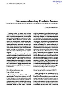
Chemical Structure and Properties of Vitamin E Family Vitamin E is composed of a family of at least eight different organic compounds. There are four tocopherols (alpha-T, beta-T, gamma-T, and delta-T) and four tocotrienols (alpha-T3, beta-T3, gamma-T3, and delta-T3). They are fat-soluble vitamins and function as antioxidants by preventing the propagation of free radicals. They also have non-antioxidant functions such as regulating cellular signal transduction pathways and preventing infertility (9-11). They all have a chroman ring (head). They are distinguished by the phytyl tail of the tocopherols and the farnesyl tail of the tocotrienols. Tocopherols have three chiral carbons. One of the chiral centers is located at the carbon 2 (C2) of the heterocyclic ring, while the other two are located on the 4th and 8th carbon of the phytyl tail (Figure 1). Each chiral center has two possible configurations, R or S (i.e. right or left configuration, respectively). Therefore, tocopherols can have 8 (23) stereoisomers, but naturally occurring tocopherol exists in the RRR configuration at all 3 chiral carbons. Synthetic tocopherol is a mixture of almost equal amounts of all the possible stereoisomers (RRR, RRS, RSS, SSS, SSR, SRR, SRS, and RSR) and is usually identified as allrac (all-racemic) or as dl-tocopherol. Tocotrienols have a chiral center at the C2 carbon in the heterocyclic ring, which can have two possible stereoisomers (i.e. R or S configuration) and three double bonds located at the 3′, 7′, and 11′ positions of the farnesyl group. The double bonds at the 3′ and 7′ positions can generate geometric isomers (E or Z isomers). Natural tocotrienols are EE in the 3′ and 7′ positions. The structures of the tocopherols and tocotrienols are shown in Figure 1.
11
For: R1 = R3 = CH3, R2 = H R2 = R3 = CH3, R1= H R1 = R2 = R3 = CH3 R1 = R2 = H, R3 = CH3
beta-tocopherol / tocotrienol gamma-tocopherol / tocotrienol alpha-tocopherol / tocotrienol delta-tocopherol / tocotrienol
Figure 1. Structures of various homologs of tocopherol and tocotrienol
12
Prostate Cancer Prevalence Prostate cancer is a disease in which cells in the prostate gland become abnormal and start to grow uncontrollably, forming tumors. Prostate cancer is second only to lung cancer as the most frequently diagnosed and leading cause of cancer death for men in the United States (12). On average, one man in six will be diagnosed with prostate cancer during his lifetime, and more than 200,000 men in the U.S. are diagnosed with prostate cancer every year. Chemotherapy and Chemoprevention Perhaps the areas of cancer research with the greatest potential for reducing cancer mortality are chemotherapy and chemoprevention. Chemotherapy is the treatment of existing cancer using specific chemical agents or drugs that are selectively destructive to malignant cells and tissues while chemoprevention is the use of chemical agents, drugs, or food supplements to prevent the future development of cancer. Epidemiological observations, experimental evidence from animal carcinogenesis models, knock-out models, in vitro studies with cancer cell lines, and clinical trials have shown the efficacy of this approach. One of the principal goals of cancer chemotherapeutic or chemopreventive agents is the elimination of damaged or malignant cells through cell cycle inhibition or induction of programmed cell death (apoptosis), leaving normal cells unaffected (13). Knowing the molecular mechanisms involved in inducing cancer cell death is of vital importance because it gives us an idea of the specific proteins regulated and this can lead to the design of more specific drugs that can selectively target cancer cells. A major goal of chemotherapy and chemoprevention is to design agents with a very high level of specificity. Proteomics provides the optimal tool for the study of these molecular mechanisms. 13
Vitamin E as a Chemotherapeutic and Chemopreventive Agent Vitamin E has been under intensive study as a chemotherapeutic and/or chemopreventive agent for many types of cancers (1). Multiple primary and secondary intervention trials of vitamin E for the prevention of various cancers are currently underway; these studies should define the role vitamin E plays in the prevention and treatment of various cancers (e.g. oral cavity, pharynx, oesophagus, prostate, colon, lung, breast, skin, cervix, and bladder) (14). Various chemical modifications of vitamin E have also been made and used as chemopreventive agents for various types of cancers. There is increasing evidence that vitamin E succinate is the most effective antitumor analogue of vitamin E (1). Cell Culture Studies Cell culture studies show that antioxidant vitamins (like vitamin E) and some phytochemicals selectively induce apoptosis in cancer cells but not in normal cells and also prevent angiogenesis and metastasis (15). Collectively, these studies suggest a potential role for antioxidants like vitamin E as supplements in cancer therapy. While tocopherols have been found in many different plant species and tissues, and their biosynthesis, physiology, and distribution have been studied in detail, little is known about the physiology and distribution of tocotrienols (9). Although chemically very similar, tocotrienols have been shown to display significantly more potent apoptotic activities than tocopherols in preneoplastic, neoplastic, and highly malignant cells grown in culture at treatment doses that show no adverse effects on normal cell growth or function (10). The inhibitory effects of the various forms of vitamin E on cell growth follow the order: alpha-T