Vertigo in the hyperviscosity syndrome JAMES C. ANDREWS, MD, LARRY A. HOOVER, MD, REGINA S. LEE, AB. and VICENTE HONRUBIA, MD. Los Angeles, California
Diseases that cause hyperviscosity of the blood can result in otologlc symptom+ especially vertigo. These symptoms are predominantly produced by peripheral vestibular Involvement. The pathophysiology probably involvesvascular obstruction in the venules. Specific changes in the microvasculature are presumed to be comparable to those that occur with hyperviscosity disorders In the retina. Maintenanceof a normal blood viscosity will prevent damage to the ear, as well as other organs. Addltlonally, reduction of the blood viscosity can greatly improve otologic symptoms. Case sfudies and a review of the pertinent literature are included. (OTOLARYNGOL HEAD NECK SURG 1988;98:144.)
T h e hyperviscosity syndrome describes a variety of symptoms and physical signs attributed directly to an abnormally elevated blood viscosity. These may include headache, vertigo, nystagmus, hearing loss, visual disturbances, retinal vein congestion, retinal hemorrhages, and mucosal hemorrhages. Congestive heart failure and renal impairment can occur as The hyperviscosity syndrome is associated with polycythemia (primary or secondary), macroglobulinemia, and hypergammaglobulinemia, as well as a variety of other diseases. The recognition of otologic symptoms is considered important in the diagnosis of the hyperviscosity syndrome. In one of the earliest reports of polycythemia, Vaquez4 included vertigo and tinnitus among the primary findings. Similarly, Osle? noted vertigo in four of nine patients he diagnosed with polycythemia. Larger series have shown a 30% to 44% incidence of vertigo and a 3% incidence of tinnitus with p~lycythemia.~.’ In a review of 182 patients with Waldenstrom’s macroglobulinemia, vestibular symptoms were noted in nine and hearing loss in seven.”Other authors have reported vertiginous symptoms in as many as 20% of patients with Waldenstrom’s macroglobulinemia.’ Of six patients with leukemia that resulted in hyperviscosity syn-
~~~
~
~
From the Division of Head and Neck Surgery, UCLA Medical CenterLos Angeles (Drs. Andrews and Honrubia), and the Division of Head and Neck Surgery, Olive View-UCLA Medical Center (Dr. Hoover). Presented at the Annual Meeting of the American Neurotology Society, Denver, Colo., April 25, 1987. Submitted for publication April 25, 1987; accepted Aug. 28, 1987. Reprint requests: Larry A. Hoover, MD, Division of Head and Neck Surgery, UCLA Medical Center, Los Angeles, CA 90024.
144
drome, three were shown to have balance disorders and three were noted to have d e a f n e ~ s.Sickle ~ cell anemia crisis is considered a hyperviscosity phenomenon, with red blood cells that form a sludge and obstruct the microvasculature. In a study of 83 Jamaican patients with sickle cell anemia (12 to 39 years of age), there was a 22% incidence of 25-decibel (dB) or greater hearing loss as compared to a 4% incidence in 83 agematched control subjects.” In many of these hyperviscosity disorders, a reduction of the blood viscosity will somewhat resolve and often abate these otologic symptoms.’.’”’2 CASE STUDIES Case 1. A 27-year-old man with recent onset of episodic vertigo was referred to the UCLA Head and Neck Clinic. His medical history was significant for tetralogy of Fallot repaired by a Pott’s anastomosis when he was 4% months old. Subsequent to this, pulmonary hypertension, which resulted in polycythemia, had developed. The patient had undergone periodic phlebotomies since he was 18 years old in attempt to maintain his hematocrit between 50% and 60%. He had experienced the onset of vertigo while he was hiking in the mountains. His symptoms were severe enough to incapacitate him. He was taken to an emergency room where administration of intravenous fluid resolved his vertigo. After resuscitation by fluid, his hematocrit was 68.7%. The patient later related that he had spent a great deal of time in the sun that day and had become dehydrated. After this incident, the patient experienced several more episodes of vertigo, all of which were secondary to a relatively elevated hematocrit. To a certain degree, he could even monitor his own hematocrit by these vertiginous symptoms. On two occasions, neurotologic examination of the patient was performed just before he underwent phlebotomy. A physical examination of his head and neck was normal. A hearing test of the patient revealed a symmetric mild sensorineural
Volume 98 Number 2 February 1988
Vertigo in the hyperviscosily syndrome 145
RIGHT EAR 125
FREClU E N C V IN Hz
250
500
1000
2000
4000
8000
I
1
1
I
I
1
125
250
500
I
I
LEFT EAR 1000
I
2000
4000
I
1
8000
0 10 20 30
e
m
40
.-
E
(Y
b
-1
100
I
I
I
I
I
110
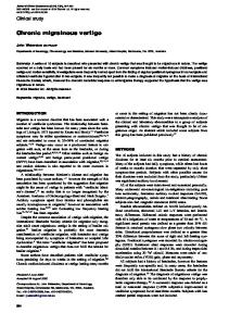
Fig. 1. Air conduction audiogramsfrom each of the patients in the case studies. None of the patients had a conductive hearing loss.
R. 30
R. 44 rL
10 deg. 1.0
I 1 sec. -
Fig. 2. Electronystagmographicdata of the caloric tests from the patient in case 2 reveal a normal response to right external ear canal irrigattonwith 30" (R. 30) and 4 4 O (R. 44) water. Lefl external ear canal irrigation with ice water (L. 0) failed to elicit a response.
hearing loss (Fig. 1). Testing of vestibular function revealed minimal caloric responses bilaterally, with maximum slow component velocities of induced nystagmus less than 2 degreedsecond, even after ice-water stimulation of the external ear canals. Positional, rotatory, and optokinetic tests were entirely normal. These test results were interpreted as suggestive of a bilateral peripheral vestibular disorder. Avoidance of dehydration and periodic phlebotomy helped control this patient's problem. Case 2. A 58-year-old man had a 2-year history of intermittent episodes of vertigo, each lasting as long as 3 days at
a time. Physical examination of the patient was significant for a spontaneous right-beating horizontal nystagmus. When his hearing was tested, it was revealed that he had a symmetric mild high-frequency sensonneural loss (Figure 1); speech reception thresholds measured 12 dB hearing loss, with a speech discrimination of 98% for each ear. Studies of vestibular function demonstrated a spontaneous right-beating nystagmus in the dark and an absent caloric response to external ear canal stimulation with 30" and 44"water and icetemperature water on the left side (Fig. 2). Sinusoidal rotatory testing showed that induced nystagmus was asymmetric and
OtolaryngologyHead and Neck Surgery
146 ANDREWS et al.
80
I
VOR
80
I
VOR
0
-80
.
””_
80
.
.
I
I
801VVOR
.
.
. .
.
.
.
.
.
.
.
.
.
.
.
~.
.
1
1
OKN
1
0
0
-804 0
. .
. .
. .
. .
!
- . .
~.
20 Time (seconds)
.
~
~
1 40
Fig. 3. Studies of vestibular function with slow eye velociiy in degreeslsecond [deg/sec),plotted against time in seconds, from the patient in case 2. Note the reduced response and asymmetry in the VOR. The OKN was slightly reduced, whereas the WOR appeared normal.
decreased (Fig. 3). The vestibular ocular reflex response (VOR) to rotation was specifically greater to clockwise than to counterclockwise rotation in this patient. Optokinetic nystagmus (OKN) was slightly reduced. In interpretation of these results, a right-beating spontaneous nystagmus that was inhibited by fixation and a unilaterally absent response to caloric stimulation on the left were indicative of a peripheral left vestibular paresis. Studies of rotatory function were consistent with stimulation of the horizontal semicircular canal of the remaining right labyrinth and showed better response to ampullopetal than to ampullofugal endolymph flow. This would be the result of an acute peripheral left vestibular lesion (in this man) with minimal compensatory recovery. It was believed that the decreased OKN was compatible with either the normal changes of aging or possibly with an overall decreased excitability of the central vestibular pathways and was thus without localizing value. The additional finding of suppression of rotation-induced nystagmus by visual fixation and a normal visual vestibular ocular reflex (VVOR) contraindicate a central vestibular cause of his vertigo. In this patient’s laboratory evaluation it was discovered
-80 0
20
40
Time (seconds)
Fig. 4. Studies of vestibular function from the patient in case 3. The VOR was quite minimal, yet WOR and OKN were normal.
that he had an abnormally elevated hematocrit of 67%. Studies of his bone marrow helped establish a diagnosis of polycythemia Vera. Treatment was begun; phlebotomies were performed every 2 to 3 months-and later with P”-in efforts to maintain more normal hematocrit. Although the patient felt better, he continued to experience a mild vertiginous feeling. Case 3. A 56-year-oId woman with recently diagnosed Waldenstrom’s macroglobulinemia was referred for evaluation and treatment. Some of her symptoms included unsteady gait, episodic vertigo, and tinnitus. Her vertigo lasted from a few minutes to several hours, and could be brought on by plasmapheresis treatments among other things. The significant physical finding in her neurotologic examination was a left beating peripheral spontaneous nystagmus . Measurement of her hearing revealed a bilateral and asymmetric sensorineural loss, significantly worse in the right ear (Fig. 1). The speech reception threshold in her ear was 38 dB hearing loss (with a discrimination ability of 96%); her left ear had 20 dB hearing loss (with a discrimination ability of loo%), which indicated her hearing loss was probably of cochlear origin. Studies of vestibular function were significant for a spontaneous left beating nystagmus in the dark, with a slow component velocity of 4 to 5 degreeslsecond. Caloric examination could
Volume 98 Number 2
Vertigo in the hyperviscosity syndrome 147
Febrwry 1988
Table 1. Summary of the neurotologic examination in the case studies
Hyperviscosity disorder Otologic symptoms Physical findings Audio Caloric testing Optokinetic testing VOR VVOR
Case 1
Case 2
Case 3
Polycythemia (secondary) Episodic vertigo Normal Mild symmetric SNHL Bilateral minimal function Normal Normal Normal
Polycythemia Vera Episodic vertigo Right spontaneous nystagmus Symmetric HFSNHL Left paresis Symmetric reduction Symmetric reduction Normal
Waldenstrom's globulinemia Unsteady gait, vertigo, tinnitus Left spontaneous nystagmus Asymmetric right SNHL Bilateral minimal function Normal Symmetric reduction Normal
~~~
~~
SNHL,Sensorineural hearing loss; HFSNHL. high-frequency sensorineural hearing loss; VOR. vestibular ocular reflex; VVOR. visual vestibular ocular reflex
increase the patient's left spontaneous nystagmus, with infusion of 30" water into the right external ear canal and 44" water into the left ear canal. The maximum slow component velocity of these caloric responses was 7 to 9 degreeslsecond. The spontaneous nystagmus was completely inhibited by stimulation of the left ear canal with 30" water infusion and the right ear canal with 44"water infusion. Thus the caloric response was minimal at 2 to 4 degreeslsecond-well below the normal range in our 1aborat0ry.I~The woman had an almost complete absence of sinusoidal rotationally induced nystagmus-as seen in the VOR test-whereas the VVOR and OKN tests were normal (Fig. 4). Her vestibular testing was believed to represent a bilateral peripheral vestibular loss, with some remaining function probable in the left ear. Ophthalmologic examination of this patient for blurred vision revealed papilledema, engorged retinal vessels, and retinal hemorrhages. All of this was consistent with the retinopathy of Waldenstrom's macroglobulinemia. If similar changes were occumng in this patient's inner ear, there was probably involvement of the auditory and vestibular apparatus on the right, but only of the vestibular organ on the left.
DISCUSSION
Vertigo, as a primary symptom in the hyperviscosity syndrome, is directly related to the state of hyperviscosity. This is supported by the finding that symptomatic improvement occurs with a reduction of blood hyperviscosity, regardless of underlying cause. For this reason, a vascular cause for the symptom of vertigo in these patients is implicated. In assessing a vascular origin of vertigo, both central and peripheral causes must be considered. As with vertebrobasilar insufficiency, vertigo could originate from the labyrinth, the eighth nerve, the vestibular nuclei or all three, since these structures all derive their blood supply from the vertebrobasilar circulation.l4 Blood flow disturbances may also affect the cerebellum and result in vertigo. The differentiation between central and peripheral causes of vertigo can be deduced by interpretation of
the patient's medical history, physical examination, and tests of audiologic and vestibular function (summarized in Table 1 and Fig. 1). In our patients, the experience of episodic vertigo began suddenly and with no other neurologic symptoms; this is indicative of a peripheral lesion. The physical finding of a unilateral spontaneous nystagmus inhibited by fixation in two of our patients (cases 2 and 3) suggested a peripheral vestibular lesion. Caloric reflexes were abnormal in our patients; cases 1 and 3 showed minimal responses to all temperatures tested bilaterally and case 2 demonstrated a unilateral vestibular paresis. Optokinetic reflex abnormality was present in one patient (case 2), but was not determined to have any localizing diagnostic value. Rotational testing was normal in one patient (case 1) and showed a significantly diminished response in the other two patients (cases 2 and 3). Good auditory function was maintained in two patients (cases 1 and 2), whereas one patient had an asymmetric hearing loss, probably of cochlear origin (case 3). Although it seems that peripheral vestibular dysfunction dominates in producing vertigo in patients with the hyperviscosity syndrome, central causes probably contribute as well. The pathophysiology of hyperviscosity is generally directed towards disturbance in the blood rheology. Since the blood supply to the ear is an extension of the circulation to the brain, these structures should show similar blood flow properties. In a study of cerebral blood flow in various states of hyperviscosity, Brown and Marshall16were able to determine that regulatory mechanisms allowed normal transport of oxygen to the brain. Patients with polycythemia exhibited reduced cerebral blood flow, but this was compatible with an increased oxygen-carrying capacity of their blood. In contrast, patients with a paraproteinemic cause of their hyperviscosity maintained a cerebral blood flow appropriate for their blood oxygenation. Hildesheimer et al." performed an experimental study on the effect of hyperviscosity on hearing in
148 A N m etol.
guinea pigs. One ear of each animal was perfused with polycythemic blood, while its other ear served as a control. Similar to the studies in cerebral blood flow, it was found that, although blood flow was reduced in the ear perfused with polycythemic blood, scala media oxygenationand hearing-as measured by click-evoked action potentials-were maintained. If the oxygen content of the blood was also reduced to a Po,of 30 mmHg, however, this significantly decreased both the hearing and the oxygen levels in the scala media in the experimental ear. The specific changes that occur in the central microcirculation can be inferred by what occurs in the retina. Also, in regard to vascular supply, the eye and the ear are similar in that they are both arterial end organs that derive their blood supply from the central nervous system. The typical ophthalmologic vasculature findings in states of hyperviscosity include diminished blood flow in the conjunctival vessels, markedly distended and tortuous retinal veins, and multiple retinal hemorrhages.2*’8 These findings are similar to what occurs in central retinal vein occlusion. In fact, hyperviscosity has been reported as a cause of central retinal vein occlusion.’9 Reduction of the blood viscosity can increase the flow in the conjunctival vasculature and reverse the retinal venous distention.” Symptomatic visual improvement occurs as Venous obstruction of the labyrinth has been well characterizedby Kimura and Per1man.21.22 They studied guinea pigs in which the inferior cochlear veins had been unilaterally obstructed. The vein of the vestibular aqueduct was left intact, however, for technical considerations. Postoperatively, these animals were vertiginous and showed a nystagmus towards the ear that had not been operated on. Cochlear microphonics in these animals revealed severe hearing loss. Temporal bone histopathology revealed dilation of the vein of the vestibular aqueduct as well as surrounding capillaries. Edema of the epithelium was early noted. Within a few hours of venous obstruction, hemorrhage into the epithelium and perilymphatic and endolymphatic spaces occurred. Hemorrhage was more common in the cochlear duct. Much later, hemorrhage was followed by fibrosis and ossification. Sensory cells were also particularly affected with hair-cell damage, followed by ganglion cell degeneration. There is some human temporal bone evidence to support the idea that the hyperviscosity syndrome leads primarily to venous and capillary obstruction. In a report of a 10-year-old boy who died during a sickle cell crisis, the temporal bones were described.= Significant findings included the generalized clumping of sickle cells in the venous channels (with notable engorgement
OtolaryngologyHead and Neck Surgery
of the stria vascularis) and degeneration of the hair cells in the organ of Corti. No comment was made concerning the vestibular structures. A temporal bone report of a patient with longstanding cryoglobulinemia revealed that more of the chronic changes occurred with hyperviscosity.” In all cochlear turns, the organ of Corti was missing, whereas (surprisingly) the spiral ganglion cells were relatively preserved. The stria vascularis was atrophic. A proteinaceous exudate, within the scala media, with fibrin-like crystals was described; this could possibly have been the sequelae of bleeding into this space. Fibrosis and ossification had occurred in the scala tympani of the basal turn of the cochlea. Blood vessels were decreased in general and the diameter of the inferior cochlear vein seemed reduced. In the vestibular organ, there was an eosinophilic precipitate in the region of the saccule. Again, this may have represented the long-term results of previous bleeding into the saccule. Additionally, there was ossification of the vestibule and the semicircular canals and a marked loss of fibers in the vestibular nerve. The changes described in these two reports on temporal bone seem consistent with venous and capillary obstruction, in that the degree of organ degeneration, fibrosis, and ossification is less severe than that which is seen with internal auditory artery occlusion.25Thus, the sequence of events may well be increased blood viscosity that will lead to obstruction in the venules and capillaries and result in inadequate blood flow and hypoxia of the ear. This, in association with an often intrinsic coagulation defect in many hyperviscosity disorders, may lead to acute hemorrhage within the organ. Later, fibrosis and ossification occur. In the patient whose blood viscosity is fluctuating, these changes may occur more slowly and intermittently. The inner ear is supplied by the internal auditory artery, an end artery which branches to form the common cochlear artery that supplies primarily the cochlea and saccule, and the anterior vestibular artery that supplies the semicircular canals and utricle.26Similarly, venous drainage from the cochlea and saccule is provided by the vein of the cochlear aqueduct, whereas the utricle and semicircular canals drain through the vein of the vestibular aqueduct.% Theoretically, with this pattern of vascular supply, a disease process that obstructs blood flow in the smaller vessels may preferentially involve the vestibular or auditory structures. In our patients, vestibular dysfunction has arisen probably as a result of diminished blood flow through the vein of the vestibular aqueduct and the anterior vestibular artery. The patient in case 3 did show an appreciable unilateral hearing loss that indicated this ear also
Volume 98 Number 2 February 1988
had involvement of the auditory organ by obstructed blood flow through the vein of the cochlear aqueduct and the anterior cochlear artery. Some caution must be used in this symptomatic interpretation of otologic blood flow abnormalities, as the pattern of vascular supply to the inner ear is not that exact and there is collateral flow.*’ Physicians who evaluate patients with vertigo should be aware that diseases that result in hyperviscosity syndrome are likely to produce vertigo as well as other otologic symptoms. Testing of vestibular function will lead to more accurate diagnosis and better understanding of the disease process. Adequate maintenance of normal blood viscosity will prevent damage to the ear and other organs. In addition, otologic symptoms produced by the state of hyperviscosity can be greatly improved by reduction of the blood viscosity. REFERENCES
1. Baer MR, Stein RS, Dessypris EN. Chronic lymphocytic leukemia with hyperleukocytosis: the hyperviscosity syndrome. Cancer 1985;56:2865-9. 2. Fahey JL, Barth WF, Solomon A. Serum hyperviscosity syndrome. JAMA 1965;192:464-7. 3. Preston FE, Sokol RJ, Lilleyman JS, Winfield DA, Blackburn EK. Cellular hyperviscosity as a cause of neurological symptoms in leukemia. Br Med J 1978;1:476-8. 4. Vaquez MH. Ser une forme speciale de cyanose s’accompagnant d’hyperglobulie excessive et persistante. C R Soc Biol 1892; 44:384-8. 5. Osler W. Chronic cyanosis with polycythemia and enlarged spleen: a new clinical entity. Am J Med Sci 1903;126:187-201. 6. Calabresi P, Meyer 00. Polycythemia Vera. I. Clinical and laboratory manifestations. Ann Intern Med 1959;50: 1182-1202. 7. Silverstein A, Gilbert H, Wasserman LR. Neurologic complications of polycythemia. Ann Intern Med 1962;57:909-15. 8. Logothetis J, Silverstein P, Coe J. Neurological aspects of Waldenstrom’s macroglobulinemia. Arch Neurol 1960;3:564-73. 9. Todd GB, Serjeant GR, Larson MR. Sensori-neural hearing loss in Jamaicans with SS disease. Acta Otolaryngol 1973;76 268-72.
Vertigo in the hyperviscosity syndrome 149
10. Davis EC, Nilo ER. Hearing improvement induced by phlebotomy in polycythemia. Laryngoscope 1965;75:1847-52. 11. Wilkinson P, Davidson W, Sommarepe A. Turnover of I T labeled autologous macroglobulin in Waldenstrom’s macroglobulinemia. Ann Intern Med 1966;65:308-16. 12. Solomon A, Fahey JL. Plasmapheresis therapy in macroglobulinemia. Ann Intern Med 1963;58:789-800. 13. Baloh RW, Honrubia V. Clinical neurophysiology of the vestibular system. Philadelphia: F.A. Davis Co., 1979:136. 14. Baloh RW. The essentials of neurotology. Philadelphia: F.A. Davis Co., 1984:122-4. 15. Dintenfass L. Blood viscosity, hyperviscosity, and hyperviscosaemia. Lancaster, England: MTP Press Ltd., 1985113-29. 16. Brown MM, Marshall J. Regulation of cerebral blood flow in response to changes in blood viscosity. Lancet 1985;8429: 604-9. 17. Hildesheimer M, Rubinstein M, Nuttal AL, Lawrence M. Influence of blood viscosity on cochlear action potentials and oxygenation. Hear Res 1982;8:187-98. 18. Mausolf FA, Mensher JH. Experimental hyperviscosity retinopathy. Ann Ophthalmol 1973;5:205-9. 19. Trope GE, Lowe GDO, McArdle BM, et al. Abnormal blood viscosity and hemostasis in long-standing retinal vein occlusion. Br J Ophthalmol 1983;67:137-42. 20. Schwab PJ, Okun E, Fahey JL. Reversal of retinopathy in Waldenstrom’s macrcglobulinemia by plasmapheresis. Arch Ophthalmol 1960;64:515-21. 21. Kimura R, Perlman HB. Extensive venous obstruction of the labyrinth. A. Cochlear changes. AM Otol Rhinol Laryngol 1956;65:332-50. 22. Kimura R, Perlman HB. Extensive venous obstruction of the labyrinth. A. Vestibular changes. Ann Otol Rhinol Laryngol 1956;65:620-38. 23. Morgenstein KM, Manace ED. Temporal bone histopathology in sickle cell disease. Laryngoscope 1969;79:2172-80. 24. Nomura Y, Tsuchida M, Mori S, Sakurai T. Deafness in cryoglobulinemia. Ann Otol Rhinol Laryngol 1982;91:250-5. 25. Perlman HB, Kimura R, krnandez C. Experiments on temporary obstruction of the internal auditory artery. Laryngoscope 1959;69:591-613. 26. Schuknecht HF. Pathology of the ear. : Harvard University Press, 1974:23-96. 27. Igarashi M, Alford BR, Konishi S, Shaver EF, Guilford FR. Functional and histopathological correlations after microembolism of the peripheral labyrinthine artery in the dog. Laryngoscope 1969;79:603-23.