A COMPARISON BETWEEN MALE AND FEMALE STRENGTH TO BODY MASS RATIOS AND VARUS/VALGUS KNEE ANGLES DURING JUMP LANDINGS
A Thesis by TRACIE LYNN HAINES
Submitted to the Graduate School Appalachian State University in partial fulfillment of the requirements for the degree of MASTER OF SCIENCE
May 2010 Department of Health, Leisure, and Exercise Science
A COMPARISON BETWEEN MALE AND FEMALE STRENGTH TO BODY MASS RATIOS AND VARUS/VALGUS KNEE ANGLES DURING JUMP LANDINGS
A Thesis by TRACIE LYNN HAINES May 2010
APPROVED BY:
__________________________________ Jeffrey M. McBride Chairperson, Thesis Committee
__________________________________ N. Travis Triplett Member, Thesis Committee
__________________________________ Rebecca A. Battista Member, Thesis Committee
__________________________________ Paul L. Gaskill Chairperson, Department of Health, Leisure and Exercise Science
__________________________________ Edelma D. Huntley Dean, Research and Graduate Studies
ii
Copyright by Tracie Lynn Haines 2010 All Rights Reserved
iii
ABSTRACT
A COMPARISON BETWEEN MALE AND FEMALE STRENGTH TO BODY MASS RATIOS AND VARUS/VALGUS KNEE ANGLES DURING JUMP LANDINGS (May 2010) Tracie Lynn Haines B.S., The College of New Jersey M.A., Appalachian State University Chairperson: Jeffrey M. McBride
The reason for the presence of valgus knee angles during jumping in certain individuals is unclear. It appears to be more prevalent in females but has been observed in male subjects as well. Valgus knee position has been attributed to an increase in knee injury. The purpose of this investigation was to compare valgus/varus knee angles during various jumps and lower body strength levels between males and females relative to body mass. Seventeen recreationally active females (age: 21.94±2.59 years; height: 167.32±5.20 cm; mass: 64.42±8.39 kg; % body fat: 26.89±6.26; squat 1RM: 66.18±19.47 kg; S:BM: 1.03±0.28) and thirteen recreationally active males (age: 21.69±1.65 years; height: 176.85±7.26 cm; mass: 72.39±9.23 kg; % body fat: 13.15±5.18; squat 1RM: 115.77±30.40 kg; S:BM: 1.59±0.31) performed a one repetition maximum in the squat (1RM) and three of each of the following jumps: counter-movement jump (CMJ), 30 cm
iv
drop jump (DJ30), 45 cm drop jump (DJ45) and 60 cm drop jump (DJ60). Knee angles were analyzed through videography, concentric forces were collected from a force plate, and body composition was analyzed by dual energy X-ray absorptiometry to allow for squat to body mass ratio (S:BM) and squat to fat-free mass (S:FFM) to be calculated. Significant differences (p ≤ 0.05) were found between male and female 1RM, male and female S:BM, and male and female S:FFM. Significant differences were found between male and female varus/valgus knee positions during maximum flexion (MF) of the right leg in the CMJ, DJ30, DJ45 and DJ60. Significant differences were found between male and female varus/valgus knee positions during maximum flexion of the left leg in the CMJ, DJ30, DJ45 and DJ60. Peak concentric force data was only found to be significant between males and females at CMJ, but not at DJ30, DJ45, or DJ60. Correlations between varus/valgus knee angles and S:BM for all for jumps displayed moderate, nonsignificant relationships (CMJ, r = 0.445; DJ30 r = 0.448; DJ45 r = 0.449; DJ60 r = 0.439). In conclusion, males and females have significantly different lower body strength levels and varus/valgus knee position when landing from jumps. The discrepancy in strength level could be an important factor in knee position when landing.
v
ACKNOWLEDGEMENTS
This thesis was completed through the support of the Office of Student Research Grant, the Graduate Senate Association Senate Research Grant, the Graduate Student Research Grant through the Cratis D. Williams Graduate School of Appalachian State University and the North Carolina BeActive Grant. I would like to genuinely thank Jeffrey McBride, my mentor and my friend. I thank you for teaching me the art of research and the science of biomechanics. I would like to acknowledge and thank my thesis committee, Travis Triplett and Becki Battista, for their guidance, support, and friendship. This thesis would not have been possible without the hardworking and dedicated graduate assistants in the Neuromuscular Lab, especially Tyler Kirby. I would like to thank Michael Landram for all of his support and for making me want to be a better scientist. Lastly, I thank my family from the bottom of my heart; Mom, Dad, Jamie and MP, for all of your unconditional love, support, and for always believing in me and my ideas, no matter how outrageous they may seem.
vi
TABLE OF CONTENTS
Page ABSTRACT...............................................................................................................iv ACKNOWLEDGEMENTS.......................................................................................vi LIST OF TABLES.....................................................................................................viii LIST OF FIGURES ...................................................................................................ix INTRODUCTION .....................................................................................................1 METHODS ................................................................................................................10 Subjects ..........................................................................................................10 Study Design..................................................................................................10 1RM Strength Testing....................................................................................11 Jumping Procedures .......................................................................................11 Videography ..................................................................................................11 Data Analyses ................................................................................................12 Statistical Analyses .......................................................................................12 RESULTS ..................................................................................................................14 DISCUSSION ............................................................................................................15 REFERENCES ..........................................................................................................18 TABLES ....................................................................................................................23 FIGURES...................................................................................................................24 APPENDIX A – Institutional Review Board Documentation...................................34 VITA ..........................................................................................................................41
vii
LIST OF TABLES
Table 1: Subject Anthropometrics and Strength Measurements...............................23
viii
LIST OF FIGURES
Figure 1: Representation of varus and valgus knee angles .......................................24 Figure 2: Differences in squat 1RM..........................................................................25 Figure 3: Differences in S:BM and S:FFM...............................................................26 Figure 4: Varus/valgus knee position of right leg.....................................................27 Figure 5: Varus/valgus knee position of left leg.......................................................28 Figure 6: Concentric peak force of jumps.................................................................29 Figure 7: Correlation of S:BM and varus/valgus knee position in CMJ...................30 Figure 8: Correlation of S:BM and varus/valgus knee position in DJ30..................31 Figure 9: Correlation of S:BM and varus/valgus knee position in DJ45..................32 Figure 10: Correlation of S:BM and varus/valgus knee position in DJ60................33
ix
INTRODUCTION
The reason for the presence of valgus knee angles during jumping in certain individuals is unclear (Shultz, 2008). It appears to be more prevalent in females, but has been observed in male subjects as well (Russell, Palmieri, Zinder, & Ingersoll, 2006; Jacobs, Uhl, Mattacola, Shapiro, & Rayens, 2007; Wallace, Kernozek, Mikat, Wright, Simons, & Wallace, 2008; Schmitz, Shultz, & Nguyen, 2009). Possible reasons may be musculoskeletal design flaws, lack of strength in gluteal muscles, muscle strength imbalances or deficiency in general lower body strength relative to body mass (Bonci, 1999; Cahue, Dunlop, Hayes, Song, Torres, & Sharma, 2004; Russell, Palmieri et al., 2006; Myer, Chu, Brent, & Hewett, 2008; Wallace, Kernozek et al., 2008; Varadarajan, Gill, Freiberg, Rubash, & Li, 2009). The concern given to valgus knee angles during jumping is due to the possible relationship of this phenomenon to anterior cruciate ligament injury (Ireland, 1999; Russell, Palmieri et al., 2006; Myer, Chu et al., 2008; Schmitz , Ficklin, Shimokochi, Nguyen, Beynnon, Perrin, & Shultz, 2008; Shimokochi & Shultz, 2008; Wallace, Kernozek et al., 2008; Schmitz, Shultz et al., 2009). Therefore, the purpose of this investigation was to compare valgus/varus knee angles during various jumps and lower body strength levels between males and females relative to body mass.
1
Strength is defined as the maximal force that a muscle or muscle group can generate at a specified velocity (Baechle & Earle, 2008). There are many biomechanical factors involved in strength such as neural control, cross-sectional area (CSA), and fiber type. Neural control affects the maximal force output of a muscle by establishing the amount of motor units that will be recruited and the rate at which they fire. An increase in the amount of motor units recruited and a faster firing rate will result in a larger force output. Early increases in strength can be attributed to better synchronization of neural control than an increase in the CSA of a muscle. The force a muscle can exert is directly correlated to its CSA. Human skeletal muscle generates a maximum of between 16 and 30 Newtons of force per square centimeter of muscle cross section (McArdle & Katch, 2007). Muscle fibers can be generally categorized as Type I (slow twitch) or Type II (fast twitch) by their contractile and metabolic properties. Type I fibers have a slow speed of action, a slow speed of relaxation, high resistance to fatigue and have a low force capacity (McArdle & Katch, 2007). In contrast, Type II fibers have a fast speed of action, a fast speed of relaxation, a low to moderate resistance to fatigue and have a high force capacity. Recruitment of Type II fibers results in a more powerful contraction and increases force production. There are apparent sex differences in strength measures. When comparing absolute strength, males have considerably greater strength than females in all muscle groups tested, with females scoring about 50% lower than males for upper-body strength and about 30% lower for lower-body strength (McArdle & Katch, 2007). Males on average possess greater CSA of muscle fiber with approximately 45% of their body as muscle mass while female posses 36% (Brooks, Fahey, & Baldwin, 2005). Sex
2
differences in body composition play a large role in strength divergence with females requiring 12% of essential body fat and males only 3% of essential body fat. This large discrepancy in body composition allows males to have a higher portion of their bodies capable of generating force. Females have the same spectrum of muscle fiber types as males with Type I, Type II and all of their subgroups. However, sex differences have been shown in the ratio of muscle fiber sizes in untrained males and females (Zatsiorsky & Kraemer, 1995). Males displayed a significantly greater CSA of muscle fibers, with the trend of males possessing more Type II fibers and females Type I fibers. Consistently throughout the literature it has been shown that males outperform females of the same age in measures of strength (Zatsiorsky & Kraemer, 1995; Thomas, Kraemer, Spiering, Volek, Anderson, & Maresh, 2007; Walsh, Bohm, Butterfield, & Santhosam, 2007; Laffaye & Choukou, 2009; Papaiakovou, Giannakos, Michailidis, Patikas, Bassa, Kalopisis, Anthrakidis, & Kotzamanidis, 2009). Thomas and colleagues (2007) tested 19 male and 14 female NCAA Division I soccer players to assess power output at different percentages of their squat 1RM. Results showed the squat 1RM was significantly different between males and females (M, 121.0±22.5 kg; F, 83.2±12.6 kg). Handgrip strength, a valuable measurement to document progression of muscle strength, also revealed significant differences in strength between males and females. An investigation of 300 subjects found significant differences between male and female hand grip strength levels in both dominant hand (M, 38.9±10.0 kg; F, 22.0±5.7 kg) and nondominant hand (M, 36.4±10.0 kg; F, 19.5±5.6 kg) (Budziareck, Pureza Duarte, & Barbosa-Silva, 2008). The use of sprint time as a measure of strength has also been used to determine gender and chronological age differences (Papaiakovou, Giannakos et al.,
3
2009). Three-hundred sixty children between 7-18 years of age were tested in the 30m sprint. For all age groups males performed better on average than females, with significant differences in running speed between genders being found at 16, 17, and 18 years of age (M, 7.01±0.19 m/s; F, 5.99±0.16 m/sec). It has been reported that the average female has 54 to 73% of the maximal vertical jump and 75% or the maximal standing long jump of the average man (Zatsiorsky & Kraemer, 1995). To further investigate, Laffaye and Choukou (2009) examined gender differences in the CMJ, 30cm DJ, and 60cm DJ using skilled volleyball players and found that despite the test, males significantly outperformed females in jump height (M, 46.6±7.5 cm; F, 36.0±5.4 cm) and in mean power (M, 56.9±26.0 W/kg; F, 42.4±19 W/kg). Likewise, Walsh, Böhm, Butterfield, and Santhosam (2007) examined the CMJ, CMJ without arm swing (CMJW), static jump (SJ), and the SJ without arm swing (SJW). The effect of arm swing was being investigated due to the upper body strength discrepancies with females having an average of 55.8% of the upper extremity strength of males (NASA, 1978). Despite the negation of arm swing, males had a significantly higher jump height than females for every jump (CMJ: M, 53.0±7.0 cm; F, 39.0±7.0 cm; CMJW: M, 43.0±6.0 cm; F, 31.0±5.0 cm) (Walsh, Bohm et al., 2007). Therefore, it has been continually demonstrated that males outperform females in strength and power movements. A squat one repetition maximum (1RM) is defined as the heaviest resistance that can be lifted for one complete repetition and is used to determine a person’s lower body maximal strength. The value can be expressed in absolute value terms in kilograms or as a relative value to body mass (S:BM) or fat free mass (S:FFM). Nuzzo and colleagues
4
found that squat 1RM expressed relative to body mass is significantly correlated with CMJ performance (Nuzzo, McBride, Cormie, & McCaulley, 2008). Relative values may be utilized to account for body fat differences in males and females, with females having 12% essential body fat and males having 3% (Brooks, Fahey et al., 2005). The squat 1RM relative to body weight has been shown to be a predictor of CMJ performance. Nuzzo and colleagues (2008) found significant correlations (p ≤ 0.05) between relative squat 1RM and CMJ peak power, peak velocity and jump height (r = 0.676, r = 0.731, r = 0.690). Valgus and varus are terms to describe frontal-plane knee positions (Shultz, 2008). Valgus is a medial deviation of a joint, and varus is a lateral deviation of a joint (Whiting & Zernicke, 2008). Both males and females have exhibited a knee valgus position during squatting and while landing from a jump (Russell, Palmieri et al., 2006; Schmitz, Ficklin et al., 2008; Wallace, Kernozek et al., 2008; Dempsey, Lloyd, Elliott, Steele, & Munro, 2009). Females have been shown to display more knee valgus than males in squatting and landing (Russell, Palmieri et al., 2006; Jacobs, Uhl et al., 2007; Wallace, Kernozek et al., 2008; Quatman & Hewett, 2009; Schmitz, Shultz et al., 2009). Wallace and colleagues (2008) compared the squat 1RM and vertical jumping ability to determine if a relationship existed between knee frontal plane motions between the two movement patterns. Fourteen out of twenty-two subjects, both male and female, displayed a more valgus than varus position during maximal squatting. When examining landing mechanics during a single-leg drop jump, it was also found that valgus knee angles were higher in females (Russell, Palmieri et al., 2006). At initial contact with the ground from a 60 cm single-leg drop jump, females landed in a more valgus position than
5
males (F, -0.65±3.32°; M, 3.85±4.03°). When subjects reached their point of maximal knee flexion during landing, both males and females returned to a varus position, however females were in a more “relative” valgus position than males (F, 3.13±6.84°; M, 15.26±9.41°). The presence of valgus knee angles has also been observed in sidestep cutting. Dempsey and colleagues examined the knee valgus loading during different sidestep cutting techniques (Dempsey, Lloyd et al., 2009). The load on the knee joint from peak valgus in males was present in both planned (-0.38±0.26 Nm·kg-1·m-1) and unplanned (-0.40±0.23 Nm·kg-1·m-1) cutting. Dempsey’s research was only conducted on male subjects and did not include female participants. In contrast, differences between males and females in knee valgus position have not been observed pre-pubertal (Myer, Chu et al., 2008; Schmitz, Shultz et al., 2009). Knee position from a drop jump, while valgus, was comparable between males (R, 11.9±8.2°; L, 14.3±9.3°) and females (R, 12.9±7.4°; L, 10.0±6.4°) in the pre-maturation phase. As maturation progressed, differences were observed as males tended to land in less of a valgus position (R, 9.6±10.9°; L, 8.8±7.9°) than females (R, 15.1±8.4°; L, 15.9±9.2°). Schmitz (2009) expressed that a possible reason for the differences may lie in the strength level of males and females. The reason(s) for the incidence of knee valgus position in certain individuals is still unclear and debated. Investigations have observed the relationship between this occurrence and musculoskeletal design flaws, lack of strength in gluteal muscles, muscle strength imbalances or deficiency in general lower body strength relative to body mass (Bonci, 1999; Cahue, Dunlop et al., 2004; Russell, Palmieri et al., 2006; Myer, Chu et al., 2008; Wallace, Kernozek et al., 2008; Varadarajan, Gill et al., 2009). As previously
6
mentioned, Wallace and colleagues (2008) examined the relationship between the squat 1RM and vertical jumping knee position. Females tended to land from a vertical jump in a more valgus position than their male counterparts, however, in this investigation the correlation coefficients for squat strength verses knee valgus angle were low for both males (R2 = 0.09) and females (R2 = 0.06). Limitations in the study existed within the subjects and the analysis. Only strong subjects were recruited with men being required to squat at least 1.5 times their body mass and females having to maximally squat at least 1.0 times their body mass. This confines the subject pool to only strong subjects and may not have implications for the majority of the general population. Also, when comparing maximal strength to knee valgus angles, absolute values were used instead of relative values to the subjects’ body mass. Russell and colleagues (2006) examined the effect of the gluteus medius (GM), hypothesizing that a weaker GM which helps stabilize the pelvis and abduct the femur, would be associated with a valgus knee position during landing from a single-leg drop jump. GM activation did not differ between males and females, therefore was not a factor in greater valgus landing in females. Strength in hip abduction was also studied by Wallace and colleagues (2008) between the squat 1RM and vertical jumping knee position. Hip abduction strength correlated better with knee valgus angles in females (R2 = 0.51) but did not with males (R2 = 0.10). The role of hip abduction strength has ambiguous findings and is still being investigated. Kiriyama and colleagues (2009) researched gender differences during single-legged drop landings and the role of external and internal rotation strength of the shank (Kiriyama, Sato, & Takahir, 2009). A lack of external rotation strength was hypothesized to create greater
7
valgus knee angles. It was found that females had significantly lower external and internal rotation strength values than males. The reason for studying knee valgus angles during dynamic activities is that increased knee valgus angle may play a role in the incidence of anterior cruciate ligament (ACL) injury (Ireland, 1999; Russell, Palmieri et al., 2006; Beynnon & Shultz, 2008; Myer, Chu et al., 2008; Schmitz, Ficklin et al., 2008; Wallace, Kernozek et al., 2008; Schmitz, Shultz et al., 2009; Shimokochi, Yong Lee, Shultz, & Schmitz, 2009). When the knee is in a valgus position the hips are adducted and internally rotated and the tibia is externally rotated (Jacobs, Uhl et al., 2007). The ACL is located in the knee; originating at a point anterior to the intercondylar area of the tibia and inserting at the posterior part of the medial surface of the lateral condyle of the femur. Its function is to limit hyperextension of the knee and to prevent anterior sliding of the tibia on the femur (Tortora & Grabowski, 2003). Russell and colleagues reported that increasing valgus position by 5° from a neural alignment can increase the load on the ACL by six times (Russell, Palmieri et al., 2006). When the stress reaches a certain threshold where it can not be supported, the ACL will tear and possibly other supporting structures will tear. Video analysis by Krosshaug and colleagues (2007) of 39 ACL injuries has provided evidence that there are two principal loading patterns attributing to ACL injuries (Krosshaug, Nakamae, Boden, Engebretsen, Smith, Slauterbeck, Hewett, & Bahr, 2007). Injury as a result of knee valgus collapse (a combination of knee valgus, hip internal rotation and tibial rotation) was one of the two predominant loading patterns for noncontact ACL injury. Besides the acute injury of an ACL tear, valgus knee position has
8
also be attributed to more chronic injury such as osteoarthritis (Cahue, Dunlop et al., 2004; Sharma, 2007). As shown in previous literature, there is a lack of consensus as to the reason(s) behind valgus knee angles. There appears to be a lack of research on the general lower body strength of a subject relative to body mass, as other studies have used absolute strength (Jacobs, Uhl et al., 2007; Wallace, Kernozek et al., 2008). Limitations have also risen due to the strength of the subject pool with subjects being able to squat 1.0-1.5 times their body mass (Wallace, Kernozek et al., 2008). Therefore, the purpose of this investigation was to compare valgus/varus knee angles during various jumps to relative lower body strength levels between males and females. It was hypothesized that squat 1RM differences in males and females may account for the valgus/varus knee positions observed.
9
METHODS
Subjects Seventeen recreationally active females (21.94±2.59 years) and thirteen recreationally active males (21.69±1.65 years) participated in this study (Table 1). Recruitment of subjects was limited to those between the ages of 18-28 years old, who have had no prior lower body injuries within the previous two years, and know how to properly execute a back squat. The participants were notified of the potential risks and gave written consent, as approved by the Institutional Review Board for the Protection of Human Subjects at Appalachian State University.
Study Design Participants reported to the Neuromuscular Laboratory for one session lasting approximately 90 minutes. The first half of the session included a five-minute dynamic warm-up before performing three CMJ, three DJ at 30 cm (DJ30), three DJ at 45 cm (DJ45), and three DJ at 60 cm (DJ60) in a randomized fashion. The second half consisted of a 1RM squat and a dual energy X-ray absorptiometry (DEXA) scan (Hologic, Inc., Discovery QDR Series, Bedford, MA). Participants performed all jumps on a force plate (AMTI, BP6001200, Watertown, MA) and were recorded via standard video (JVC, Model GR-AX700, Wayne, NJ) during the whole session. The entire session was
10
completed barefoot to negate various shock absorption and landing patterns caused by different footwear (Shimokochi, Yong Lee et al., 2009).
1RM Strength Testing A 1RM squat was used to assess dynamic strength values. Subjects performed warm-up sets as dictated by their estimated 1RM (8x30%, 5x50%, 3x70%, 1x90%). Depth was predetermined at 90° of knee flexion by a goniometer and was monitored each repetition by principle investigator and laboratory researchers. After the warm-up sets; participants performed one repetition to 90° of knee flexion until a 1RM was established. Rest periods of three to five minutes were given between trials. This protocol has been previously utilized in this laboratory (McBride, Blow, Kirby, Haines, Dayne, & Triplett, 2009).
Jumping Procedures Participants performed all jumps with their hands securely on their hips to negate arm swing and blockage of markers (Shimokochi, Yong Lee et al., 2009). The CMJ were initiated by the principle investigator giving the subject a verbal consent to jump when they were ready. The DJs were set at 30 cm, 45 cm and 60 cm height from the force plate. When instructed, subjects stepped off of the box and performed a maximal jump immediately upon landing. Participants were instructed to perform a maximal effort jump every trial.
Videography Preparation for 2D videography required participants to be dressed in dark, tight clothing to create contrast. White anatomical markers were placed on the right and left 11
greater trochanter, lateral condyle and lateral malleolus. A video recorder was set up 10 feet away facing the subject in the frontal view. The camera sampled at 60Hz. A calibration matrix was set up within the frame of view and was measured to convert pixels into real world units.
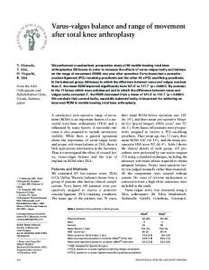
Data Analyses Video was analyzed using Peak Motus videography program (Vicon Peak Motus, Denver, CO). Three static trials of each subject were analyzed and averaged to determine the individual’s static or ‘neutral’ position. Every jump was analyzed to determine knee angle at maximal flexion (MF) where the subject was at his/her lowest squatting position from landing. Knee angles were determined by taking the static position and subtracting the MF. Negative values exhibited a valgus position and positive values exhibited a varus position (Figure 1). (Static Position º) – (Max Flexion º) = Knee Angle º The force plate collected data at 1000Hz and was analyzed with a customized LabVIEW analysis program (LabVIEW, National Instruments, Version 7.1, Austin, Texas). Peak concentric force (PCF) was measured for all of the jumps. DEXA scans were performed and analyzed as instructed by the Hologic Discovery QDR Series Manual. A whole body scan was completed to determine each subject’s percent body fat and fat free mass. This allowed for determination of S:FFM.
Statistical Analyses Statistical analysis was performed using SPSS software (version 14.0, SPSS, Inc., IL). Independent t-tests were used to determine significant differences between male and
12
female squat 1RM, S:BM, S:FFM, MF and PCF. Pearson product correlations were performed to determine the relationship between knee position and S:BM in each jump. The significance level was set at p ≤ 0.05.
13
RESULTS
Significant differences (p ≤ 0.05) were found between male and female 1RM, male and female S:BM, and male and female S:FFM (Figures 2,3). Significant differences were found between male and female MF of the right leg in the CMJ, DJ30, DJ45 and DJ60 (Figure 4). Significant differences were also found between male and female MF of the left leg in the CMJ, DJ30, DJ45 and DJ60 (Figure 5). Peak concentric force data was only found to be significant between males and females at CMJ, but not at DJ30, DJ45, or DJ60 (Figure 6). Pearson product correlations between knee angles and S:BM for all for jumps displayed moderate relationships (CMJ, r = 0.445; DJ30 r = 0.448; DJ45 r = 0.449; DJ60 r = 0.439) (Figures 7-10).
14
DISCUSSION
The primary findings of this investigation are 1) that females have significantly lower squat to body mass ratios than males, and 2) females land from jumps in a significantly more valgus knee position than males. These findings are concurrent with the previous literature that valgus knee position is more prevalent in females than males (Russell, Palmieri et al., 2006; Jacobs, Uhl et al., 2007; Wallace, Kernozek et al., 2008; Schmitz, Shultz et al., 2009). It is not surprising that females have significantly lower strength levels in 1RM, S:BM, and S:FFM than males. Therefore, strength may be a factor in valgus knee position, which increases the load on the ACL. As shown in Figures 2 and 3, males were significantly stronger than females in 1RM, S:BM, and S:FFM. These findings are in line with previous literature of males possessing more strength than females in both relative and absolute measures (Zatsiorsky & Kraemer, 1995; Thomas, Kraemer et al., 2007; Walsh, Bohm et al., 2007; Laffaye & Choukou, 2009; Papaiakovou, Giannakos et al., 2009). Part of the discrepancy in strength levels may lie in the natural differentiation in body composition between males and females (Brooks, Fahey et al., 2005). This however, would have been corrected when using S:FFM. Positive, moderate relationships between all jumps were displayed when correlating squat to body mass ratios and varus/valgus knee position when pooling both males and females together. It appears that strength may play a role in varus/valgus knee
15
position. The moderate correlations do not conclude or eliminate strength levels as a factor in knee position; however, further investigations into the topic should be explored to isolate the role of strength from other possible factors. In both the right and left legs and in every jump (CMJ, DJ30, DJ45 and DJ60), males landed in a significantly more varus position than females. Females landed in a significantly more ‘relative’ valgus position than males. These findings are in agreement with the previous literature of females having a more prevalent valgus knee position when jumping (Russell, Palmieri et al., 2006; Jacobs, Uhl et al., 2007; Wallace, Kernozek et al., 2008; Schmitz, Shultz et al., 2009). On average, the females were still in a varus knee position during their maximum flexion when landing from the jumps. However, this knee position is determined from the individuals’ static position which does not rule out the possibility that the females’ static knee position could have been in a valgus position to start. It has been concluded from previous research that a valgus knee position puts an individual at a higher incidence of knee injury, especially to the ACL (Russell, Palmieri et al., 2006). This investigation indicated that females land from jumps in a more relative valgus position. This knee position could therefore be putting females at a higher risk for knee injury. The occurrence of ACL injury in males and females has been extensively researched and documents the higher incidence in females (Russell, Palmieri et al., 2006; Jacobs, Uhl et al., 2007; Wallace, Kernozek et al., 2008; Quatman & Hewett, 2009; Schmitz, Shultz et al., 2009). From the results, it may be possible that increasing one’s strength levels through higher S:BM and S:FFM could play an potential role in reducing the valgus knee position
16
when landing from jumps. Previous literature has failed to relate these discrepancies in strength as a primary factor in valgus/varus knee position. Correlations between strength levels and knee position in this investigation were found to be moderate, but not significant enough to conclude a direct relationship between the two variables. Possible increases in strength may reduce valgus knee position and therefore, the incidence of ACL and other knee injuries. The findings from this investigation warrant the need for more examination into the mechanisms of how strength levels play a role in valgus/varus knee position during jump landings.
17
REFERENCES
Baechle, T. R. & Earle R. W. (2008). Essentials of Strength Training and Conditioning (2nd ed.). Champaign, IL: Human Kinetics.
Beynnon, B. D. & Shultz S. J. (2008). Anatomic alignment, menstrual cycle phase, and the risk of anterior cruciate ligament injury. J Athl Train, 43 (5), 541-542.
Bonci, C. M. (1999). Assessment and evaluation of predisposing factors to anterior cruciate ligament injury. J Athl Train, 34 (2), 155-164.
Brooks, G. A., Fahey T. D. & Baldwin, K.M. (2005). Exercise Physiology: Human Bioenergetics and Its Application. Boston: McGraw Hill.
Budziareck, M. B., Pureza Duarte R. R. & Barbosa-Silva, M.C. (2008). Reference values and determinants for handgrip strength in healthy subjects. Clin Nutr, 27 (3), 357362.
18
Cahue, S., Dunlop D., Hayes, K., Song, J., Torres, L. & Sharma, L. (2004). Varus-valgus alignment in the progression of patellofemoral osteoarthritis. Arthritis Rheum, 50 (7), 2184-2190.
Dempsey, A. R., Lloyd D. G., Elliott, B.C., Steele, J.R. & Munro, B.J. (2009). Changing sidestep cutting technique reduces knee valgus loading. Am J Sports Med 37 (11), 2194-2200.
Ireland, M. L. (1999). Anterior cruciate ligament injury in female athletes: epidemiology. J Athl Train, 34 (2), 150-154.
Jacobs, C. A., Uhl T. L., Mattacola, C.G., Shapiro, R. & Rayens, W.S. (2007). Hip abductor function and lower extremity landing kinematics: sex differences. J Athl Train, 42 (1), 76-83.
Kiriyama, S., Sato, H. & Takahira, N. (2009). Gender differences in rotation of the shank during single-legged drop landing and its relation to rotational muscle strength of the knee. Am J Sports Med, 37 (1), 168-174.
Krosshaug, T., Nakamae, A., Boden, B.P., Engebretsen, L., Smith, G., Slauterbeck, J.R., Hewett, T.E. & Bahr, R. (2007). Mechanisms of anterior cruciate ligament injury in basketball: video analysis of 39 cases. Am J Sports Med, 35 (3), 359-367.
19
Laffaye, G. & Choukou, M.A. (2009). Gender bias in the effect of dropping height on jumping performance in volleyball players. J Strength Cond Res, 0 (0), 1-6.
McArdle, W. D. & Katch V. L. (2007). Exercise Physiology: Energy, Nutrition, and Human Performance. Philadelphia: Human Performance.
McBride, J. M., Blow, D., Kirby, T.J., Haines, T.L., Dayne, A.M. & Triplett, N.T. (2009). Relationship between maximal squat strength and five, ten, and forty yard sprint times. J Strength Cond Res, 23 (6), 1633-1636.
Myer, G. D., Chu D. A., Brent, J.L. & Hewett, T.E. (2008). Trunk and hip control neuromuscular training for the prevention of knee joint injury. Clin Sports Med, 27 (3), 425-448.
NASA (1978). Anthropometric Source Book Volume I: Anthropometry for Designers. NASA Reference Publication.
Nuzzo, J. L., McBride J. M., Cormie, P. & McCaulley, G.O. (2008). Relationship between countermovement jump performance and multijoint isometric and dynamic tests of strength. J Strength Cond Res, 22 (3), 699-707.
Papaiakovou, G., Giannakos A., Michailidis, C., Patikas, D., Bassa, E., Kalopisis, V., Anthrakidis, N. & Kotzamanidis, C. (2009). The effect of chronological age and
20
gender on the development of sprint performance during childhood and puberty. J Strength Cond Res, 23 (9), 2568-2573.
Quatman, C. E. & Hewett T. E. (2009). The anterior cruciate ligament injury controversy: is "valgus collapse" a sex-specific mechanism? Br J Sports Med, 43 (5), 328-335.
Russell, K. A., Palmieri R. M., Zinder, S.M. & Ingersoll, C.D. (2006). Sex differences in valgus knee angle during a single-leg drop jump. J Athl Train, 41 (2), 166-171.
Schmitz, R. J., Ficklin T. K., Shimokochi, Y., Nguyen, A.D., Beynnon, B.D., Perrin, D.H. & Shultz, S.J. (2008). Varus/valgus and internal/external torsional knee joint stiffness differs between sexes. Am J Sports Med, 36 (7), 1380-1388.
Schmitz, R. J., Shultz S. J. & Nguyen, A.D. (2009). Dynamic valgus alignment and functional strength in males and females during maturation. J Athl Train, 44 (1), 26-32.
Sharma, L. (2007). The role of varus and valgus alignment in knee osteoarthritis. Arthritis Rheum, 56 (4), 1044-1047.
Shimokochi, Y. & Shultz S. J. (2008). Mechanisms of noncontact anterior cruciate ligament injury. J Athl Train, 43 (4), 396-408.
21
Shimokochi, Y., Yong Lee, S. Shultz, S.J & Schmitz, R.J. (2009). The relationships among sagittal-plane lower extremity moments: implications for landing strategy in anterior cruciate ligament injury prevention. J Athl Train, 44 (1), 33-38.
Shultz, S. J. (2008). ACL injury in the female athlete: a multifactorial problem that remains poorly understood. J Athl Train, 43 (5), 455.
Thomas, G. A., Kraemer W. J., Spiering, B.A., Volek, J.S., Anderson, J.M. & Maresh, C.M. (2007). Maximal power at different percentages of one repetition maximum: influence of resistance and gender. J Strength Cond Res, 21 (2), 336-342.
Tortora, G. J. & Grabowski S. R. (2003). Principles of Anatomy and Physiology. New York: John Wiley & Sons, Inc.
Varadarajan, K. M., Gill T. J., Freiberg, A.A., Rubash, H.E. & Li, G. (2009). Gender differences in trochlear groove orientation and rotational kinematics of human knees. J Orthop Res, 27 (7), 871-878.
Wallace, B. J., Kernozek T. W., Mikat, R.P., Wright, G.A., Simons, S.Z. & Wallace, K.L. (2008). A comparison between back squat exercise and vertical jump kinematics: implications for determining anterior cruciate ligament injury risk. J Strength Cond Res, 22 (4), 1249-1258.
22
Walsh, M. S., Bohm, H., Butterfield, M. M. & Santhosam, J. (2007). Gender bias in the effects of arms and countermovement on jumping performance. J Strength Cond Res, 21 (2), 362-366.
Whiting, W. C. & Zernicke R. F. (2008). Biomechanics of Musculoskeletal Injury. Champaign, IL: Human Kinetics.
Zatsiorsky, V. M. & Kraemer W. J. (1995). Science and Practice of Strength Training. Champaign, IL: Human Kinetics.
23
Table 1: Subject Anthropometrics and Strength Measurements (Mean ± SD)
Age, years Height, cm Body Mass, kg Body Fat, % Squat 1RM, kg S:BM, ratio
Males n = 13 21.69 ± 1.65 176.85 ± 7.26 72.39 ± 9.23 13.15 ± 5.18 115.77 ± 30.40 1.59 ± 0.31
Females n = 17 21.94 ± 2.59 167.32 ± 5.20 64.42 ± 8.39 26.89 ± 6.26 66.18 ± 19.47 1.03 ± 0.28
24
Figure 1: Representation of valgus and varus knee angles.
25
Figure 2: Differences in squat 1RM between males and females. *Significant (p ≤ 0.05) difference between males and females.
26
4
S:BM S:FFM
Ratio
3
*
*
2 1 0 Female
Male
Figure 3: Differences in S:BM and S:FFM in males and females. *Significant (p ≤ 0.05) differences between males and females in S:BM and S:FFM.
27
Right Leg
Varus
80 60
CMJ DJ30 DJ45 DJ60
40
* * * *
20 0
Valgus
-20 -40 -60 -80 Female
Male
Figure 4: Valgus/varus knee position of the right leg in males and females. *Significant (p ≤ 0.05) difference between males and females in CMJ, DJ30, DJ45 and DJ60.
28
Left Leg
Varus
80 60
CMJ DJ30 DJ45 DJ60
40
** **
20 0
Valgus
-20 -40 -60 -80 Female
Male
Figure 5: Valgus/varus knee position of the left leg in males and females. *Significant (p ≤ 0.05) difference between males and females in CMJ, DJ30, DJ45 and DJ60.
29
Concentric Peak Force (N)
4000
CMJ DJ30 DJ45 DJ60
3000
*
2000 1000 0 Female
Male
Figure 6: Concentric peak force of jumps between males and females. *Significant (p ≤ 0.05) difference between males and females in the CMJ.
30
100
CMJ
Varus
80
60
40
Valgus
20
0
-20 0.6
0.8
1.0
1.2
1.4
1.6
1.8
2.0
2.2
2.4
S:BM
Figure 7: Correlation of S:BM and varus/valgus knee position in CMJ of all subjects.
31
100
DJ30
Varus
80
60
40
Valgus
20
0
-20 0.6
0.8
1.0
1.2
1.4
1.6
1.8
2.0
2.2
2.4
S:BM
Figure 8: Correlation of S:BM and varus/valgus knee position in DJ30 of all subjects.
32
DJ45
100
Varus
80
60
40
Valgus
20
0
-20 0.6
0.8
1.0
1.2
1.4
1.6
1.8
2.0
2.2
2.4
S:BM
Figure 9: Correlation of S:BM and varus/valgus knee position in DJ45 of all subjects.
33
DJ60
Varus
100
80
60
40
Valgus
20
0
-20
-40 0.6
0.8
1.0
1.2
1.4
1.6
1.8
2.0
2.2
2.4
S:BM
Figure 10: Correlation of S:BM and varus/valgus knee position in DJ60 of all subjects.
34
APPENDIX A
Institutional Review Board Documentation
35
Office Use Only _____-_____
APPALACHIAN STATE UNIVERSITY REQUEST FOR REVIEW OF HUMAN PARTICIPANTS RESEARCH Please type and submit one copy to the Chairperson, IRB, c/o Graduate Studies and Research, John E. Thomas Building. 1. Date: 8 / 5 / 09 2. Project Title: Relationship between squat strength and knee position during drop jumps 3. Principal Investigators: Jeffrey M. McBride 4. Phone: 828-262-6333 5. E-mail address:
[email protected] 6. Academic Department/Unit: Health, Leisure and Exercise Science 7. Relationship to Appalachian State University (Τ): Staff_____ Faculty__X___ Graduate Student____ Undergraduate Student_____ 8. If student, name of faculty mentor: ______________________ 9. Faculty mentor’s e-mail address: _______________ 10. This is (Τ): specific project_X____
grant proposal_____
other_______
11. Funding agency/sponsor (if applicable):_____________________ 12. Projected data collection dates: 08/17/09 - 08/16/10 13. Have the investigators completed training in the use of humans in research? Yes_X__ No___ I have read Appalachian State University’s Policy and Procedures on Human Subjects Research and agree to abide them. I also agree to report and significant and relevant changes in procedures and instruments as they relate to participants to the Chairperson of the Institutional Review Board PI ______________ Faculty Mentor
___________ Date ___________ Date
_________________ Co-investigator _________________ Co-investigator
________ Date ________ Date
36
1. Purpose of proposed research. The study of biomechanics is a vital component for athletic performance and injury prevention. It has been proposed that when an individual squats or lands from a jump in a valgus position (knees inward); it places them at an increased risk for knee injury. Previous studies have compared knee angle kinematics between males and females. However, previous studies have lacked to compare how squat strength compares to landing mechanics, regardless of sex. The purpose of this study is to compare an individual’s maximal strength with their landing mechanics. 2. Briefly describe your subject population. Will any individuals be excluded solely on the basis of gender, race, color, or any other demographic characteristic? If so, please explain. Subjects will include fifteen male and fifteen female recreational and competitive athletes between the ages of 18 and 25. Subjects will be recruited primarily through graduate assistants and will not be excluded solely on the basis of race, color, or any other demographic characteristic. 3. Give a brief description of your research procedures as they relate to the use of human participants. This description should include, at least, the following: · Procedures Participants in this experiment will visit the Neuromuscular Laboratory in the Holmes Convocation Center on one occasion, lasting approximately 90 minutes. Subjects will be prepped with reflective markers on their lower body for capturing video data. After completing a proper warm-up, the subjects will complete five counter movement jumps (CMJ), five single-leg CMJs (right and left leg), and five depth jumps (DJ) from 30cm, 45cm, and 60cm in a randomized order. All exercises will be performed barefoot to account for the variation in shock absorption and landing position with different footwear. A twenty-minute rest period will be given. Subjects will then complete a one repetition maximum squat (three warm up sets of lighter weights will be performed leading up to one to three attempts at their maximum). Subjects will be given ample rest periods between sets. A force plate and videography system will be utilized to measure kinematics during the trials. Data will be collected onto a computer for further analysis. · Name and description of data gathering instrument (attach copy, if applicable) AMTI Force Plate Vicon Nexus Video System LabView Analysis Software
37
· How will the data be collected? (e.g., audio, video, written records) Please see “Procedures” above · Sample size Thirty · How long will the procedures take? One session, lasting approximately 90 minutes · What, if any, relationship exists between the researcher(s) and the participants? None · What, if any, relationship exists between the researcher(s) and the agencies (e.g., schools, hospitals, homes)? None · Attach statement of approval from any agencies (e.g., schools, hospitals, homes) that will be involved with recruitment of participants or data collection. N/A 4. Is deception involved? YES _____ NO __X__ If yes, please describe. 5. Do the data to be collected relate to any illegal activities (e.g. drug use, abuse, assault)? YES_____ NO__X___ If yes, please explain. 6. The benefits of this activity to the participants must outweigh the potential risks. To this end, please: a. Describe the benefits to the individual participants and to society. Subjects will receive their individual results from the study and comparative norms of their athletic performance. Subjects will be informed of their valued participation in expanding the body of knowledge in the area of exercise science. Subjects will not receive monetary compensation for their participation.
38
b. Describe the potential risks to any individual participating in this project. Please explain any possible risks of psychological, legal, physical, or social harm. What provisions have been made to insure that appropriate facilities and professional attention necessary for the health and safety of the participants are available and will be utilized? There are no inherent risks involved with this investigation except for the potential of muscle pulls or strains associated with the testing common to any type of physical activity. Testing will be monitored by qualified personnel with first aid and CPR certification. Standardized procedures for physical activity testing will be followed as previously performed in our laboratory. 7. Please describe how participants will be informed of their rights and how informed consent will be obtained and documented. Attach a copy of the consent form and any materials used in the recruitment of participants. A copy of the consent form will be reviewed with each subject and once voluntary consent is given through a signature the forms will be kept in a locked file cabinet in the Neuromuscular Laboratory. 8. The confidentiality of all participants must be maintained. To this end, please respond to the following. a. How will the confidentiality of participants be maintained? Subjects will be referred to only by subject code and all data and results will be kept in a locked file cabinet in the Neuromuscular Laboratory. Individual data will not be reported in results of final publication. b. How will confidentiality of data be maintained? All data and results will be kept in a locked file cabinet in the Neuromuscular Laboratory. c. Describe the process of final disposition of the data. How long will the data be stored and how will they be destroyed? Data will be analyzed and be reported in manuscript format and submitted to a peerreviewed journal for publication. Data will be stored for a period of five years. Papers will be shredded and data files deleted at this time. d. How are participants protected from the future harmful use of the data collected in this protocol? Data will be stored for a period of five years. Papers will be shredded and data files deleted at this time.
39
For office use only
APPALACHIAN STATE UNIVERSITY Informed Consent for Participants in Research Projects Involving Human Subjects Title of Project: Relationship between squat strength and knee position during drop jumps Investigator(s): Jeffrey M. McBride I. PURPOSE The study of biomechanics is a vital component for athletic performance and injury prevention. This biomechanical study will examine how squat strength relates to knee position during jumping. The purpose of this study is to compare an individual’s maximal strength with their jumping mechanics. II. PROCEDURES As a participant in this experiment you will be asked to visit the Neuromuscular Laboratory on one occasion for approximately 90 minutes. You will be required to engage in physical activity such as maximal squatting and maximal jumping. III. RISKS There are minimal risks associated with your involvement in this study. However, there is the possibility of muscle strains or pulls common to any type of physical activity. IV. BENEFITS You will be given results identifying the maximum amount of weight you can properly squat and how your jumping mechanics compare with the norms for your age group. V. EXTENT OF ANONYMITY AND CONFIDENTIALITY Anonymity of subjects will be maintained at all times during and after your involvement in this study. Individual data collected will remain confidential and will not be disclosed in any published document or shared with anyone but the experimenters. VI. COMPENSATION You understand that you may choose to discontinue participation in the study at any time and will not be held to completion of the study against your will. No funds have been set aside for compensation for any injury or illness resulting from participation in this study. VII. FREEDOM TO WITHDRAW Subjects are free to withdraw from the study at any time without penalty. Subjects are free not to answer any questions or respond to experimental situations that they choose without penalty. There may be circumstances under which the investigator may determine that a subject should not continue as a subject. 40
VIII. APPROVAL OF RESEARCH This research project has been approved, as required, by the Institutional Review Board of Appalachian State University. _____________________________ IRB Approval Date
_____________________________ Approval Expiration Date
IX. SUBJECT’S RESPONSIBILITIES I voluntarily agree to participate in this study. I have the following responsibilities: • •
Complete one session lasting 90 minutes involving physical activity Perform to the best of my ability to ensure accurate data
X. SUBJECT’S PERMISSION I have read and understand the Informed Consent and conditions of this project. I have had all my questions answered. I hereby acknowledge the above and give my voluntary consent: _________________________________________________Date__________ Subject signature ________________________________________________ Date __________ Witness (Optional except for certain classes of subjects)
Should I have any questions about this research or its conduct, I may contact: Dr Jeffrey M. McBride Investigator
828-262-6333 Telephone
[email protected] e-mail
Ms. Julie Taubman Administrator, IRB Graduate Studies and Research Appalachian State University Boone, NC 28607
828-262-7981 Telephone
[email protected] e-mail
41
VITA
Tracie Lynn Haines was born in Pitman, New Jersey, on July 8th, 1983. After graduating from Pitman High School, she attended The College of New Jersey and graduated with honors with a Bachelor of Science in Health and Exercise Science. After three years of working at Velocity Sports Performance, she accepted a graduate assistantship at Appalachian State University in August 2008. Tracie was awarded her Masters of Science from Appalachian State University in May 2010. Tracie’s plans include teaching at the collegiate level and pursing a Doctorate in Exercise Science. Tracie’s parents are Thomas Haines and Deborah Thompson who reside in Southern New Jersey.
42