ProSono
Copyright 2006
Uterine Pathology Uterine Anomalies Congenital anomalies (See section on Mullerian duct anomalies) DES exposure: “ T” - shaped uterus Constricting bands within the uterine cavity Hypoplastic uterus Intrauterine polypoid defects and synechiae. Synechiae are adhesions found within the uterine cavity. Acquired anomalies Asher man’ ssy ndr ome:obl i t er at i onoft heendomet r i alc av i t yasar esul t of excessive or traumatic uterine instrumentation. Endometrial tissue is replaced with collagen resulting in development of uterine adhesions or synechiae. Clinical presentation: amenorrhea or hypomenorrhea; infertility; recurrent spontaneous abortions.
Benign Disorders of the Uterus Pyometra and hematometra: pus and/or blood filling the uterine cavity. Conditions are typically associated with outflow obstruction of the genital tract such as cervical or vaginal atresia, acquired cervical stenosis, imperforate hymen; and transverse vaginal septum. Clinical presentation: primary amenorrhea; dysmenorrhea; endometriosis, infertility. Endometritis: an inflammatory reaction of the endometrium. May be acute or chronic. Most common causes include: STD infections; instrumentation procedures that introduce organisms into the endometrial cavity. Others Include: Teratoma Lymphangioma Polypoid adenomas
Uterine Pathology
(1)
ProSono
Copyright 2006
Leiomyomas (fibroids, myomas) Incidence: Frequent and commonly appearing benign uterine tumor of muscle cell origin. In one large study, 75% of women had some degree of myomatous changes in the uterus; only 20-30% have clinically detectable myomas; most are asymptomatic. More common in: Black women: three times higher than in Caucasians Older women: incidence increases as a woman moves through reproductive years. Peri-menopausal status Obese women. Pathology: benign muscle tumor. Estrogens and progesterone stimulate myomatous tissue proliferation. Gross appearance is that of a white, swirling compact cellular tumor that creates a pseudocapsule by its expansion into the myometrium. Myomas may occupy any position within the uterus or Mullerian ductal system and there development will distort the uterine shape accordingly. Clinical signs and symptoms: Four major presenting symptoms: Abnormal uterine bleeding Heavy periods Alteration in normal menstrual flow Pelvic mass: palpable on pelvic exam Sensations of pressure in the pelvis Frequency of urination Dyspareunia (painful intercourse) Pain: may be caused by torsion or degeneration of a fibroid. Clinical complications may include: Torsion: the twisting of a pedunculated myoma on its pedicle. Results in interruption of blood flow to the mass and typically causes sudden onset, acute pelvic pain with tenderness localized to the myoma. If the torsion is not reduced, ischemia of the myoma results. Prolapse: extrusion of a submucosal myoma through the vagina. Degeneration: inadequate perfusion to a fibroid can cause chronic ischemic changes that result in breakdown of the tissue (necrosis). This degeneration of the mass typically results in liquefaction of the central portion of the fibroid. Calcific changes can also occur as calcium salts precipitate within the tumor. Ultrasound changes may reflect these alterations in gross appearance of the fibroid. Pregnancy related complications: during pregnancy, myomas can change due to the altered hormonal status. About 20% increase in size; 20% decrease in size and 60% remain unchanged.
Uterine Pathology
prgmea.com
(2)
ProSono
Copyright 2006
Dystocia: difficult vaginal delivery. Malpresentation of the fetus may occur if the myoma lies in the lower uterine segment. Lower uterine segment and/or cervical myomas may prevent normal cervical dilatation. Placental abruption: if a portion of the placenta is implanted over a submucous myoma, there may be defective implantation resulting in an increased risk of premature separation of the placenta from the uterine wall (placental abruption). Gross pathologic appearances: Myomas are usually multiple, discrete, and spherical, or irregularly lobulated. Although myomas have a false capsular covering, they are clearly demarcated from the surrounding myometrium and can be easily and cleanly enucleated from the surrounding tissue. On gross examination in transverse section, they are buff-colored, rounded, smooth, and usually firm. Generally they are lighter in color than the myometrium. When a fresh specimen is sectioned, multiple, whorling tissue planes can be identified.
Location of fibroids. Fibroids can occur anywhere in the uterus, cervix, or broad ligament but are most commonly found in the uterine corpus. Description of the location of a fibroid is based on its relationship to uterine layer and anatomical part. P –pedunculated: arising from a stalk I –intramural: interstitial location within the myometrium M –submucosal: lying directly beneath the endometrium and frequently projecting into the uterine cavity; most commonly produces symptoms, i.e., bleeding S –subserous: (subserosal) lying beneath the outer peritoneal surface of the uterus L–interligamentous: occurring within the broad ligament C –cervical Exophytic: growing out of and away from the uterus.
Uterine Pathology
prgmea.com
(3)
ProSono
Copyright 2006
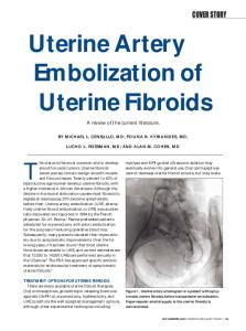
Sonographic appearances: specific sonographic appearance depends on the size of the fibroid and type of degeneration present. Most common sonographic findings include: Well-circumscribed, hypoechoic masses Increased attenuation within the mass Calcification within or surrounding the mass Distortion of normal uterine contour Extrinsic compression of the posterior bladder wall.
* Focal subserous fibroids ()
Hyperattenuation in a focal fibroid (*)
Adenomyosis Incidence: With routine histological examination, adenomyosis is found in approximately 5-10% of postmenopausal women and 15% of women under the age of 40. It is associated with uterine fibroids 50% of the time and with endometriosis 14mm
Uterine Pathology
prgmea.com
(5)
ProSono
Copyright 2006
Uterine (endometrial) polyps Incidence: common in the endometrial cavity particularly at ages 29-59; greatest incidence occurs after age 50. Pathology: a mass of endometrial tissue that projects out or away from the surface of the endometrium. They consist of an excessive localized growth of endometrial tissue with a stromal core and epithelial and mucosal tissue surrounding it. May be single or multiple and range in size from small, 1mm excrescences to masses that fill or distend the uterine cavity. Most commonly arise in the fundal region May undergo malignant change Clinical signs and symptoms: Often asymptomatic Vaginal bleeding; either inter-menstrual flow or heavy periods (menorrhagia). Infertility Occasionally cause postmenopausal bleeding Usually discovered incidentally during D&C Gross pathologic appearances: Smooth, red or brown ovoid body with a velvety texture. Sonographic appearances: Non-specific thickened endometrium, usually focal but occasionally diffuse Discrete mass in endometrium, possibly with a vascular stalk demonstrated with color Doppler May be indistinguishable form endometrial hyperplasia Hysterosonography is ideal for demonstrating polyp size and location Endometrial Carcinoma Incidence: Endometrial carcinoma is the most common type of gynecologic malignancy. It usually occurs in women 60 - 70 years of age. Pathology: There are three histologic types of uterine cancer: Adenocarcinoma MOST COMMON Adenoacanthoma Adenosquamous carcinoma Risk factors include: Obesity Hypertension Diabetes mellitus Strong familial history of uterine cancer
Uterine Pathology
(6)
ProSono
Copyright 2006
Natural history: Initially the tumor mass grows into the uterine cavity. Myometrial invasion is the first indication of continued spread of the disease. Without treatment, the malignancy may spread to the cervix, adnexa, fallopian tubes and ovaries. Distant metastases may occur if the pelvic lymphatic system is infiltrated. Clinical Signs: Vaginal bleeding; post-menopausal Hypermenorrhea, intermenstrual flow in patients still having periods Pain as the result of uterine distention Sonographic Findings: Alteration in size, shape and sonographic texture of the uterine parenchyma Increased uterine size Thickening of endometrial echoes ( >5 mm) especially in a postmenopausal woman (varies with patient's hormone status) Fluid in the endometrial cavity
Uterine Pathology
(7)
ProSono
Copyright 2006
References: Scott JR, et al. Danf or t h’ sObst et r i csandGy necol ogy8th ed. Lippincott Williams and Wilkins, Philadelphia. 1999. DeCherny AH (ed). Current Obstetric and Gynecologic Diagnosis and Treatment 8th ed Lange Medical Books, 1994. Berman, M (ed). Diagnostic Medical Sonography: A Guide to Clinical Practice 2nd ed Lippincott, Philadelphia, 1997.
Uterine Pathology
(8)