Downloaded from www.ajronline.org by 37.44.207.53 on 01/28/17 from IP address 37.44.207.53. Copyright ARRS. For personal use only; all rights reserved
Uterine Artery Embolization for the Treatment of Adenomyosis: Clinical Response and Evaluation with MR Imaging Gary P. Siskin 1 Mitchell E. Tublin Brian F. Stainken Kyran Dowling Eric G. Dolen
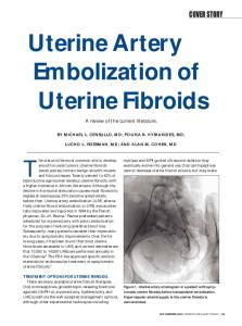
OBJECTIVE. This study was performed to evaluate the MR imaging appearance and clinical response of patients undergoing uterine artery embolization for the treatment of menorrhagia due to adenomyosis. MATERIALS AND METHODS. A retrospective review of 15 patients with adenomyosis and menorrhagia who underwent uterine artery embolization was performed. The diagnosis of adenomyosis was based on established MR imaging criteria. Clinical response was assessed at a minimum of 3 months after embolization. Follow-up MR imaging was performed 6 months after embolization. RESULTS. Of the 15 patients in this study, five had diffuse adenomyosis without evidence of uterine fibroids, one had focal adenomyosis without evidence of uterine fibroids, and the remaining nine had adenomyosis with one or more fibroids. At follow-up, 12 (92.3%) of the 13 patients reported significant improvement in presenting symptoms and quality of life. One patient continued experiencing menorrhagia, and one patient experienced amenorrhea during the 5 months of follow-up after embolization. MR imaging in nine patients, performed at a mean of 5.9 months after embolization, revealed significant reductions in median uterine volume (42%), median fibroid volume (71%), and mean–junctional-zone thickness (11 mm; 33%; p < 0.5). Six of the nine patients had subendometrial regions of decreased T2 signal intensity after embolization. CONCLUSION. Uterine artery embolization is a promising nonsurgical alternative for patients with menorrhagia and adenomyosis. Significant improvement in presenting symptoms and in quality of life is associated with decreases in uterine size and junctional zone thickness. Larger prospective studies are needed to establish the safety and efficacy of this procedure for patients with adenomyosis.
A
Received October 2, 2000; accepted after revision February 2, 2001. 1 All authors: Department of Radiology, A-113, Albany Medical College, 47 New Scotland Ave., Albany, NY 12208. Address correspondence to G. P. Siskin.
AJR 2001;177:297–302 0361–803X/01/1772–297 © American Roentgen Ray Society
AJR:177, August 2001
denomyosis, a benign uterine disease, is characterized by the ectopic growth of endometrial glands and stroma into the myometrium and by diffuse enlargement of the uterus [1]. The reported prevalence of adenomyosis in specimens obtained at hysterectomy has ranged from 8.8% to 31% [2, 3]. A prospective clinical diagnosis of adenomyosis is difficult to make because the most common presenting symptoms, including abnormal uterine bleeding and dysmenorrhea, are similar to those seen with other benign uterine disorders [4]. At the present time, hysterectomy is the definitive treatment for adenomyosis [5]. However, the applicability of nonoperative therapies for benign uterine disease, ranging from hormonal therapy to endometrial ablation, continues to be the focus of investigation in the gynecology community.
Uterine artery embolization has been shown to be an effective treatment for uterine bleeding caused by a variety of conditions, including postpartum hemorrhage, postoperative hemorrhage, cervical pregnancy, and arteriovenous malformations [6–8]. Recently, uterine artery embolization has emerged as a treatment option for patients with symptomatic uterine fibroids. The procedure results in the reduction of both uterine and fibroid volume and in significant improvements in associated symptoms and quality of life [9–13]. In our interventional radiology practice, we noted that many patients referred for uterine artery embolization on the basis of initial sonographic screening studies and clinical symptoms suggestive of fibroids were, in fact, shown to have adenomyosis on preembolization MR imaging of the pelvis. We expected
297
Downloaded from www.ajronline.org by 37.44.207.53 on 01/28/17 from IP address 37.44.207.53. Copyright ARRS. For personal use only; all rights reserved
Siskin et al. this finding, given the frequent coexistence of adenomyosis and fibroids, their often similar clinical symptoms, and the potential difficulties in identifying adenomyosis at screening sonography [4]. The purpose of this study was to report the clinical response and MR imaging appearance of the uterus in patients who underwent uterine artery embolization for the treatment of menorrhagia in the presence of adenomyosis. Materials and Methods We performed a retrospective analysis of all patients with adenomyosis who underwent uterine artery embolization at our institution, after receiving institutional review board approval. All patients included in this study were initially diagnosed with uterine fibroids by their gynecologists. The diagnosis was based on office sonography and clinical symptoms (abnormal uterine bleeding, dysmenorrhea, and bulk-related symptoms such as abdominal distention, frequent urination, and dyspareunia). On the basis of this diagnosis, all patients presented to our office for a consultation regarding the appropriateness of performing uterine artery embolization for uterine fibroids. To date, 224 patients have undergone uterine artery embolization for the treatment of symptomatic uterine fibroids at our institution. Our study population consisted of 15 patients who had a diagnosis of adenomyosis based on MR imaging performed before uterine artery embolization. These patients had a mean age of 46.5 years (range, 37–56 years). All patients presented with abnormal uterine bleeding, 11 (73.3%) of 15 patients presented with dysmenorrhea, and seven (46.7%) of 15 patients presented with bulk-related symptoms, including abdominal distention and bladder compression with frequent urination. Three patients (20%) underwent a trial of hormonal therapy with a gonadotropin-releasing– hormone antagonist before embolization. No patient in this study population underwent myomectomy or endometrial ablation before embolization. Twelve of 15 patients had at least one child, with seven patients reporting a minimum of one miscarriage. Of note, one patient had a history of protein S and possibly protein C deficiency that was of uncertain clinical significance. All patients underwent preprocedural and 6month follow-up MR imaging of the pelvis. MR imaging included axial and sagittal echo-train (fast spin-echo T2-weighted) and axial gradient T1-weighted with and without fat saturation images. Images were acquired with a Signa 1.5-T system (General Electric Medical Systems, Milwaukee, WI). A torso phased-array coil, 5-mm– thick sections with a 2.5-mm gap, a 256 × 256 matrix, and 2 excitations were used for all series. All studies were reviewed both prospectively and retrospectively by two radiologists, with a consensus report ultimately generated after formal review. Uterine and index myoma volume (length × height × width × 0.5233) were calculated for each
298
study patient. A maximal–junctional-zone measurement was then obtained with hand calipers. Adenomyosis was considered present if any portion of the junctional zone was greater than 12 mm in thickness. The presence of myometrial high-signal foci was considered ancillary evidence of adenomyosis. After the presence of adenomyosis was diagnosed with MR imaging, therapeutic options were discussed with all patients. Embolization was included as an option because of its success at addressing identical symptoms in patients with uterine fibroids [9–13]. Whereas none of the patients in the study population had contraindications for surgery, they all strongly preferred avoiding surgical treatment if another option was available. After their options were reviewed, all patients selected embolization as their treatment of choice; no patient in our population with MR imaging findings consistent with adenomyosis decided to undergo hysterectomy. Written informed consent was obtained from all patients before embolization. The technique and protocol used for embolization were the same as those used during uterine artery embolization for the treatment of symptomatic uterine fibroids. The right common femoral artery was used for arterial access in all patients. After obtaining a nonselective pelvic arteriogram (Fig. 1A), we catheterized the left uterine artery, using a 5-French Cobra Glidecatheter (Terumo; Boston Scientific, Watertown, MA) and a 0.035-inch Glidewire (Terumo; Boston Scientific). Once the catheter position was confirmed (Fig. 1B), Contour polyvinyl alcohol particles (Interventional Therapeutics, Fremont, CA) measuring 355–500 µm in diameter were injected until stasis of flow was achieved. The right uterine artery was then selectively catheterized (Fig.1C), and additional particles were injected until the vessel was occluded. In patients in whom the Cobra catheter was flow-limiting when positioned in one or both uterine arteries, a Tracker-325 catheter (Target; Boston Scientific) was used in a coaxial fashion to deliver the polyvinyl alcohol particles. After bilateral uterine artery embolization, completion angiography was performed in all patients to document arterial occlusion and to determine if significant collateral supply to the uterus was present. Polyvinyl alcohol particles measuring 355–500 µm in diameter were selected on the basis of their effectiveness shown during uterine fibroid embolization [12]. We performed embolization on an outpatient basis in all patients, using a protocol described previously [14]. All patients were contacted by telephone 2 or 3 times during the first 7–10 days of the immediate postprocedure recovery period. Patients were then seen in the clinic at 1 month for a follow-up appointment. At the time of follow-up imaging, a questionnaire was given to each patient to assess the changes in their presenting symptoms and quality of life. This questionnaire is used to assess the degree of clinical improvement in all patients undergoing uterine artery embolization at our institution. Patients were presented with a list of items assessing quality of life before and after embolization; vari-
ables included the ability to perform activities of daily life and to socialize outside the home, overall energy level, the degree of interest in and pain experienced during sexual intercourse, and the degree of pain or cramping experienced during menstruation. Patients were asked to rank the degree of severity or impairment, using a scale of 1–10 (with 1 representing “no severity or impairment” and 10 representing “severe severity or impairment”). We performed statistical analysis, using the Wilcoxon’s rank sum test, to assess the significance of the changes in responses to the questionnaire before and several months after embolization. In addition, summary descriptive statistical tests and the Wilcoxon’s sum rank test were used to assess the significance of reductions in uterine volume, fibroid volume, and junctional zone thickness after embolization.
Results
Pelvic MR imaging in all patients revealed a junctional zone thickness greater than 12 mm. The mean–junctional-zone thickness before embolization was 30.4 mm (range, 13–70 mm). Of the 15 patients making up the study population, five had diffuse adenomyosis without evidence of uterine fibroids, one had focal adenomyosis without evidence of uterine fibroids, and nine had adenomyosis (diffuse or focal) with one or more uterine fibroids. Bilateral uterine artery embolization was successfully performed in all patients. The mean duration of the procedure in this group of 15 patients was 60.9 min (range, 44–87 min). In four patients, embolization was performed with a 5-French Cobra Glidecatheter, and in 11 patients, a microcatheter was required because of the occlusive nature of the Cobra catheter in one or both uterine arteries. The expected postprocedure symptoms, including pelvic pain, nausea, and fever, occurred in all patients and were treated according to a previously described protocol with medications including meperidine hydrochloride (Demerol; Abbott Laboratories, Chicago, IL), ketorolac tromethamine, hydrocodone bitartrate and acetaminophen (Lortab; UCB Pharma, Atlanta, GA), and prochlorperazine (Compazine; SmithKline Beecham, Philadelphia, PA) in all patients [15]. Clinical response was assessed by a questionnaire that addressed quality-of-life variables before and after embolization. Questionnaire follow-up at a minimum of 3 months after embolization was available in 13 patients. One patient was lost to follow-up after the embolization procedure. The mean length of clinical followup was 8.2 months (range, 3–16 months) after embolization. The variables assessed on this questionnaire, the median response values before and after embolization, and the statistical
AJR:177, August 2001
Downloaded from www.ajronline.org by 37.44.207.53 on 01/28/17 from IP address 37.44.207.53. Copyright ARRS. For personal use only; all rights reserved
Uterine Artery Embolization in Adenomyosis significance of the responses are listed in Table 1. We noted statistically significant improvement in the ability to perform activities of daily life and to socialize outside the home, overall energy level, the degree of pain experienced during sexual intercourse, and the degree of pain or cramping experienced during menstruation when comparing the responses obtained before and after embolization (p < 0.05). No significant changes were noted in the level of interest in sexual intercourse after embolization. On the basis of data obtained from this questionnaire, we found that there was also statistically significant improvement in the number of days patients experienced bleeding during the menstrual cycle and in the amount of time between changes of sanitary napkins or tampons during the menstrual period (p < 0.05). Imaging follow-up was available in nine patients at a mean duration of 5.9 months (range, 4.25–7.25 months) after embolization. In these nine patients, the mean–uterine-volume measurement decreased from 455.4 to 230.6 cm3. The median percentage of reduction in uterine volume was 42% (range, 24.4–65.0%; p < 0.05). The mean volume of the dominant fibroid identified in the six patients with fibroids and adenomyosis on MR imaging follow-up decreased from 56.5 to 27.0 cm3. The median percentage reduction in the 11 fibroids seen in six patients was 71% (range, 18.7–100%; p < 0.05). Mean thickness of the junctional zone in the nine patients with MR imaging follow-up decreased from 31 to 20 mm. The median percentage reduction in junctional zone thickness was 33% (range, 0–60%; p < 0.05). Six patients with MR imaging follow-up showed re-
TABLE 1
Self-Assessment by 13 Patients of Changes in Quality of Life After Uterine Artery Embolization
Variables Ability to perform activities of daily life Ability to socialize outside the home Overall energy level Interest in sexual intercourse Pain during sexual intercourse Pain or cramping during menstruation
Beforea Aftera
p
7
2