Acta Neurochir (2011) 153:142–147 DOI 10.1007/s00701-010-0718-2
CLINICAL ARTICLE
Use of a collagen biomatrix (TissuDura®) for dura repair: a long-term neuroradiological and neuropathological evaluation Ciro Parlato & Giuseppe di Nuzzo & Marianna Luongo & Raffaele Stefano Parlato & Marina Accardo & Luigi Cuccurullo & Aldo Moraci
Received: 24 February 2010 / Accepted: 11 June 2010 / Published online: 12 July 2010 # Springer-Verlag 2010
Abstract Purpose The aim of this study was to evaluate the clinical, neuroradiological, and neuropathological outcomes of patients treated with equine collagen foil (TissuDura®) as a dura mater substitute during cranial and spinal neurosurgical procedures. Materials and Methods All patients treated at the Department of Neurosurgery of the Second University of Naples with TissuDura between 2005 and 2009 were included. Dural reconstruction was performed using TissuDura, overlaid 1 cm over the dural defect with additional fixation using fibrin glue. No surgical sutures were used. Patients underwent postoperative contrast-enhanced magnetic resonance scans at 1 week, 1 month, and 1 year after surgery to detect any cerebrospinal fluid (CSF) leaks, infections, inflammations, or CSF circulation in the surgical region. Results Dural reconstruction was performed in 74 patients, including 50 patients with tumors, two with C2 neurinoma, two with acoustic neurinoma, six with Chiari I malformation, two with severe head injury, and 12 requiring spinal Electronic supplementary material The online version of this article (doi:10.1007/s00701-010-0718-2) contains supplementary material, which is available to authorized users. C. Parlato : G. di Nuzzo : M. Luongo : R. S. Parlato : A. Moraci Department of Neurosurgery, Second University of Naples, Naples, Italy M. Accardo : L. Cuccurullo Department of Public Health, Section of Pathology, Second University of Naples, Naples, Italy C. Parlato (*) Viale Colli Aminei 21, 80121 Naples, Italy e-mail:
[email protected]
surgery. Clinical and neuroradiological findings were normal and no signs of graft rejection or CSF leaks at postoperative follow-up were observed. In two cases of atypical meningioma, re-operation of the dural reconstruction was performed after 1 year. No adherences between brain and neodura were detected, and histopathological investigations demonstrated dural regeneration. Conclusions Following dural reconstructions with TissuDura without surgical sutures, no local toxicity or complications were observed for up to 1 year. TissuDura demonstrated elasticity, non-reactivity, and good adaptability. The overlay technique using fibrin glue was simple and fast. Future studies and longer follow-up are needed to confirm the efficacy of TissuDura. Keywords Dural repair . Dural substitute . Collagen biomatrix . TissuDura
Introduction After many cranial and spinal neurosurgical procedures, reconstruction of the dura mater is needed. Watertight closure of dural defects is required, as an inadequate closure of the dura at the end of neurosurgical procedures exposes the patient to cerebrospinal fluid (CSF) leaks, infections, hypertensive pneumocephalus, pseudomeningocele, and other complications that can lead to a longer period of hospitalization. Over the past decades, various types of material have been evaluated to determine the ideal dural replacement technique, including autografts, allografts, xenografts, and synthetic grafts. However, many of these products have been associated with postoperative complications, some of which were serious, such as hemorrhages, development of
Acta Neurochir (2011) 153:142–147
corticomeningeal adhesions, and inflammatory reactions [1, 2]. Friction is the major complication of polytetrafluoroethylene duraplasty because of the strong surface tension and poor adaptability [3]. Autologous duraplasties are accompanied by potential donor-site complications and are inappropriate for large defects [3]. Acellular dermal grafts prepared from cadaver human skin do not suffer from these drawbacks, and these matrices have been reported to promote the formation of new dural tissue by providing a scaffold for the growth of local cells while the matrix itself is fully reabsorbed over time [4–11]. However, use of human cadaver skin is associated with the development of atrophy or adhesion with the brain surface [1, 4–11]. Furthermore, relationships between iatrogenic Creutzfeldt– Jakob disease and implanted human cadaveric dura grafts have recently been reported [12]. Similarly, implanted bovine xenografts carry a risk of transmission of bovine spongiform encephalopathy [4, 6]. TissuDura (TissuDura®; Baxter, Vienna, Austria) is composed of colloidal collagen from equine Achilles tendon and is inactivated with sodium hydroxide and chloriduric acid. Previous reports have shown an absence of local and systemic toxicity and a low incidence of adherences and inflammation with TissuDura, making it a viable option for dural substitution [13–15]. Here, we present our experience using this equine dural substitute in a series of patients who required dural graft implantation during various cranial and spinal neurosurgical procedures. Unlike previous reports, where a number of patients required sutures for fixation of the collagen matrix, the neurosurgical procedures described here were performed without the need for suturing, reducing surgery times, and allowing TissuDura to be applied to anatomically difficult locations.
Methods All cases of dural reconstruction performed at our department of neurosurgery of the Second University of Naples between 2005 and 2009 were included. TissuDura was rehydrated in physiological saline to obtain a transparent film that could be cut into the desired shape. Patches of the collagen matrix were cut with scissors to the appropriate size for the dural defects with an overlap of 1 cm. The patch was then placed over the dural defect, overlapping at the margins. The patch was fixed using fibrin glue. To avoid postoperative fibrosis, minimal amounts of fibrin glue were used in the repair of the spinal dural defects. Surgical sutures were not used during dural reconstruction. Multiple layers of TissuDura were used when we performed duroplasty in three cases of bis-chloronitrosourea (BCNU) wafer-treated recurrent glioblastomas, and in two cases of
143
atypical meningiomas, reoperated for recurrence. All the patients underwent postoperative neuroradiological evaluation with contrast-enhanced magnetic resonance (MR) scans at 1 week, 1 month, and 1 year after surgery, to detect any CSF leaks, infections, inflammations and CSF circulation in the surgical region after dural reconstruction. Histopathological and ultra structural investigations were also carried out in patients who required re-operation.
Results Patients Dural reconstruction was performed in 74 patients. These included 50 cases of tumors (31 in the frontoparietal paramedian region and 19 in the parieto–occipital paramedian region), two cases of C2 neurinoma, two cases of acoustic neurinoma, six cases of Chiari I malformation, two cases of severe head injury and 12 cases of spinal surgery. Reconstruction procedure No patient required removal of the graft, and the overlay technique with the use of fibrin glue was simple and fast. The time needed for the neurosurgical procedures was reduced. TissuDura facilitated the application of small patches in anatomically difficult locations during spinal and posterior cranial fossa surgery. The dural reconstruction in the posterior cranial fossa during surgical treatment of the Chiari I malformation was particularly easy and fast. Video 1 demonstrates the overlay technique used in cerebellopontine angle surgery following removal of acoustic neurinoma (electronic supplementary material). Of the 74 patients on which dural reconstruction was performed, in three cases of BCNU wafer-treated recurrent glioblastomas, we performed a multilayer TissuDura approach during the re-operation. Follow-up At follow-up, we did not observe any signs of graft rejection or CSF leaks in any of our patients, and no other complications occurred. We observed the reorganization of dura and normal CSF circulation (data not shown). In the two cases of recurrent atypical meningioma, reoperation of the dural reconstruction was performed after 1 year. We observed no adherences between the brain and neodura. Neodura formation allowing fibroblast ingrowth and collagen formation was observed (Fig. 1). In these cases, histopathological and ultra structural findings from the previously implanted TissuDura showed fibroblasts and new normal dural tissue. The collagen matrix was fully
144
Acta Neurochir (2011) 153:142–147
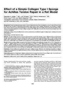
Fig. 1 a, b, c Intraoperative images showing the first duroplasty in a patient affected by atypical meningioma. d Dural reconstruction with TissuDura with no postoperative CSF leaks
degraded and replaced by natural collagen. TissuDura was not recognized as a foreign material and no tissue reactions were observed (Figs. 2, 3). In the two cases of C2 neurinoma, in which dural reconstruction of the lateral recess of the dural sac was performed, postoperative examinations showed the complete removal of neurinoma without CSF leaks (Fig. 4).
Discussion
Fig. 2 Neurohistopathological photomicrographs of dural specimen from the neodura. The neodura is composed of fibroblasts and newly deposed reticulate collagen fibers with the typical undulate aspect of the fibers and nuclei. H & E, ×200
Fig. 3 Ultrastructural photomicrographs of dural specimen from the neodura. Typical undulate aspect of the fibers and the nuclei confirmed the migrated fibroblasts. The collagen matrix was fully degraded and replaced by native collagen neodura after 12 months
In this case series of 74 patients, dural reconstructions with TissuDura, without use of surgical sutures were performed using fibrin glue for additional fixation. The application of
Acta Neurochir (2011) 153:142–147
145
Fig. 4 a, b Preoperative and c, d postoperative MR showing the complete removal of C2 neurinoma, and e, f the postoperative outcome without CSF leaks
TissuDura was simple and fast, resulting in reduced surgical time. At 12 months follow-up, no local toxicity or complications such as CSF leaks, adherences or inflammation were observed in any of our patients. TissuDura appeared to have all the features of the ideal dura substitute: elasticity, non-reactivity, and good adaptability [13–15]. Unlike other techniques evaluated for dural repair, such as
autografts, allografts, xenografts, and synthetic grafts, TissuDura is not associated with complications such as friction, atrophy, hemorrhages, or development of corticomeningeal adhesions. Furthermore, there is limited risk of disease transmission due to the equine origin of TissuDura and the inactivation step with sodium hydroxide and chloriduric acid.
146
We used TissuDura in a wide range of spinal and cranial neurological procedures. In our two cases of dural reconstruction of the lateral recess of the dural sac, after removal of the C2 neurinoma, postoperative examinations showed the complete removal of neurinoma without CSF leaks. Using a multilayer-TissuDura approach, dural reconstruction was effective in all three of our patients affected by BCNU wafer-treated recurrent glioblastomas with no CSF leaks detected. In the literature, CSF leaks are reported in around 5% of BCNU wafer-treated patients [16]. Dural reinforcement with TissuDura allowed our patients to subsequently undergo radiotherapy and chemotherapy. Usually, dural reconstruction during spinal surgery must be meticulously sutured to avoid CSF leaks because of the increased hydrostatic pressure in the supine or upright position; nevertheless, we performed the reconstructions without sutures and at follow-up we did not observe any CSF leakages. The use of autologous fat transplants is recommended as a rapid, effective means for repair of dural tears or defects that are inaccessible or unsuitable for standard suture technique [17]. These techniques are also applicable for reinforcement of suboccipital and spinal dural suture lines [17]. TissuDura may be a viable alternative to these techniques. Hida et al. developed a dura-substitution technique using bio absorbable fabric and fibrin glue [18]. In 160 patients who underwent dura repair using this polyglycolic acid– fibrin sheet method, ten (6.3%) experienced subcutaneous CSF leakage [18]. The authors concluded that this technique represents a novel alternative to artificial dural substitutes that were available at that time [18]. Recently, Narotam et al. have shown that collagen matrix is suitable for use in the posterior fossa where it can be applied as an onlay graft, without the inconvenience and time-consuming process of suturing [19]. After application, the collagen matrix provides a chemical signal for fibroblast infiltration, which commences after 3–4 days and becomes established over the next 10–14 days. The fibroblasts use the pores in the collagen matrix as a scaffold to lay down new collagen. Over 6–8 weeks, the collagen matrix is resorbed as the new dura is constituted. This represents a more developed collagen-based dural graft compared with a collagen sponge; it was associated with a good safety profile, as well as being effective, easy to use, and time efficient in posterior fossa surgery [19]. However, meticulous layered wound closure, the detection and effective control of hydrocephalus, and the use of closed suction wound drainage were required to minimize potential complications related to the use of collagen matrix duraplasty in the posterior fossa [19]. In our patients, the overlay technique for dural reconstructions using TissuDura was easy and fast and allowed good results in dural repair during posterior
Acta Neurochir (2011) 153:142–147
fossa and spinal surgery without the use of closed suction. To avoid postoperative fibrosis, we used a minimal amount of fibrin glue in the repair of the spinal dural defects. Studies of intracranial implantation in adult sheep showed that implantation of collagen biomatrix did not result in inflammation, CSF leaks, or impaired wound healing [20]. Microscopic assessment of graft incorporation 2 weeks postoperatively showed loosening of the homogeneous structure of the collagen graft with invasion of lymphocytic and monocytic components. After a period of 4 weeks, lymphocytic and monocytic exudates were present and polymorphonuclear giant cells were common with numerous fibroblasts. Eight weeks postoperatively, inflammatory infiltrates had regressed further. After 24 weeks, there was a further regression of inflammatory infiltrates and continuity between the highly collagenous endogenous dura and the newly formed collagen fibers of neodura [20]. In another case report, histological examination of a tissue sample taken 40 days after implantation of a collagen matrix revealed that the graft had been replaced by significant ingrowth of the native dura [14]. Similarly, in our two patients with recurrent meningiomas who required re-operation, long-term follow-up using histopathological and ultra structural techniques demonstrated re-absorption of the TissuDura and regeneration of dura mater. To our knowledge, this is the first report with documented histopathological and ultra structural imaging showing dura regeneration after TissuDura graft insertion. Our ultra structural observations showed an absence of inflammatory infiltrate and migration of fibroblasts, resulting in the neodura regeneration. The collagen matrix was fully degraded and replaced by native collagen neodura after 12 months.
Conclusions In this case series of 74 patients, TissuDura demonstrated all aspects of the ideal dura substitute: elasticity, nonreactivity, good adaptability, and ease and speed of application. Dural reconstructions were performed without surgical sutures, and no incidences of local toxicity or complications such as CSF leaks, adherences, or inflammation were observed. Moreover, the overlay technique with the use of fibrin glue was simple and quick to perform. Histopathological and ultra structural images demonstrated dura regeneration after TissuDura graft insertion in patients with recurrent meningioma after 12 months follow-up. Future studies with longer follow-up periods are required to confirm the efficacy of TissuDura for dura repair. Acknowledgements Editorial assistance was provided by AlphaPlus Medical Communications Ltd (UK).
Acta Neurochir (2011) 153:142–147 Disclosures/conflicts of interest Patients gave consent for treatment and use of figures and cases. None of the authors has any financial interest in the materials and methods used, nor in any of the manufacturers mentioned in this report. Financial support for the production of this manuscript was provided by Baxter BioSurgery, Italy.
References 1. Alleyne CH Jr, Barrow DL (1994) Immune response in hosts with cadaveric dural grafts. Report of two cases. J Neurosurg 81:610– 613 2. Robertson SC, Menezes AH (1997) Hemorrhagic complications in association with silastic dural substitute: pediatric and adult case reports with a review of the literature. Neurosurgery 40:201–205, discussion 205-206 3. Islam S, Ogane K, Ohkuma H, Suzuki S (2004) Usefulness of acellular dermal graft as a dural substitute in experimental model. Surg Neurol 61:297–302, discussion 303 4. Anson JA, Marchand EP (1996) Bovine pericardium for dural grafts: clinical results in 35 patients. Neurosurgery 39:764–768 5. Chaplin JM, Costantino PD, Wolpoe ME, Bederson JB, Griffey ES, Zhang WX (1999) Use of an acellular dermal allograft for dural replacement: an experimental study. Neurosurgery 45:320– 327 6. Filippi R, Schwarz M, Voth D, Reisch R, Grunert P, Perneczky A (2001) Bovine pericardium for duraplasty: clinical results in 32 patients. Neurosurg Rev 24:103–107 7. Laun A, Tonn JC, Jerusalem C (1990) Comparative study of lyophilized human dura mater and lyophilized bovine pericardium as dural substitutes in neurosurgery. Acta Neurochir (Wien) 107:16–21 8. Maher CO, Anderson RE, McClelland RL, Link MJ (2003) Evaluation of a novel propylene oxide-treated collagen material as a dural substitute. J Neurosurg 99:1070–1076 9. Narotam PK, Jose S, Nathoo N, Taylon C, Vora Y (2004) Collagen matrix (DuraGen) in dural repair: analysis of a new
147
10.
11.
12.
13.
14.
15.
16.
17.
18.
19.
20.
modified technique. Spine (Phila Pa 1976) 29:2861–2867, discussion 2868-2869 Narotam PK, Reddy K, Fewer D, Qiao F, Nathoo N (2007) Collagen matrix duraplasty for cranial and spinal surgery: a clinical and imaging study. J Neurosurg 106:45–51 Warren WL, Medary MB, Dureza CD, Bellotte JB, Flannagan PP, Oh MY, Fukushima T (2000) Dural repair using acellular human dermis: experience with 200 cases: technique assessment. Neurosurgery 46:1391–1396 Hoshi K, Yoshino H, Urata J, Nakamura Y, Yanagawa H, Sato T (2000) Creutzfeldt–Jakob disease associated with cadaveric dura mater grafts in Japan. Neurology 55:718–721 Biroli F, Fusco M, Bani GG, Signorelli A, Esposito F, de Divitiis O, Cappabianca P, Cavallo LM (2008) Novel equine collagenonly dural substitute. Neurosurgery 62:273–274, discussion 274 Gazzeri R, Neroni M, Alfieri A, Galarza M, Faiola A, Esposito S, Giordano M (2009) Transparent equine collagen biomatrix as dural repair. A prospective clinical study. Acta Neurochir (Wien) 151:537–543 Stendel R, Danne M, Fiss I, Klein I, Schilling A, Hammersen S, Pietilae T, Janisch W, Hopfenmuller W (2008) Efficacy and safety of a collagen matrix for cranial and spinal dural reconstruction using different fixation techniques. J Neurosurg 109:215–221 Sabel M, Giese A (2008) Safety profile of carmustine wafers in malignant glioma: a review of controlled trials and a decade of clinical experience. Curr Med Res Opin 24(11):3239–3257 Black P (2000) Cerebrospinal fluid leaks following spinal or posterior fossa surgery: use of fat grafts for prevention and repair. Neurosurg Focus 9:e4 Hida K, Yamaguchi S, Seki T, Yano S, Akino M, Terasaka S, Uchida T, Iwasaki Y (2006) Nonsuture dural repair using polyglycolic acid mesh and fibrin glue: clinical application to spinal surgery. Surg Neurol 65:136–142, discussion 142-133 Narotam PK, Qiao F, Nathoo N (2009) Collagen matrix duraplasty for posterior fossa surgery: evaluation of surgical technique in 52 adult patients. Clinical article. J Neurosurg 111:380–386 Knopp U, Christmann F, Reusche E, Sepehrnia A (2005) A new collagen biomatrix of equine origin versus a cadaveric dura graft for the repair of dural defects–a comparative animal experimental study. Acta Neurochir (Wien) 147:877–887