SICM Tuition
Biology AS
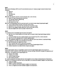
Unit 2: Exchange, Transport and Reproduction Exchange surfaces We have done a lot at GCSE, and I keep going back to it…which is great fun because I get to see how much (/little) you know…so let’s try this again. Name three ways in which exchange surfaces are adapted to diffuision. (Yes, this is part of Module 1 too…so you better be able to do this!!) Oh, and while you’re at it…name the Law that this refers to. Fick’s Law thickness of membrane / length of pathway surface area × concentration difference surface area Rate of diffusion α thickness of membrane concentration gradient There are many places in humans in which diffusion is vital. Some examples include: lungs (where the alveoli are extremely well adapted) kidney (exretory products…urine, uric acid etc.) intestine (remember things called villi?) It goes without saying (but I’ll say it anyway…) that the body is well adapted in certain places for diffusion. The three examples we looked at above are prime examples and we will look at these in detail later. Exchange processes We have already mentioned that surface area is important and this, combined with the volume of an organism very much determines the way in which an organism exchanges gases. Let us think about the way in which size affects the surface area:volume ratio. 2µ 5µ
Surface area: 24µ2 Volume: 8µ3 Surface area:volume ratio: 3:1
Surface area: 150µ2 Volume: 125µ3 Surface area:volume ratio: 1.2 : 1
As you can see, there is a huge difference. The smaller cube has a huge surface area:volume ratio. This has many affects on organisms of small size. Think about a small organism such as an amoeba or protozoa. These organisms are so small that they don’t need things such as lungs. They are able to carry out all of their gas exchange directly with the surroundings because of their vast surface area and small volume. However, this also causes problems with water loss which can lead to dehydration very easily. Humans, as with all larger organisms, require ventilation systems to perfuse all cells or a circulatory system to take the gases round: as all cells require gases such as oxygen for respiration (yes…respiration IS still important…). Of course, this is because we are larger than a couple of cells thick…however much we diet (so girls, you may as well just give up!). Page 1
SICM Tuition
Biology AS
Gas Exchange in flowering plants Lucky you…you get to study plants…God’s creations are indeed a wonder and I’m sure that this will help you understand the real wonders of creation (or, you’ll just get thoroughly bored, but will have to deal with it and learn it all anyway). We want to look at the mesophyte leaf (not only because it has a fun name…but because it’s on your syllabus) and particularly look at the idea of gas exchange within the plant. A mesophyte leaf is basically a leaf from a plant that lives in normal conditions (not too dry and not too wet). Now, once again, this is all stuff that we did at GCSE but seeing as you don’t seem to remember any of it (and who can blame you…(except me…)) we’ll go over it again.
Waxing cuticle
wax layer – transparent to let light through – reduces water loss.
Upper epidermis
for protection and structure. It is relatively transparent.
Palisade layer
tightly packed to stop light passing. Contain lots of chloroplasts to make photosynthesis most efficient.
Spongy mesophyll layer
lets oxygen leave and CO2 enter and leave. Some photosynthesis takes place but not a lot.
Lower epidermis
guard cell and stomata (pores) – allow gas exchange and water loss.
Vein
composed of two main layers. Xylem takes water from roots to the leaf for photosynthesis. Phloem takes products of photosynthesis around the plant.
Page 2
SICM Tuition
Biology AS
Stomata and gas exchange In order to photosynthesis, plants require gases to be exchanged with the surroundings. Write down the equation for photosynthesis to remind yourself of it. Carbon Dioxide
+
Water
light chlorophyll
Glucose
+
Oxygen
The way in which plants do this is by having holes for the gasses to diffuse into. These holes are called stomata and are created and altered by cells called guard cells. The guard cells have a funny shape which means that when they are turgid, they make the stomata open. When they are flaccid, they close the stomata meaning that no gas exchange takes place. The reason for them becoming turgid or flaccid is dependent on the amount of photosynthesis that takes place. When a lot of photoshynthesis takes place (e.g. in daylight), the glucose concentration in the guard cell increases. This causes water to move into the guard cell by osmosis making it turgid. This happens because water moves from an area of high water potential (outside cell) to an area of lower water potential (in cell). Explain what happens at night. Not a lot of photosynthesis. Therefore concentration of glucose decreases. Water moves out of cell via osmosis (from an area of high water (cell) potential to low water potential (outside cell)). Stomata close. Gas exchange stops. Stomata – “hole” where gases (and water vapour) can diffuse in and out of
Day
Two turgid guard cells – normally these are very close to each other
Chloroplasts absorb light for photosynthesis Cell sap gets stronger (i.e. lower water potential) Water enters guard cell via osmosis
Night
Guard cell swells and becomes turgid
No light – no photosynthesis This pulls the guard cells apart The stomata are open
Cell sap gets weaker (i.e. higher water potential) No water enters Guard cells are flaccid Page 3
Stomata are closed
SICM Tuition
Biology AS
Gas Exchange in Humans There’s no need to tell you that the lungs are the organs by which humans take in and expel gases…so let’s go straight into the detail. The thorax (chest), is bound by the ribs and the muscular sheet known as the diaphragm. Within this space, the lungs are present. Air passes into a series of tubes which are designed to not only moisten and warm the air that comes in, but also protect the body by filtering out some dust etc. There are three subsections of these tubes: -
Trachea
-
Bronchi
-
Bronchioles
The walls of the trachea and bronchi are composed of an epithelial layer as well as connective tissue, smooth muscle and cartilage along with a tough outer coat. Bronchioles differ in the fact that they lack cartilage. Cartilage supports the trachea and bronchi and stops them from collapsing. In the trachea, the cartilage forms C-shaped rings that keep the tube open during inspiration. Smooth muscle lies at the back of the tube. The reason for the cartilage not being all the way around is that the oesophagus (gullet / food pipe!) is at the back of the trachea and the smooth muscle allows expansion of the oesophagus as food travels down. In the bronchi, the cartilage is arranged in plates within smooth muscle. The smooth muscle also helps keep the tubes open. The diameter of the tubes is regulated by the automatic part of the nervous system. This ensures adequate diameter for suitable gas flow. The trachea, bronchi and bronchioles are lined with epithelial cells. These epithelial cells are made up of different types: ciliated and goblet cells. Goblet cells produce and secrete mucus into the respiratory tract and ensures that there is a layer of mucus along the trachea to trap dust and bacteria. Ciliated cells have a protective function. They sweep the mucus along so that it does not block the trachea and so that it can be renewed. Generally, the mucus moves along to the oesophagus where it is swallowed and destroyed by the acidity of the stomach. Alveoli are air sacs and they make up th mass of the lungs. They are attached to the end of the bronchioles via alveolar ducts. Although each alvelous is very small, there are loooaaaaaads of them (about 700 MILLION!!) which provides a massive surface area of about 70-90m2! They are covered in blood vessels that bring in deoxygenated blood via the pulmoary artery. Page 4
SICM Tuition
Biology AS
The vast blood supply helps to provide a sufficient concentration gradient for the gases which need to be exchaged. For example, the partial pressure of oxygen in the air in the alveolus will be higher than the pO2 in the blood and so oxygen will move into the blood via diffusion. If you exercise more, you use up more oxygen and so there is less oxygen in the blood. This means that there is a steeper concentration gradient in the lungs: more diffusion into blood!
Page 5
SICM Tuition
Ventilation -
Inspiration -
Biology AS
main muscles to be considered include the intercostal muscles and the diaphragm
external intercostal muscles contract ribs move upwards and outwards diaphragm contracts: moving downwards incrased thorax volume reduced pressure (below outside air pressure) air enters - inflating the lung
Expiration (do this one yourself!) no active muscle contractions external intercostal muscles relax ribs move downwards and inwards diaphragm relaxes: moving upwards decrased thorax volume increased pressure (above outside air pressure) air forced out - deflating the lung -
in forced breathing, expiration is active and internal intercostal muscles and abdominal muscles contract – incresing the pressure in the thorax.
Friction between the lings and the inner surace of the wall of the thorax is reduced by the pleural fluid which lies between the two pleural membranes that surround the lungs (see diagram on previous page). Breathing control Normally, respiratory movements are involuntary. They are controlled by the rhythmic discharges of the medulla in the brain. From the inspiratory control centre, impulses travel down the phrenic nerves to the diaphragm and down the intercostal nerves to the external intercostal muscles causing them to contract. When the alveoli are stretched, the stretch receptors send impulses to the inspiratory control centre inhibiting inspiration and causing the muscles to relax. Therefore expiration takes place. Normally, expiration is passive. Sometimes it can be forced – by sending impulses to the internal intercostal muscles rom the expiratory contro centre.
Page 6
SICM Tuition
Biology AS
Draw a diagram showing how breathing is controlled by the body. Lungs deflated
Inhibitory impulses no longer fired nerve impulses fired
EXPIRATION
Diaphragm and intercostal muscles relax 5 no impulses
2 Breathing Centre
Inhibition of inspiration
6 expiratory activiy (air out)
4
Inhibitory impulses fired from stretch receptors in lungs
Vagus Phrenic nerve nerve INSPIRATION
Lungs inflated
1 stimulation
CO2 from respiratory activity
Diaphragm and intercostal muscles contract
3 inspiratory activiy (air in)
Increase in CO2 in blood speeds up frequence of nerve impulses. Decrease in CO2 in blood slows fown frequence of nerve impulses Aorta and carotid arteries contain chemoreceptors which detect changes in CO2 levels. pH levels also change with CO2: as CO2 increases, pH decreases (as carbonic acid is formed).
Tidal volume = volume of air bbreated in and out of lungs at rest (typical value ≈ 0.5dm3) Ventilation rate = volume of breath × number of breaths per minute (units = litres/min)
Page 7
SICM Tuition
Biology AS
Digestion and Absorption Right…well, not only did you do this at GCSE (cough cough) but we also did this two weeks ago. So…seeing as I don’t feel like doing any work, you can do it all…
Label the trachea on the diagram. Also label the epiglottis (it’s not really on the diagram..but guess!). What does the epiglottis do? The food (bolus) passes into the throat and the epiglottis should close, preventing the food entering the trachea.
epiglottis
Which two parts make up the small intestine? i.
Duodenum
ii.
Ileum
Which two parts make up the large intestine? i.
Colon
ii.
Rectum
Digestion begins in the mouth. Saliva is released from the salivary gland and mixes the food, softening it, dissolving it and making it easier to swallow. Saliva also contains the enzyme amylase, which begins to break down starch into maltose. The physical action of chewing (mastication) breaks food into smaller pieces. This makes it easier to swallow, and increases the surface area for enzymes to work on. The food (bolus) passes into the throat and the epiglottis should close, preventing the food entering the trachea. The bolus travels down the oesophagus to the stomach by a process called peristalsis. This is how it travels through the rest of the alimentary canal.
Page 8
SICM Tuition
Biology AS
Stomach cardiac sphincter before stomach o prevents anything getting out (and in)
muscular wall of gullet
circular muscle contracting
-
holding area for food as small intestine can only deal with small amounts so you don’t need to eat small bits constantly you can eat lots and it’s held in stomach
-
food longitudinal muscle will contract here
digestion: o pepsin: protein → polypeptides
-
kills bacteria using HCl
-
“chyme” is name of release into duodenum (also what you vomit)
Most absorption takes place in the small intestine. This is adapted very well for its function.
Peristaltic movement is initiated by circular smooth muscles contracting behind the chewed material to prevent it from moving back into the mouth, followed by a contraction of longitudinal smooth muscles which pushes the digested food forward.
Small intestine Duodenum contain Brunner’s glands pancreas gives out pancreatic juices o containing alkaline mucus (to neutralise acid conditions from stomach) and some enzymes o Brunner’s glands give out enzymes and mucus -
bile o o o o o o
produced in the liver by breakdown of haemoglobin contains salts and pigments in alkaline solution emulsifies fats and oils larger globules of fat reduced to smaller globules for larger surface area NOT an enzyme (does not contain protein)
Ileum no Brunner’s glands (this is how to tell the difference) where most re-absorption takes place large surface area: o many folds o mucosa and submucosa are folded o villi present in mucosa: � small finger-like projections to increase surface area o all epithelial cells in ileum have even more villi called microvilli
Page 9
SICM Tuition
Biology AS
Give three ways in which the small intestine is made good for absorption? - It is long so that there is time for digestion to take place. - It is folded which makes a greater surface area. - It has a good blood supply so the digested food can be absorbed quickly by the blood. - It has thin walls so that diffusion can take place – short diffusion pathway. - It has lots of villi, which give more surface area. - It has various enzymes, which break down various foods. Large intestine -
-
caecum / appendix: no function in humans colon: ascending, horizontal and descending: o remains of digestion turned into faeces o water removed faeces are stored in rectum for short period o if not enough roughage, constipation occurs as roughage is indigestible and initiates peristalsis
-
anal sphincter o controlled by autonomic nervous system o but there is a degree of voluntary control
-
rectum o possible to maintain faeces o if not possible, it would be easy to follow a trail...adaptation
Digestion of Carbohydrates Fill in the following table stating where carbohydrates are broken down and by which enzymes: Where it is produced
Secretion
Enzyme(s)
What it does
Salivary Gland
Saliva
Salivary amylase
Breaks down Starch into Maltose
Pancreas
Pancreatic juices
Pancreatic Amylase
Breaks down starch into Maltose
Walls of the Ileum
Intestinal juice
Maltase
Breaks down Maltose into Glucose
Walls of the Ileum
Intestinal juice
Sucrase
Breaks down Sucrose into Fructose and Glucose
Carbohydrates are broken down into disaccharides (which you should know about from module 1…..) by the enzymes in the mouth and in the pancreas. They are then broken down into monosaccharides on the cell surface membrane of the gut (by maltase and sucrase). These monomers are then absorbed by the cells lining the gut and taken through into the blood vessels. Page 10
SICM Tuition
Biology AS
Absorption This takes place in the ileum through small finger-like projections called villi. Muscle layers to allow peristalsis Blood vessels to remove digested food molecules
Bolus
Folded inner wall of ileum. Increases surface area for absorption Lumen – hollow part of gut
On the surface of the inner folded wall are millions of villi, which greatly increase the surface area for absorption. Digested food Molecules move into villus by diffusion
Lacteal
Wall of villus – only one cell thick
Network of capillaries
Low food molecule concentration Lymph Vessel
-
Amino acids and glucose diffuse into the capillaries and are taken away in the blood. Fatty acids and glycerol diffuse into the lacteal and are taken away into the lymph vessel – they are added to the blood again later.
Advantages -
Thin membrane means a short diffusion pathway for materials. Therefore absorbs food molecules quickly.
-
Vast network of blood capillaries take food material quickly.
-
Substances taken away quickly so that it maintains a steep concentration gradient. Page 11
SICM Tuition
Biology AS
6102 Unit 2B: Exchange, transport and reproduction Unit 2 incorporates the following sections from the Subject Criteria: 3.9 Exchange with the environment 3.10 Mass transport 3.11 Adaptations to the environment 2.B1 Exchanges with the environment - understand what materials need to be exchanged; respiratory - gases; nutrients; excretory products Exchange processes understand the relationship of size and surface area to volume - ratio; - understand the features of exchange surfaces which aid passive - and active transport; - understand the special features of gas exchange surfaces; - understand the need for ventilation mechanisms. Gas exchange in protozoa - understand how gas exchange is achieved in a protozoan. Gas exchange in flowering plants - describe the external and internal structure of a mesophyte leaf; - understand the structure and roles of stomata and the mechanism - of stomatal opening in terms of changes in ion concentrations - leading to changes in turgidity; understand how gas exchange is - achieved. Gas exchange in humans - recall the structure of the thorax; understand the mechanism of - ventilation, including the role of the pleural membranes; - understand how breathing is controlled; understand vital capacity - and tidal volume; - recall the structure of alveoli and understand their role in gas - exchange; explain the function of surfactants; know that breathing - is controlled by the respiratory centre in the brain. - Practical work should include the use of simple respirometers. Digestion and absorption - describe the structure of the alimentary canal in relation to - digestion and absorption; - describe mastication and movement of food along the gut; - describe the histology of the ileum wall; understand the sources - and effects of secretions concerned with the digestion of - carbohydrates.
Page 12