DIABETES-INSULIN-GLUCAGON-GASTROINTESTINAL
Mild Endoplasmic Reticulum Stress Augments the Proinflammatory Effect of IL-1 in Pancreatic Rat -Cells via the IRE1␣/XBP1s Pathway Michela Miani, Maikel L. Colli, Laurence Ladrière, Miriam Cnop, and Decio L. Eizirik Laboratory of Experimental Medicine (M.M., M.L.C., L.L., M.C., D.L.E.) and Division of Endocrinology (M.C.), Erasmus Hospital, Université Libre de Bruxelles, B-1070 Brussels, Belgium
The prevalence of obesity and type 1 diabetes in children is increasing worldwide. Insulin resistance and augmented circulating free fatty acids associated with obesity may cause pancreatic -cell endoplasmic reticulum (ER) stress. We tested the hypothesis that mild ER stress predisposes -cells to an exacerbated inflammatory response when exposed to IL-1 or TNF-␣, cytokines that contribute to the pathogenesis of type 1 diabetes. INS-1E cells or primary rat -cells were exposed to a low dose of the ER stressor cyclopiazonic acid (CPA) or free fatty acids, followed by low-dose IL-1 or TNF-␣. ER stress signaling was inhibited by small interfering RNA. Cells were evaluated for proinflammatory gene expression by RT-PCR and ELISA, gene reporter activity, p65 activation by immunofluorescence, and apoptosis. CPA pretreatment enhanced IL-1- induced, but not TNF-␣induced, expression of chemokine (C-C motif) ligand 2, chemokine (C-X-C motif) ligand 1, inducible nitric oxide synthase, and Fas via augmented nuclear factor B (NF-B) activation. X-box binding protein 1 (XBP1) and inositol-requiring enzyme 1, but not CCAAT/enhancer binding protein homologous protein, knockdown prevented the CPA-induced exacerbation of NF-B-dependent genes and decreased IL-1-induced NF-B promoter activity. XBP1 modulated NF-B activity via forkhead box O1 inhibition. In conclusion, rat -cells facing mild ER stress are sensitized to IL-1, generating a more intense and protracted inflammatory response through inositol-requiring enzyme 1/XBP1 activation. These observations link -cell ER stress to the triggering of exacerbated local inflammation. (Endocrinology 153: 3017–3028, 2012)
T
ype 1 diabetes (T1D) is a chronic autoimmune disease triggered by the interaction between genetic and environmental factors (1–3). The prevalence of T1D is increasing at an alarming rate, with a particularly marked increase in countries such as Poland, Romania, and the Czech Republic. It is expected that new cases of T1D in young European children will double between 2005 and 2020 (4). This seems to be a global phenomenon, with an increase in new cases of T1D also observed in the United States, Australia, New Zealand, and Canada (5– 8). This
fast increase of T1D incidence suggests the effect of environmental rather than genetic factors (4). Modern Western society has easy access to high-fat and high-caloric food. This, coupled to lack of physical activity, favors the development of obesity in children and adults (9). Obesity is usually associated with type 2 diabetes (9), but there is also a correlation between body mass index in children and risk for T1D (10, 11). Obesity increases the demand for insulin secretion, secondary to peripheral insulin resistance (9). In parallel, high
ISSN Print 0013-7227 ISSN Online 1945-7170 Printed in U.S.A. Copyright © 2012 by The Endocrine Society doi: 10.1210/en.2011-2090 Received December 8, 2011. Accepted April 4, 2012. First Published Online April 23, 2012
Abbreviations: CCL2, Chemokine (C-C motif) ligand 2; CHOP, C/EBP homologous protein; CPA, cyclopiazonic acid; CXCL1, chemokine (C-X-C motif) ligand 1; DMSO, dimethylsulfoxide; ER, endoplasmic reticulum; FACS, fluorescence-activated cell sorting; FBS, fetal bovine serum; FFA, free fatty acid; FoxO1, forkhead box O1; GAPDH, glyceraldehyde-3phosphate dehydrogenase; IB, inhibitor B; iNOS, inducible nitric oxide synthase; IRE1␣, inositol-requiring enzyme 1␣; KD, knockdown; NF-B, nuclear factor B; siControl, control siRNA; siRNA, small interfering RNA; siXBP1, siRNA against XBP1; T1D, type 1 diabetes; THP, thapsigargin; UPR, unfolded protein response; XBP1, X-box binding protein 1; XBP1s, XBP1 spliced.
For editorial see page 2949
Endocrinology, July 2012, 153(7):3017–3028
endo.endojournals.org
The Endocrine Society. Downloaded from press.endocrine.org by [${individualUser.displayName}] on 15 January 2017. at 07:40 For personal use only. No other uses without permission. . All rights reserved.
3017
3018
Miani et al.
ER Stress Favors -Cell Inflammation
Endocrinology, July 2012, 153(7):3017–3028
concentrations of circulating free fatty acids (FFA), present in obese insulin-resistant patients, impair -cell function and survival (12). Increased FFA, coupled to the augmented demand of insulin synthesis, pose a heavy demand on the -cell endoplasmic reticulum (ER), the organelle where synthesis and folding of insulin takes place. This may lead to ER stress and its consequent adaptive response known as the unfolded protein response (UPR) (12). The overall goals of the UPR are to reduce the arrival of newly synthesized proteins in the ER, to increase the folding capacity of the organelle, and to facilitate the extrusion of misfolded proteins. In case of excessive and/or prolonged ER stress, the UPR triggers the mitochondrial pathway of apoptosis in -cells (12, 13). The three main branches of the UPR are regulated by membrane-associated ER proteins, namely activating transcription factor-6, RNAactivated protein kinase-like eukaryotic initiation factor 2␣ kinase, and inositol-requiring enzyme 1␣ (IRE1␣) (Supplemental Fig. 1, published on The Endocrine Society’s Journals Online web site at http://endo.endojournals.org) (9, 12, 13).
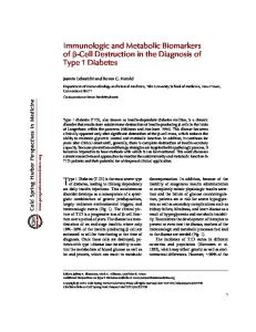
Recent studies have shown that the UPR can modulate the inflammatory response (14, 15). Thus, ER stress enhances interferon- production by macrophages after engagement of toll-like receptors (16), and macrophages facing ongoing ER stress are hyperresponsive to toll-like receptor stimulation, leading to sustained production of proinflammatory cytokines and chemokines (17). In line with these observations, ER stress enhances polyIC (a byproduct of viral infection)-induced innate immunoresponse in dendritic cells (18). Because of the strong inflammation present in pancreatic islets during early T1D (insulitis) (1), the potential connection between ER stress and inflammation is of particular interest, because it could provide a mechanistic link between the growing incidence of obesity and T1D in young individuals. The inflammatory mediators involved in insulitis are produced by the infiltrating immune cells and by the -cells themselves (1). Previous findings by us (1, 19) and others (20) suggest that the transcription factor nuclear factor B (NFB) has a central role in the regulation of -cell inflammatory responses, including synthesis of several chemokines and cytokines (21, 22). NF-B is a family of proteins that are usually associated and retained in the cytosol by the inhibitory protein inhibitor B (IB) (23). Upon stimulation with proinflammatory cytokines or lipopolysaccharide, a cascade of intracellular events leads to the activation of the IB kinase complex that phosphorylates and targets IB to proteasome degradation. NF-B then translocates into the nucleus and modulates the expression of proinflammatory genes (23). Against this background, we have presently studied whether mild ER stress modulates the proinflammatory responses of pancreatic -cells exposed to low doses of the proinflammatory cytokines IL-1 or TNF-␣. We observed that preconditioning -cells FIG. 1. The combination of low-dose CPA and IL-1, but not TNF-␣, enhances NF-B activity. with the ER stressor cyclopiazonic acid A, INS-1E cells were transfected with the NF-B reporter and a pRL-CMV plasmid used as (CPA) or FFA boosted IL-1 but not internal control. Forty-eight hours after transfection, cells were pretreated with DMSO (white TNF-␣-mediated NF-B activation, bars) or CPA (6.25 M, gray bars) for 6 h and then exposed to cytokines (750 U/ml TNF-␣ or 0.5 U/ml IL-1) for 6 h or left untreated (control condition). Luciferase was assayed, and the leading to increased expression of values obtained corrected by the internal control. Results are the mean ⫾ SEM of five NF-B target genes involved in the inindependent experiments; #, P ⬍ 0.05 and ###, P ⬍ 0.001 vs. DMSO (control) and ***, P ⬍ flammatory response, e.g. inducible ni0.001 as indicated; ANOVA. AU, Arbitary unit. B, The same experimental design with CPA or DMSO pretreatment and combination with cytokines (as in A) was used to evaluate mRNA tric oxide synthase (iNOS), Fas, chemoexpression of Fas, CCL2, CXCL1, IB␣, and iNOS by real-time RT-PCR. Values were corrected kine (C-C motif) ligand 2 (CCL2), and by the housekeeping gene GAPDH. Results are the mean ⫾ SEM of four to five independent chemokine (C-X-C motif) ligand 1 experiments; #, P ⬍ 0.05; ##, P ⬍ 0.01; and ###, P ⬍ 0.001 vs. DMSO; ***, P ⬍ 0.001 as indicated, ANOVA. (CXCL1). X-box binding protein 1
The Endocrine Society. Downloaded from press.endocrine.org by [${individualUser.displayName}] on 15 January 2017. at 07:40 For personal use only. No other uses without permission. . All rights reserved.
Endocrinology, July 2012, 153(7):3017–3028
endo.endojournals.org
3019
(XBP1) and IRE1␣, but not CCAAT/enhancer binding protein homologous protein (CHOP) knockdown (KD), prevented this exacerbation. XBP1 KD increased forkhead box O1 (FoxO1) protein expression, and FoxO1 KD further increased NF-B activation and the expression of its target genes. These observations indicate that ER stress potentiates NF-B activation in -cells via the IRE1␣/ XBP1 branch of the ER stress response, providing evidence for cross talk between a specific branch of the UPR and -cell inflammatory responses.
Pancreatic islets were isolated from male Wistar rats (Charles River Laboratories, Brussels, Belgium), treated following the guidelines of the Belgian Regulations for Animal Care and with approval from the local Ethical Committee. Rat islets were dispersed and -cells purified by FACS (FACSAria, BD Bioscience, San Jose, CA) (25, 26). The preparations used contained 93 ⫾ 2% (n ⫽ 9) -cells. -Cells were cultured in Ham’s F-10 medium containing 10 mM glucose, 2 mM glutamine, 50 M 3-isobutylL-methylxanthine, 0.5% fatty acid-free BSA (Roche, Indianapolis, IN), 5% FBS, 50 U/ml penicillin, and 50 g/ml streptomycin. The same medium but without FBS was used during the treatments described below.
Materials and Methods
Cell treatment, NO, CXCL1, and CCL2 measurements
Culture of INS-1E cells and fluorescence-activated cell sorting (FACS)-purified rat -cells The rat insulinoma cell line INS-1E [kindly provided by C. Wollheim (Centre Medical Universitaire, Geneva, Switzerland) and used between passages 60 and 71] was cultured in RPMI 1640 GlutaMAX-I, containing 5% heat-inactivated fetal bovine serum (FBS), 10 mM HEPES, 1 mM Na-pyruvate, and 50 M 2-mercaptoethanol (24).
Cytokine concentrations were selected based on dose-response studies with recombinant murine TNF-␣ (range of 50 – 1000 U/ml; Innogenetics, Ghent, Belgium) or recombinant human IL-1 (range of 0.005–10 U/ml; R&D Systems, Abingdon, UK), aiming to find the lowest dose that induced consistent NF-B stimulation. These are arbitrary concentrations, because there are no available data on the actual cytokine concentrations present in the islets during the early- or preinsulitis period. INS-1E cells and rat primary -cells were pretreated with 6.25
FIG. 2. ER stress potentiates and prolongs expression of IL-1-induced NF-B target genes. A, INS-1E cells were pretreated for 6 h with CPA (6.25 M, full line) or DMSO (dotted line) and then exposed to IL-1 (0.5 U/ml) or left untreated. Cells were collected at the indicated time points (in hours, x-axis), mRNA expression was assayed by real-time RT-PCR for iNOS, CCL2, Fas, IB␣, and CXCL1 and corrected for the housekeeping gene GAPDH. Results are the mean ⫾ SEM of six independent experiments; *, P ⬍ 0.05 and **, P ⬍ 0.01 vs. DMSO plus IL-1 at the respective time points, paired t test. B, INS-1E cells were transfected with iNOS (left) or NF-B (right) reporter together with the pRL-CMV plasmid as internal control. Forty-eight hours after transfection, cells were treated as in A. After 12 h of exposure to IL-1, cells were assayed for luciferase activity; values were corrected by the value of the internal control. Results are the mean ⫾ SEM of three independent experiments; *, P ⬍ 0.05 and ***, P ⬍ 0.001, ANOVA. AU, Arbitrary unit.
The Endocrine Society. Downloaded from press.endocrine.org by [${individualUser.displayName}] on 15 January 2017. at 07:40 For personal use only. No other uses without permission. . All rights reserved.
3020
Miani et al.
ER Stress Favors -Cell Inflammation
M CPA (Sigma-Aldrich, Steinheim, Germany), a concentration previously shown by us in dose-response studies to induce mild ER stress (27). CPA was dissolved in dimethylsulfoxide (DMSO); cells were cultured with CPA or DMSO (0.03%) alone for 6 h and then exposed to IL-1 (0.5 U/ml) or TNF-␣ (750 U/ml). Alternatively, INS-1E cells were preincubated with IL- (0.5 U/ml) for 6 h and then treated with CPA (6.25 M) or DMSO, or exposed to CPA and IL- together for 12 h. We also used 0.06 M thapsigargin (THP) (Sigma, St. Louis, MO) to induce ER stress (28). For FFA exposure, INS-1E cells were cultured in medium containing 0.85% FFA-free BSA (Fraction V; Roche) and 1% FBS. Oleate and palmitate (Sigma-Aldrich) were dissolved in 90% ethanol (29 –32) and used in a 1:1 ratio at a final concentration of 0.6 mM. This FFA mixture induces mild ER
Endocrinology, July 2012, 153(7):3017–3028
stress without triggering -cell apoptosis (29). After 24 h of exposure to the FFA mixture or 1.2% ethanol, INS-1E cells were treated with 0.5 U/ml IL-1 for 6 h. The same experimental design was used to study oleate (0.3 mM) or palmitate (0.3 mM) preconditioning followed by IL-1 treatment. For the experiments with recombinant FasL, INS-1E cells were pretreated with 6.25 M CPA for 6 h, treated with IL-1 for 6 h, and then exposed to recombinant human soluble FasL (0.1 g/ml; Alexis, Läufelfingen, Switzerland) and enhancer (1 g/ml; Alexis) for 18 h. The selected concentrations for FasL and the enhancer induce apoptosis in rat -cells (33). The cytokines and FasL were added to CPA/DMSO or FFA/ethanol-containing medium. In some experiments, the culture medium was collected for nitrite (a surrogate measure of NO production) determination (34), and for CXCL1 (R&D Systems) and CCL2 measurement by ELISA (Invitrogen, Paisley, UK). Apoptosis was determined using the nuclear dyes Hoechst 33342/propidium iodide (25, 35, 36).
mRNA extraction and real-time RT-PCR mRNA was extracted from INS-1E cells and rat primary -cells and reverse transcribed as described (25). Expression of target genes was determined by real-time RT-PCR using SYBR Green (25, 37) and comparison with a standard curve (38). Expression values were corrected by the housekeeping gene glyceraldehyde-3-phosphate dehydrogenase (GAPDH) and normalized by the highest value of each experiment considered as 1. GAPDH expression is not modified under the present experimental conditions (21, 28). Primer sequences are described in Supplemental Table 1. XBP1 spliced (XBP1s) expression was evaluated by real-time RT-PCR as previously described (39, 40).
RNA interference
FIG. 3. FFA or THP up-regulate IL-1-induced NF-B activation in -cells. INS-1E cells were transfected with the NF-B reporter and a pRL-CMV as internal control. A, After 48 h, cells were pretreated with DMSO or THP (0.06 M) or CPA (6.25 M) for 6 h and then exposed to IL-1 (0.5 U/ml) for 16 h. Luciferase activity was assayed, and the values obtained were corrected by the internal control. Results are the mean ⫾ SEM of three independent experiments; #, P ⬍ 0.05; ##, P ⬍ 0.01; and ###, P ⬍ 0.001 vs. DMSO; §§§, P ⬍ 0.001 vs. THP; $$$, P ⬍ 0.001 vs. CPA; ***, P ⬍ 0.001 as indicated, ANOVA. B, Cells were exposed to oleate plus palmitate 1:1 (0.6 mM) or ethanol 1.2% (Et) for 24 h and then treated with IL-1 (0.5 U/ml) for 6 h. Luciferase activity was assayed, and the values obtained were corrected by the internal control. C, The same experimental conditions as in B were applied, cells were harvested, and mRNA expression was assayed for iNOS, CCL2, CXCL1, and Fas and corrected by the housekeeping gene GAPDH. Results are the mean ⫾ SEM of four independent experiments; #, P ⬍ 0.05 and ###, P ⬍ 0.001 vs. Et; **, P ⬍ 0.01 and ***, P ⬍ 0.001 as indicated, ANOVA. AU, Arbitrary unit.
Specific small interfering RNA (siRNA) were used for the silencing of CHOP, IRE1␣, XBP1, and FoxO1 (Supplemental Table 2). We have previously shown (40) and confirmed here that similar biological effects are observed after knocking down XBP1 with another siRNA. Allstars Negative Control siRNA (QIAGEN, Venlo, The Netherlands) was used as negative control. -Cells were transfected overnight with 30 nM of siRNA mixed with Lipofectamine RNAiMAX (Invitrogen, Carlsbad, CA) (41). The negative control siRNA (siControl) does not affect -cell gene expression or insulin release, compared with nontransfected cells (41, 42). Cells transfected with siRNA were used for experiments 24 – 48 h after transfection.
Promoter reporter assay INS-1E cells were transfected using Lipofectamine 2000 (Invitrogen). The constructs used were the pRL-CMV (Promega, Madison, WI), as an internal control, and the pNF-B-Luciferase (BD Biosciences, Palo Alto, CA), the piNOS-1002 luc (35, 43) or the pFas-811 luc (44). Forty-eight hours after transfection, cells were treated with TNF-␣ or
The Endocrine Society. Downloaded from press.endocrine.org by [${individualUser.displayName}] on 15 January 2017. at 07:40 For personal use only. No other uses without permission. . All rights reserved.
Endocrinology, July 2012, 153(7):3017–3028
IL-1 for 6 h. Alternatively, cells were exposed to CPA or THP or to the combination of oleate plus palmitate for 6 or 24 h, and then IL-1 was added for 12 or 16 h. Luciferase activities were studied with the Dual-Luciferase Reporter Assay System (Promega) (35). The values obtained were corrected for the luciferase values of the internal control plasmid pRL-CMV and normalized by the highest value of each experiment considered as 1.
endo.endojournals.org
3021
antimouse IgG was used as secondary antibody (Lucron Bioproducts, De Pinte, Belgium). Immunoreactive bands were revealed using the SuperSignal West Femto chemiluminescent substrate (Thermo Scientific, Rockford, IL), detected using a ChemiDoc XRS⫹ and quantified with the Image Lab software (Bio-Rad, Hercules, CA).
Statistical analysis Immunofluorescence and Western blotting INS-1E cells were cultured in polylysine-coated glass slides (BD Biosciences). Cells were fixed with 4% paraformaldehyde and permeabilized with 70% acetone-30% methanol. Slides were incubated for 1 h with goat serum, followed by overnight incubation at 4 C with rabbit anti-p65 (Santa Cruz Biotechnology, Inc., Santa Cruz, CA) as described (42). p65⫹ nuclei were evaluated as the hallmark of NF-B activation (36). Results are from at least 500 cells per experimental condition and expressed in percentage of positive cells/total cells. For Western blotting, cells were treated as described (45). Primary antibodies against FoxO1 (Cell Signaling, Danvers, MA) and ␣-tubulin (Sigma), as control for protein loading, were used. Horseradish peroxidase-conjugated donkey antirabbit or
Data are expressed as mean ⫾ SEM. Comparisons were performed by two-tailed paired Student’s t test or by ANOVA followed by Student’s t test with Bonferroni correction, as indicated. A P value of less than 0.05 was considered as significant.
Results Low doses of cytokines induce NF-B activation and p65 nuclear translocation A dose response with TNF-␣ and IL-1 was done to determine the threshold for NF-B activation (Supplemental Fig. 2A), based on which the concentrations of 750 U/ml TNF-␣ and 0.5 U/ml IL-1 were chosen for subsequent experiments. The promoter reporter studies (Supplemental Fig. 2A) were confirmed by immunofluorescence for the nuclear translocation of p65, the main cytokineinduced NF-B family member in -cells (35). The selected doses were compared with higher cytokine concentrations used in previous studies (22, 35). Both TNF-␣ at 750 U/ml and IL-1 at 0.5 U/ml induced clear NF-B translocation to the nucleus (Supplemental Fig. 2B), although with somewhat lower intensity than 1000 U/ml TNF-␣ and 10 U/ml IL-1.
FIG. 4. CPA enhances IL-1-induced iNOS, CCL2, CXCL1, and Fas mRNA expression and NO production in FACS-purified primary -cells. A, Rat -cells were pretreated with CPA (6.25 M) or DMSO for 6 h and then exposed or not to IL-1 (0.5 U/ml) for the time points indicated on the x-axis. Cells were collected, mRNA expression was assayed for iNOS, CCL2, Fas, and CXCL1 and corrected for the housekeeping gene GAPDH. Results are the mean ⫾ SEM of five independent experiments; #, P ⬍ 0.05; ##, P ⬍ 0.01; and ###, P ⬍ 0.001 vs. DMSO; *, P ⬍ 0.05 and **, P ⬍ 0.01 as indicated, ANOVA. B, The same protocol as in A was used, but -cells were exposed to IL-1 or not for 24 h. The medium was collected and assayed for nitrite. Results are the mean ⫾ SEM of four independent experiments; **, P ⬍ 0.01 as indicated, paired t test.
Pretreatment with CPA sensitizes INS-1E cells and primary -cells to IL-1-induced NF-B activation and expression of proinflammatory genes The sarcoplasmic/ER Ca2⫹-ATPase blocker CPA was used to induce ER stress in -cells. The low dose of CPA (6.25 M) selected induces UPR markers but has limited impact on cell viability (27). Both TNF-␣ and IL-1 activated the NF-B reporter (Fig. 1A). CPA induced a mild increase in reporter activity and potentiated IL-1-induced, but not TNF-␣-induced, NF-B activation (Fig. 1A). Preexposure with IL-1 followed by CPA treatment (Supplemental Fig. 3A) or a coadministration of CPA plus IL-1 (Supplemental Fig. 3B) also enhanced NF-B activation. We next quantified the mRNA expression of five well-known
The Endocrine Society. Downloaded from press.endocrine.org by [${individualUser.displayName}] on 15 January 2017. at 07:40 For personal use only. No other uses without permission. . All rights reserved.
3022
Miani et al.
ER Stress Favors -Cell Inflammation
Endocrinology, July 2012, 153(7):3017–3028
NF-B target genes in -cells, i.e. iNOS (43), CCL2 (46), CXCL1 (22), Fas (44), and IB␣ (35). In keeping with the reporter assay (Fig. 1A), CPA pretreatment synergized with IL-1 but not with TNF-␣ (Fig. 1B). This cross talk between CPA-induced ER stress and IL-1-induced
NF-B activation resulted in a marked increase in iNOS, CCL2, CXCL1, and Fas mRNA expression, whereas no change was observed for IB␣, which inhibits NF-B activation (Fig. 1B). The expression of another gene implicated in the negative feedback regulation of the NF-B pathway, namely A20, was not potentiated by CPA plus IL-1 compared with IL-1 alone (data not shown). Because CPA did not augment expression of TNF-␣-induced NF-B-related genes, subsequent experiments were performed using the combination CPA-IL-1. CPA induced expression of UPR genes downstream of the three main UPR pathways, namely CHOP, BiP, and XBP1s (Supplemental Fig. 4). Expression of these genes was only mildly (in the case of CHOP) or not modified by the subsequent addition of IL-1 or TNF-␣ (Supplemental Fig. 4). A time-course analysis (Fig. 2A) confirmed that CPA pretreatment favors a higher IL-1-induced expression of cytokines and chemokines at 6 h and leads to prolonged expression of CCL2, iNOS, CXCL1, and IB␣ up to 12–24 h. Use of reporter constructs containing the rat iNOS promoter region from nucleotide ⫺1002 to ⫹132 (piNOS1002luc) (43) or six NF-B binding sites coupled to luciferase (35) showed a basal CPA-induced promoter activity and 2-fold augmentation of the stimulatory effect of IL-1 (Fig. 2B), confirming regulation at the transcriptional level. We next evaluated whether other chemical or physiological ER stressors, e.g. THP and the equimolar combinaFIG. 5. XBP1s KD prevents induction of iNOS, CCL2, CXCL1, and Fas expression. INS-1E cells tion of oleate and palmitate, or the two were transfected with siXBP1 or with siControl. A, After 48 h, cells were treated with CPA FFA alone (29), also favored IL-1-in(6.25 M) or DMSO for 6 h and then exposed or not to IL-1 (0.5 U/ml) for 6 h. Cells were collected, and mRNA expression was assayed for XBP1s, iNOS, Fas, CCL2, and CXCL1 and duced NF-B activation. Oleate plus corrected for the housekeeping gene GAPDH. Results are the mean ⫾ SEM of three palmitate induces a mild ER stress withindependent experiments; ##, P ⬍ 0.01 and ###, P ⬍ 0.001 vs. siControl DMSO; **, P ⬍ out -cell apoptosis (29), a finding con0.01 and ***, P ⬍ 0.001 as indicated, ANOVA. B, Twenty-four hours after control or XBP1 KD, INS-1E cells were transfected with iNOS, Fas, or NF-B luciferase reporters together with firmed in the present series of experithe pRL-CMV plasmid used as internal control. Twenty-four hours later, cells were treated ments (see below). Pretreatment with with CPA (6.25 M) or DMSO for 6 h and then exposed or not to IL-1 (0.5 U/ml) for 16 h. THP, oleate plus palmitate, or oleate Luciferase activity was assayed and corrected by the internal control; ###, P ⬍ 0.001 vs. siControl DMSO; **, P ⬍ 0.01 and ***, P ⬍ 0.001 as indicated, ANOVA. AU, Arbitrary unit. and palmitate separately increased by C, In the same experimental conditions as in B, p65 translocation to the nucleus was evaluated; 2-fold IL-1-induced NF-B activaHoechst was used as a nuclear marker. D, The p65⫹ nuclei observed in C were quantified and tion, confirming the observation with expressed as percentage of total cells. Results are the mean ⫾ SEM of three independent experiments; ###, P ⬍ 0.001 vs. siControl DMSO and ***, P ⬍ 0.001 as indicated, ANOVA. CPA (Fig. 3, A and B, and Supplemental
The Endocrine Society. Downloaded from press.endocrine.org by [${individualUser.displayName}] on 15 January 2017. at 07:40 For personal use only. No other uses without permission. . All rights reserved.
Endocrinology, July 2012, 153(7):3017–3028
Fig. 5A, respectively). Furthermore, the FFA markedly amplified IL-1-induced iNOS, CCL2, and CXCL1 expression (Fig. 3C). Oleate and palmitate alone increased IL-1-induced activation and augmented expression of some NF-B-regulated genes (Supplemental Fig. 5B) but to a lesser extent than the two FFA together (Fig. 3C). We confirmed that the presently used concentrations of FFA induced the ER stress markers CHOP, BiP, and XBP1s (Supplemental Fig. 6). CPA-induced exacerbation of the proinflammatory effects of IL-1 was also observed in FACS-purified primary -cells (Fig. 4A). Thus, preculture with CPA augmented IL-1-induced CCL2, CXCL1, and iNOS expression. There was also a significantly increased NO production, measured as medium nitrite accumulation, indicating that iNOS mRNA induction by CPA is functional (Fig. 4B). This was further evaluated in INS-1E cells by measuring NO and the chemokines CXCL1 and CCL2 (Supplemental Fig. 7, A–C). CPA increased by 2-fold the IL-1 effect, confirming the mRNA observations. The CPA plus IL-1-induced increase in Fas mRNA expression in INS-1E cells (Fig. 1B) was paralleled by a
endo.endojournals.org
3023
nearly 2-fold increase in apoptosis, compared with IL-1 or CPA alone, when -cells were exposed to recombinant FasL (Supplemental Fig. 7D), indicating that the overexpressed Fas is functional.
XBP1s, but not CHOP, mediates the exacerbation of the NF-B-mediated proinflammatory response The above-described experiments identified a cross talk between ER stress and NF-B pathways in -cells. We next examined which of the ER stress-related signals mediate this cross talk. Previous reports showed that the ER stress-induced transcription factor CHOP modulates cytokine expression (47). CPA, and especially CPA plus IL-1, induced CHOP expression in INS-1E cells (Supplemental Fig. 4A). This induction was largely prevented by a specific siCHOP (Supplemental Fig. 8), but the KD failed to prevent CPA plus IL-1-induced expression of iNOS, CCL2, CXCL1, and Fas. Induction of these genes by CPA plus IL-1 was identical when comparing cells transfected with siControl or siCHOP (Supplemental Fig. 8), indicating that CHOP up-regulation does not mediate the proinflammatory effects of CPA-induced ER stress. Another branch of the ER stress response that has been implicated in the regulation of inflammatory cytokines/ chemokines (16) and in the activation of innate immunity (17, 48) is the IRE1␣/XBP1 pathway. CPA activated this pathway, as evidenced by XBP1 splicing (Supplemental Fig. 4C). We next transfected INS-1E cells with a siControl or a previously validated siRNA against XBP1 (siXBP1) (40). siXBP1, but not siControl, decreased basal and CPA-induced XBP1s mRNA expression by more than 70% (Fig. 5A). siXBP1 prevented CPA plus IL-1mediated enhancement of iNOS, CCL2, CXCL1, and Fas expression in INS-1E cells (Fig. 5A). These results were confirmed in INS-1E cells by the use of a second and previously validated siRNA (40) that targets a different region of XBP1 (Supplemental Fig. 9). To confirm the results obtained at FIG. 6. IRE1␣ KD prevents induction of iNOS, CCL2, CXCL1, and Fas expression. INS-1E cells the mRNA level, we examined the prowere transfected with siRNA against IRE1␣ or with siControl. After 48 h, cells were treated with CPA (6.25 M) or DMSO for 6 h and then exposed or not to IL-1 (0.5 U/ml) for 6 h. moter activity of iNOS (see above) and Cells were collected, and mRNA expression was assayed for IRE1␣, XBP1s, iNOS, Fas, CCL2, Fas promoter reporters, both containand CXCL1 and corrected for the housekeeping gene GAPDH. Results are the mean ⫾ SEM of ing NF-B-responsive elements (44). three independent experiments; #, P ⬍ 0.05; ##, P ⬍ 0.01; and ###, P ⬍ 0.001 vs. siControl XBP1 KD prevented CPA plus IL-1DMSO; *, P ⬍ 0.05; **, P ⬍ 0.01; and ***, P ⬍ 0.001 as indicated, ANOVA.
The Endocrine Society. Downloaded from press.endocrine.org by [${individualUser.displayName}] on 15 January 2017. at 07:40 For personal use only. No other uses without permission. . All rights reserved.
3024
Miani et al.
ER Stress Favors -Cell Inflammation
Endocrinology, July 2012, 153(7):3017–3028
prevented CPA plus IL-1-induced expression of iNOS, CCL2, CXCL1, and Fas (Fig. 6). XBP1s-mediated FoxO1 inhibition potentiates IL-1-induced NF-B activation The above-described experiments suggest that ER stress cross talks with the NF-B pathway via XBP1s. Previous findings in mouse embryonic fibroblasts indicate that XBP1s directly binds to FoxO1 and targets it for proteasome degradation (49). Because FoxO1 may inhibit NF-B activation (50), we next studied FoxO1 expression in INS-1E cells transfected with XBP1 or siControl and then treated as in Fig. 5A (Supplemental Fig. 10). XBP1 KD alone increased by 2-fold FoxO1 protein expression (Supplemental Fig. 10A) without changing its mRNA expression (Supplemental Fig. 10B). We next investigated whether FoxO1 has a role in the NF-B-mediated proinflammatory response of -cells exposed to CPA plus IL-1. FIG. 7. FoxO1 KD exacerbates the induction of iNOS, CCL2, and Fas expression. A, INS-1E FoxO1 KD sensitized the cells to the cells were transfected with siRNA against FoxO1 or with siControl. After 24 h, cells were IL-1 treatment, inducing an activation transfected with NF-B luciferase reporter together with the pRL-CMV plasmid used as of the NF-B reporter comparable with internal control. Twenty-four hours later, cells were treated with CPA (6.25 M) or DMSO for the one observed after CPA plus IL-1 6 h and then exposed or not to IL-1 (0.5 U/ml) for 16 h. Luciferase activity was assayed and corrected by the internal control; ###, P ⬍ 0.001 vs. siControl DMSO; *, P ⬍ 0.05 and **, in the presence of the siControl (Fig. P ⬍ 0.01 as indicated; ANOVA. AU, Arbitrary unit. B, Control or FoxO1 KD cells were treated 7A). Pretreatment with CPA further inwith CPA (6.25 M) or DMSO for 6 h and then exposed or not to IL-1 (0.5 U/ml) for 6 h. creased NF-B activity when FoxO1 Cells were harvested and the mRNA expression was assayed for FoxO1, iNOS, CCL2, and Fas and corrected by the housekeeping gene GAPDH. Results are the mean ⫾ SEM of four was silenced. siFoxO1 decreased independent experiments; #, P ⬍ 0.05 and ###, P ⬍ 0.001 vs. siControl DMSO; **, P ⬍ 0.01 FoxO1 mRNA expression by more and ***, P ⬍ 0.001 as indicated, ANOVA. than 60% (Fig. 7B). FoxO1 KD further increased iNOS, CCL2, and Fas mRNA induced exacerbation of both promoter activities (Fig. expression when cells were treated with 5B). To evaluate whether XBP1 directly modulates CPA plus IL-1, confirming its role as a negative regulator NF-B activation, we performed a reporter assay with of the NF-B pathway in -cells. the previously described NF-B construct (35) and imThese converging findings suggest that XBP1s augmunofluorescent staining to identify p65 translocation ments the activation of the NF-B pathway and conseinto the nucleus. siXBP1, but not siControl, blocked the quent increase in the expression of proinflammatory genes exacerbation of NF-B activation, as evaluated by the at least in part via FoxO1 inhibition in pancreatic -cells. NF-B reporter (Fig. 5B, bottom panel) and nuclear translocation into the nucleus, as shown by a representative picture (Fig. 5C) and quantification of p65⫹ nu- Discussion clei (Fig. 5D). Activation of XBP1 requires splicing by IRE1␣ (12). As In the present study, we evaluated the pancreatic -cell expected, KD of IRE1␣ decreased both IRE1␣ and XBP1s response to low-dose proinflammatory cytokines after expression (Fig. 6). Similar to siXBP1 (Fig. 5A), siIRE1␣ preconditioning with mild ER stress. ER stress enhances
The Endocrine Society. Downloaded from press.endocrine.org by [${individualUser.displayName}] on 15 January 2017. at 07:40 For personal use only. No other uses without permission. . All rights reserved.
Endocrinology, July 2012, 153(7):3017–3028
the proinflammatory effect of IL-1 but not TNF-␣. This effect is mediated by the IRE1␣/XBP1s branch of the UPR via modulation of FoxO1 and the key proinflammatory transcription factor NF-B. It is thought that in T1D, a succession of environmental factors hits the -cells of genetically susceptible individuals, causing local pancreatic islet inflammation and eventually apoptosis (1, 2, 7). The exact chronology and nature of events taking place in the period preceding clinical diagnosis of T1D is not known. The slow progression of the disease suggests that the triggers and/or boosters are probably mild and subclinical events, generating a chronic proinflammatory and hostile environment for pancreatic -cells (1–3). The ER stress and the NF-B pathways have been suggested to act alone (12, 36) or in combination (14, 15) in the pathogenesis of T1D and other autoimmune diseases. Macrophages and dendritic cells preconditioned with mild ER stress hyperrespond to inflammatory signals (51), exacerbating the expression of genes and proteins involved in the innate immunoresponse (16, 17). On the other hand, a low grade ER stress may evoke a physiological oxidative (or other) stress response in some cell types that preadapts them to cope with a later oxidative stress, ER stress or inflammatory assault (52). Therefore, it could be that the observed inflammatory response is part of an adaptive response to the low-grade chronic ER stress. CPA was used in the present study to induce a mild and “pure” ER stress based on previous findings from our group (27). The observations made with CPA were confirmed by -cell exposure to the pathophysiologically relevant stimulus of oleate and/or palmitate (12). The present results indicate that sensitizing -cells with ER stress inflates their response to a subsequent mild proinflammatory cytokine exposure. Indeed, iNOS, Fas, CCL2, and CXCL1 expression were enhanced and prolonged over time in INS-1E cells and primary -cells treated with IL-1. Increased expression of iNOS and Fas sensitizes -cells to apoptosis (53), whereas the chemokines CCL2 and CXCL1 contribute to the attraction of mononuclear cells involved in the build up of insulitis (1). Interestingly, there was no increase in the expression of genes involved in the negative feedback regulation of NF-B, namely IB␣ and A20. IB␣ and A20 are activated at early time points after cytokine exposure, whereas other genes, such as the chemokines CCL2 and CCL5, are up-regulated with higher doses and at later time points (54 –56). Thus, our results indicate that CPA plus IL-1 treatment preferentially increases the expression of “late genes” involved in the proinflammatory response rather than the “early genes” contributing for the negative feedback loop. The
endo.endojournals.org
3025
mechanisms involved in these differential effects of ER stress remain to be clarified, but the net result will be an augmented and prolonged proinflammatory signal. There was no enhancement in NF-B activity or its downstream genes in -cells treated with CPA and TNF-␣, suggesting that the proinflammatory effects of ER stress in -cells are context dependent. According to our previous results in -cells, TNF-␣ induces NF-B but to a lesser extent than IL-1 (35). In the present experiments, this difference was not an issue, because the cytokine concentrations were selected to allow similar activation of NFB; this required much higher concentrations of TNF-␣ (750 vs. 0.5 U/ml IL-1). Because ER stress inhibits protein translation (12), an explanation for the weaker induction of the NF-B pathway by TNF-␣ could be a decrease in the expression of TNF-receptor 1. We did not observe, however, any inhibitory effect of CPA on the TNF-receptor 1 protein expression (data not shown). Thus, the observed difference between the impact of the UPR on TNF-␣- and IL-1-induced NF-B activation must lay in other intracellular events that remain to be clarified. Previous findings in other cell types suggest that the synergistic effect of ER stress and proinflammatory me-
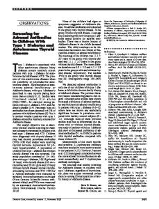
FIG. 8. Proposed model for NF-B potentiation by ER stress in -cells. Metabolic disorders, such as obesity, cause mild chronic ER stress in -cells, triggering the three UPR pathways, including the IRE1␣/XBP1 branch. Increased XBP1s sensitizes -cells to a “second hit” by IL-1, putatively produced by macrophages in response to a viral infection or other “danger ligands.” This leads to XBP1s-induced FoxO1 degradation, augmented NF-B activation, and increased local production of cytokines and chemokines, worsening inflammation (insulitis) and -cell loss.
The Endocrine Society. Downloaded from press.endocrine.org by [${individualUser.displayName}] on 15 January 2017. at 07:40 For personal use only. No other uses without permission. . All rights reserved.
3026
Miani et al.
ER Stress Favors -Cell Inflammation
diators is at least in part dependent on the splicing of XBP1 by IRE1␣ (16 –18). The outcome of this cross talk is a maximization of the innate immunoresponse to pathogens, including enhanced TNF-␣, IL-6, and interferon- expression (16 –18). Interestingly, the IRE1␣/ XBP1 pathway is activated by obesity (57), an environmental factor potentially related to T1D (10, 58). Here, we have shown that knocking down IRE1␣ or XBP1s in -cells prevents the boost in the production of chemokines (CCL2 and CXCL1) and other proinflammatory agents induced by ER stress. Studies in macrophages indicate that XBP1s directly binds to the promoter region of some immune-related genes, enhancing their expression (16, 17). Moreover, XBP1s inhibits FoxO1 (49), and FoxO1 negatively regulates the NF-B pathway (50). Our findings suggest that the ER stress-induced exacerbation of the proinflammatory response is mediated at least in part by XBP1s inhibition of the NF-B negative regulator FoxO1. Indeed, FoxO1 silencing further increases NF-B activation after CPA plus IL-1. Of note, Martinez et al. (59) found that FoxO1 contributes to fatty acid-induced clonal mouse -cell apoptosis, suggesting that FoxO1 may modulate different aspects of the response of -cells to ER stress depending on species and experimental context. We propose in Fig. 8 an overview of our main findings. Metabolic stimuli, such as obesity and insulin resistance, induce chronic ER stress in -cells, with the consequent activation of XBP1s through the IRE1␣ branch of the UPR (12, 57). This preexistent ER stress, even if mild, sensitizes -cells to low concentrations of IL-1, which may be locally released by macrophages due to viral infection or exposure to other “danger signals” (1). This will cause a more intense and protracted local production of chemokines and other inflammatory mediators via the transcription factor NF-B. The intersection between the UPR and NF-B pathways may play a relevant role in the early stages of the insulitis, aggravating and prolonging this inflammation in obese and insulin-resistant children and accelerating the development of T1D.
Acknowledgments We thank the personnel from Laboratory of Experimental Medicine, A. Musuaya, R. Makhnas, M. Pangerl, and S. Mertens, for excellent technical support. D.L.E. is the guarantor of this work, had full access to all the data, and takes full responsibility for the integrity of data and the accuracy of data analysis. Address all correspondence and requests for reprints to: Dr. Decio L. Eizirik, Laboratory of Experimental Medicine, Univer-
Endocrinology, July 2012, 153(7):3017–3028
sité Libre de Bruxelles, Route de Lennik, 808 CP618, B-1070 Brussels, Belgium. E-mail:
[email protected]. This work was supported by grants from the Fonds National de la Recherche Scientifique Belgium, the Communauté Française de Belgique-Actions de Recherche Concertées, and the European Union (projects Naimit and 〉etaBat, in the Framework Program 7 of the European Community); by the Expert Center Grant 2008.40.001 from the Dutch Diabetes Research Foundation; and by the Belgium Program on Interuniversity Poles of Attraction initiated by the Belgium State (IUAP P6/40). M.L.C. was the recipient of a scholarship from Brazilian Coordination for the Improvement of Higher Education Personnel. Disclosure Summary: The authors have nothing to disclose.
References 1. Eizirik DL, Colli ML, Ortis F 2009 The role of inflammation in insulitis and -cell loss in type 1 diabetes. Nat Rev Endocrinol 5:219 –226 2. Knip M, Veijola R, Virtanen SM, Hyoty H, Vaarala O, Akerblom HK 2005 Environmental triggers and determinants of type 1 diabetes. Diabetes 54(Suppl 2):S125–S136 3. Pozzilli P, Strollo R, Barchetta I 2009 Natural history and immunopathogenesis of type 1 diabetes. Endocrinol Nutr 56(Suppl 4): 50 –52 4. Patterson CC, Dahlquist GG, Gyürüs E, Green A, Soltész G 2009 Incidence trends for childhood type 1 diabetes in Europe during 1989 –2003 and predicted new cases 2005–20: a multicentre prospective registration study. Lancet 373:2027–2033 5. Soltesz G, Patterson CC, Dahlquist G 2007 Worldwide childhood type 1 diabetes incidence–what can we learn from epidemiology? Pediatr Diabetes 8(Suppl 6):6 –14 6. Vehik K, Hamman RF, Lezotte D, Norris JM, Klingensmith G, Bloch C, Rewers M, Dabelea D 2007 Increasing incidence of type 1 diabetes in 0- to 17-year-old Colorado youth. Diabetes Care 30: 503–509 7. van Belle TL, Coppieters KT, von Herrath MG 2011 Type 1 diabetes: etiology, immunology, and therapeutic strategies. Physiol Rev 91:79 –118 8. Vehik K, Hamman RF, Lezotte D, Norris JM, Klingensmith GJ, Dabelea D 2009 Childhood growth and age at diagnosis with type 1 diabetes in Colorado young people. Diabet Med 26:961–967 9. Hotamisligil GS 2010 Endoplasmic reticulum stress and the inflammatory basis of metabolic disease. Cell 140:900 –917 10. Viner RM, Hindmarsh PC, Taylor B, Cole TJ 2008 Childhood body mass index (BMI), breastfeeding and risk of type 1 diabetes: findings from a longitudinal national birth cohort. Diabet Med 25:1056 – 1061 11. Carlsson A, Kockum I, Lindblad B, Engleson L, Nilsson A, Forsander G, Karlsson AK, Kernell A, Ludvigsson J, Marcus C, Zachrisson I, Ivarsson SA, Lernmark A 20 June 2011 Low risk HLA-DQ and increased body mass index in newly diagnosed type 1 diabetes children in the better diabetes diagnosis study in Sweden. Int J Obes 10.1038/ijo.2011.122 12. Eizirik DL, Cardozo AK, Cnop M 2008 The role for endoplasmic reticulum stress in diabetes mellitus. Endocr Rev 29:42– 61 13. Eizirik DL, Cnop M 2010 ER stress in pancreatic -cells: the thin red line between adaptation and failure. Sci Signal 3:pe7 14. Kitamura M 2011 Control of NF-B and inflammation by the unfolded protein response. Int Rev Immunol 30:4 –15 15. Martinon F, Glimcher LH 2011 Regulation of innate immunity by
The Endocrine Society. Downloaded from press.endocrine.org by [${individualUser.displayName}] on 15 January 2017. at 07:40 For personal use only. No other uses without permission. . All rights reserved.
Endocrinology, July 2012, 153(7):3017–3028
16.
17.
18.
19.
20. 21.
22.
23. 24.
25.
26.
27.
28.
29.
30.
31.
32.
33.
signaling pathways emerging from the endoplasmic reticulum. Curr Opin Immunol 23:35– 40 Smith JA, Turner MJ, DeLay ML, Klenk EI, Sowders DP, Colbert RA 2008 Endoplasmic reticulum stress and the unfolded protein response are linked to synergistic IFN- induction via X-box binding protein 1. Eur J Immunol 38:1194 –1203 Martinon F, Chen X, Lee AH, Glimcher LH 2010 TLR activation of the transcription factor XBP1 regulates innate immune responses in macrophages. Nat Immunol 11:411– 418 Hu F, Yu X, Wang H, Zuo D, Guo C, Yi H, Tirosh B, Subjeck JR, Qiu X, Wang XY 2011 ER stress and its regulator X-box-binding protein-1 enhance polyIC-induced innate immune response in dendritic cells. Eur J Immunol 41:1086 –1097 Cnop M, Welsh N, Jonas JC, Jorns A, Lenzen S, Eizirik DL 2005 Mechanisms of pancreatic -cell death in type 1 and type 2 diabetes: many differences, few similarities. Diabetes 54(Suppl 2):S97–S107 Melloul D 2008 Role of NF-B in -cell death. Biochem Soc Trans 36:334 –339 Cardozo AK, Kruhøffer M, Leeman R, Orntoft T, Eizirik DL 2001 Identification of novel cytokine-induced genes in pancreatic -cells by high-density oligonucleotide arrays. Diabetes 50:909 –920 Ortis F, Naamane N, Flamez D, Ladrière L, Moore F, Cunha DA, Colli ML, Thykjaer T, Thorsen K, Orntoft TF, Eizirik DL 2010 Cytokines IL-1 and TNF-␣ regulate different transcriptional and alternative splicing networks in primary -cells. Diabetes 59:358 – 374 Hayden MS, Ghosh S 2004 Signaling to NF-B. Genes Dev 18: 2195–2224 Asfari M, Janjic D, Meda P, Li G, Halban PA, Wollheim CB 1992 Establishment of 2-mercaptoethanol-dependent differentiated insulin-secreting cell lines. Endocrinology 130:167–178 Rasschaert J, Ladrière L, Urbain M, Dogusan Z, Katabua B, Sato S, Akira S, Gysemans C, Mathieu C, Eizirik DL 2005 Toll-like receptor 3 and STAT-1 contribute to double-stranded RNA ⫹ IFN-␥-induced apoptosis in primary pancreatic -cells. J Biol Chem 280: 33984 –33991 Pipeleers DG, in’t Veld PA, Van de Winkel M, Maes E, Schuit FC, Gepts W 1985 A new in vitro model for the study of pancreatic ␣ and  cells. Endocrinology 117:806 – 816 Pirot P, Eizirik DL, Cardozo AK 2006 IFN-␥ potentiates endoplasmic reticulum stress-induced death by reducing pancreatic -cell defence mechanisms. Diabetologia 49:1229 –1236 Cardozo AK, Ortis F, Storling J, Feng YM, Rasschaert J, Tonnesen M, Van Eylen F, Mandrup-Poulsen T, Herchuelz A, Eizirik DL 2005 Cytokines downregulate the sarcoendoplasmic reticulum pump Ca2⫹ ATPase 2b and deplete endoplasmic reticulum Ca2⫹, leading to induction of endoplasmic reticulum stress in pancreatic -cells. Diabetes 54:452– 461 Cunha DA, Hekerman P, Ladrière L, Bazarra-Castro A, Ortis F, Wakeham MC, Moore F, Rasschaert J, Cardozo AK, Bellomo E, Overbergh L, Mathieu C, Lupi R, Hai T, Herchuelz A, Marchetti P, Rutter GA, Eizirik DL, Cnop M 2008 Initiation and execution of lipotoxic ER stress in pancreatic -cells. J Cell Sci 121:2308 –2318 Cnop M, Hannaert JC, Hoorens A, Eizirik DL, Pipeleers DG 2001 Inverse relationship between cytotoxicity of free fatty acids in pancreatic islet cells and cellular triglyceride accumulation. Diabetes 50:1771–1777 Kharroubi I, Ladrière L, Cardozo AK, Dogusan Z, Cnop M, Eizirik DL 2004 Free fatty acids and cytokines induce pancreatic -cell apoptosis by different mechanisms: role of NF-B and endoplasmic reticulum stress. Endocrinology 145:5087–5096 Zhou YP, Grill VE 1994 Long-term exposure of rat pancreatic islets to fatty acids inhibits glucose-induced insulin secretion and biosynthesis through a glucose fatty acid cycle. J Clin Invest 93:870 – 876 Liu D, Cardozo AK, Darville MI, Eizirik DL 2002 Double-stranded RNA cooperates with IFN-␥ and IL-1 to induce both chemokine expression and NF-B-dependent apoptosis in pancreatic -cells:
endo.endojournals.org
34.
35.
36.
37.
38.
39.
40.
41.
42.
43.
44.
45.
46.
47.
48.
49.
50.
51.
3027
potential mechanisms for viral-induced insulitis and -cell death in type 1 diabetes mellitus. Endocrinology 143:1225–1234 Green LC, Wagner DA, Glogowski J, Skipper PL, Wishnok JS, Tannenbaum SR 1982 Analysis of nitrate, nitrite, and nitrate in biological fluids. Anal Biochem 126:131–138 Ortis F, Cardozo AK, Crispim D, Störling J, Mandrup-Poulsen T, Eizirik DL 2006 Cytokine-induced pro-apoptotic gene expression in insulin-producing cells is related to rapid, sustained and non-oscillatory NF-B activation. Mol Endocrinol 20:1867–1879 Ortis F, Pirot P, Naamane N, Kreins AY, Rasschaert J, Moore F, Théâtre E, Verhaeghe C, Magnusson NE, Chariot A, Orntoft TF, Eizirik DL 2008 TNF-␣ and IL-1 induction of NF-B and its downstream genes has a pro-apoptotic role in pancreatic -cells. Diabetologia 51:1213–1225 Chen MC, Proost P, Gysemans C, Mathieu C, Eizirik DL 2001 Monocyte chemoattractant protein-1 is expressed in pancreatic islets from prediabetic NOD mice and in IL-1-exposed human and rat islet cells. Diabetologia 44:325–332 Overbergh L, Valckx D, Waer M, Mathieu C 1999 Quantification of murine cytokine mRNAs using real time quantitative reverse transcriptase PCR. Cytokine 11:305–312 Marchetti P, Bugliani M, Lupi R, Marselli L, Masini M, Boggi U, Filipponi F, Weir GC, Eizirik DL, Cnop M 2007 The endoplasmic reticulum in pancreatic  cells of type 2 diabetes patients. Diabetologia 50:2486 –2494 Allagnat F, Christulia F, Ortis F, Pirot P, Lortz S, Lenzen S, Eizirik DL, Cardozo AK 2010 Sustained production of spliced X-box binding protein 1 (XBP1) induces pancreatic -cell dysfunction and apoptosis. Diabetologia 53:1120 –1130 Moore F, Naamane N, Colli ML, Bouckenooghe T, Ortis F, Gurzov EN, Igoillo-Esteve M, Mathieu C, Bontempi G, Thykjaer T, Ørntoft TF, Eizirik DL 2011 STAT1 is a master regulator of pancreatic -cell apoptosis and islet inflammation. J Biol Chem 286:929 –941 Allagnat F, Cunha D, Moore F, Vanderwinden JM, Eizirik DL, Cardozo AK 2011 Mcl-1 downregulation by pro-inflammatory cytokines and palmitate is an early event contributing to -cell apoptosis. Cell Death Differ 18:328 –337 Darville MI, Eizirik DL 1998 Regulation by cytokines of the inducible nitric oxide synthase promoter in insulin-producing cells. Diabetologia 41:1101–1108 Darville MI, Eizirik DL 2001 Cytokine induction of Fas gene expression in insulin-producing cells requires the transcription factors NF-B and C/EBP. Diabetes 50:1741–1748 Colli ML, Moore F, Gurzov EN, Ortis F, Eizirik DL 2010 MDA5 and PTPN2, two candidate genes for type 1 diabetes, modify pancreatic -cell responses to the viral by-product double-stranded RNA. Hum Mol Genet 19:135–146 Kutlu B, Darville MI, Cardozo AK, Eizirik DL 2003 Molecular regulation of monocyte chemoattractant protein-1 expression in pancreatic -cells. Diabetes 52:348 –355 Goodall JC, Wu C, Zhang Y, McNeill L, Ellis L, Saudek V, Gaston JS 2010 Endoplasmic reticulum stress-induced transcription factor, CHOP, is crucial for dendritic cell IL-23 expression. Proc Natl Acad Sci USA 107:17698 –17703 Richardson CE, Kooistra T, Kim DH 2010 An essential role for XBP-1 in host protection against immune activation in C. elegans. Nature 463:1092–1095 Zhou Y, Lee J, Reno CM, Sun C, Park SW, Chung J, Lee J, Fisher SJ, White MF, Biddinger SB, Ozcan U 2011 Regulation of glucose homeostasis through a XBP-1-FoxO1 interaction. Nat Med 17:356 – 365 Salminen A, Kaarniranta K 2010 Genetics vs. entropy: longevity factors suppress the NF-B-driven entropic aging process. Ageing Res Rev 9:298 –314 O’Dea EL, Kearns JD, Hoffmann A 2008 UV as an amplifier rather than inducer of NF-B activity. Mol Cell 30:632– 641
The Endocrine Society. Downloaded from press.endocrine.org by [${individualUser.displayName}] on 15 January 2017. at 07:40 For personal use only. No other uses without permission. . All rights reserved.
3028
Miani et al.
ER Stress Favors -Cell Inflammation
52. Kolb H, Eizirik DL 2012 Resistance to type 2 diabetes mellitus: a matter of hormesis? Nat Rev Endocrinol 8:183–192 53. Eizirik DL, Mandrup-Poulsen T 2001 A choice of death: the signaltransduction of immune-mediated -cell apoptosis. Diabetologia 44:2115–2133 54. Hoffmann A, Levchenko A, Scott ML, Baltimore D 2002 The IBNF-B signaling module: temporal control and selective gene activation. Science 298:1241–1245 55. Werner SL, Kearns JD, Zadorozhnaya V, Lynch C, O’Dea E, Boldin MP, Ma A, Baltimore D, Hoffmann A 2008 Encoding NF-B temporal control in response to TNF: distinct roles for the negative regulators IB␣ and A20. Genes Dev 22:2093–2101
Endocrinology, July 2012, 153(7):3017–3028
56. Tay S, Hughey JJ, Lee TK, Lipniacki T, Quake SR, Covert MW 2010 Single-cell NF-B dynamics reveal digital activation and analogue information processing. Nature 466:267–271 57. Sha H, He Y, Yang L, Qi L 2011 Stressed out about obesity: IRE1␣XBP1 in metabolic disorders. Trends Endocrinol Metab 22:374 – 381 58. Dahlquist G 2006 Can we slow the rising incidence of childhoodonset autoimmune diabetes? The overload hypothesis. Diabetologia 49:20 –24 59. Martinez SC, Tanabe K, Cras-Méneur C, Abumrad NA, BernalMizrachi E, Permutt MA 2008 Inhibition of Foxo1 protects pancreatic islet -cells against fatty acid and endoplasmic reticulum stress-induced apoptosis. Diabetes 57:846 – 859
Members have FREE online access to the journal Hormones & Cancer. www.endo-society.org/HC
The Endocrine Society. Downloaded from press.endocrine.org by [${individualUser.displayName}] on 15 January 2017. at 07:40 For personal use only. No other uses without permission. . All rights reserved.