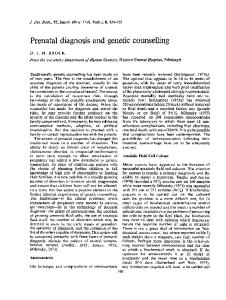
Rev. Med. Chir. Soc. Med. Nat., Iaşi – 2015 – vol. 119, no. 3
INTERNAL MEDICINE - PEDIATRICS
CASE REPORTS
TUBEROUS SCLEROSIS –PRENATAL DIAGNOSIS OF CARDIAC RHABDOMYOMA – CASE REPORT Alina-Costina Luca*, Andreea-Simona Holoc, Anca Elena Albu, C. Iordache University of Medicine and Pharmacy “Grigore T. Popa” - Iasi Faculty of Medicine Departament of Mother and Child Medicine *Corresponding author. E-mail:
[email protected] TUBEROUS SCLEROSIS – PRENATAL DIAGNOSIS OF CARDIAC PHABDOMYOMA (CASE REPORT) (Abstract). Primary cardiac tumors are rare in medical practice, with a prevalence of 0.0017-0.28% in autopsy examinations. In intrauterine life, the incidence of cardiac tumors has been reported to be of approximately 0.14%. Rhamdomyoma is the most frequent tumor formation during fetal period and childhood, accounting for 70% of all primary tumors in the fetus, neonate and infant. Diagnosing heart rhabdomyomas occurs most often by accident during fetal morphology at 22 weeks. We will present a case of a newborn diagnosed with fetal echocardiography in the 23 week of gestation having multiple cardiac tumors. Keywords: TUBEROUS SCLEROSIS, FETAL ECOGRAPHY, HYPOMELANOTIC MACULES, CARDIAC RHABDOMYOMA.
Advances in medicine and technology made it possible to diagnose a growing number of diseases, many of them in the fetal period. Cardiac tumors can be benign or malignant having as a starting point muscle layers or pericardium. These can be primary or secondary, of the metastatic type. Primary cardiac tumors are rare in medical practice, with a prevalence of 0.00170.28% at autopsy examinations. In intrauterine life, the incidence of cardiac tumors has been reported to be approximately of 0.14%. Most of them have a benign nature, only 10% of them being malignant (1, 2, 3, 4). Rhabdomyoma is the most frequent tumor formation during fetal period and in childhood, accounting for 70% of all primary tumors in the fetus, newborn and infant (5,6). It is most often located in the
ventricles but it may also originate in the atria (7, 8). Diagnosing heart rhabdomyomas occurs most often by accident during fetal morphology at 22 weeks. While the association between tuberous sclerosis and heart rhabdomyomas has been recognized, association with only one tumor formation cannot lead to the diagnosis of tuberous sclerosis. Tuberous sclerosis is a monogenic, multisystemic disease with dominant autosomal transmission, high penetration and variable expressivity, characterized by skin, cerebral and characteristic visceral manifestations. CASE REPORT We are presenting the case of a female newborn, born by caesarean section, cranial presentation, with a birth weight of 4000 g
685
Alina-Costina Luca et al.
and one minute Apgar 8 score, admitted to the Cardiology Department of ”Sf. Maria” Emergency Children's Hospital in Iaşi for seizures accompanied by perioronasal cyanosis. From the hereditary familial information, we could observe that both parents are apparently healthy and the patient’s brother was postnatally diagnosed with pyloric stenosis. Regarding pregnancy history, we found that the mother was diagnosed with chickenpox at 23 weeks of gestation. The obstetrical control performed at 26 weeks of amenorrhea, the mother did a fetal ultrasound and morphology, based on which the doctor identified echogenic images in the brain and in the heart. In the brain, there was a bilateral presence of non-homogenepus hyperechogenic images, imprecisely demarcated: at the right hemisphere in temporal lobe measuring 11/6 mm and in frontal lobe 4/4 mm and in the left hemisphere, in frontal lobe measuring 10/7 mm and 5/4 mm. In the heart, the obstetrician noted the presence of a heterogeneous hyper echogenic images, vaguely defined, with dimensions of 6/4 mm in the thickness of the right ventricular wall. He also noticed an image with the same specifications having 13/7mm (fig. 1, 2) in the thickness of the papillary muscle of the left ventricle. Therefore, based on fetal morphology, the obstetrician established the diagnosis of cardiac tumor (rhabdomyoma). Postnatally, an echocardioecography was performed that revealed multiple formations, most in the left ventricle, measuring 18/16/12 mm, with polylobate contours, with implantation to the side wall of the ventricle. In the interventricular septum, in the apex and in the thickness of the myocardium, three other formations of infra-
686
centimetric size and in the right ventricle, another oval image of 6/7 mm under septal cusp of the tricuspid.
Fig. 1. Fetal echocardiography, hyperechoic formation, non-homogenous in the right ventricle.
Fig. 2. Fetal echocardiography –presence of the two imprecisely defined hyperechogenic on the right and left ventricle. Clinical examination during last hospitalization showed that the patient had good general condition, pale skin, perioronasal cyanosis, multiple hypopigmented macules, spread on the entire body surface, the largest measuring 2/6 cm, located on the left flank. No murmur was detected by listening. The abdomen was supple, depressive, mobile with respiratory movements. In
Tuberous sclerosis –prenatal diagnosis of cardiac rhabdomyoma – case study report
terms of neuromotor activity, the child was developed normally. Biologically, we could note the presence of a mild thrombocytosis and hyposideremia. Performed electroencephalogram showed a spontaneous route with motion artifacts, with releases of ample waves in all derivations. Electrocardiography revealed a sinus rhythm with a 160-170 beats/minute, electrical axis 60 degrees, PQ =0.12s, with normal morphology, QT=0.26s (upper limit). Transfrontal ultrasound was normal, no modifications in the brain substance were found. As the child has numerous seizures, treated at that moment with Tonotil, we recommended a neurological examination that showed depigmented patches of different sizes on the trunk and limbs, epileptic spasms in extension and generalized motor seizures. Therefore, the pediatrician made the diagnosis of Bourneville tuberous sclerosis and West syndrome with lesions and recommended a CT brain scan. Since tuberous sclerosis is a multisystemic disease with dominant autosomal transmission, we also recommended that should be performed that raised again the suspicion of Bourneville tuberous sclerosis. Samples for DNA tests were collected that were transferred for confirmation to a specialized laboratory. Also, it was recommended to parents that an echocardiography should be made to see if any of them showed changes typical to tuberous sclerosis. Cardiological investigations of family members showed no evidence of cardiac tumors in none of them. The ophthalmologic examination that we recommended, including fundus exami-
nation, revealed no occurrence of tumors in the retina. Ultrasounds performed during hospitalization showed the presence of left ventricular of tumors measuring 17.5/11.1 mm and 25.2/16.7mm. On the side wall of the right ventricle, we were able to observe two growths of 4.2/1.8 mm and 3.6/1.8mm. Inside the right ventricle, a tumor of 8.1/6.7 mm was found, and at the tip of the left ventricle two tumor formations of 10.7/4.3mm and 12.9/3.1-5.3mm, respectively. The patient also showed left ventricular dysfunction and permeable oval foramen (fig. 3, 4, 5).
Fig. 3. Echocardiography - subcostal section. Shows numerous hyperchogenic formations in the right and left ventricle
Fig. 4. Echocardiography apical 4 chamber view – shows 12 tumoral formations
687
Alina-Costina Luca et al.
Fig. 5. Echocardiography, apical 4 chamber view. Tumor formations at the top of left ventricle The performed thoracoabdominal CT scan showed an expansive formation, solid density, iodophile, natively and postcontrast applied intravenously homogenous, well defined, with regular outline, which occupies the upper floor and the floor above middle mediastinum, with upper sizes 24/65/45 mm having no effect on the mass and with anterior relation to anterior chest wall; posterior relation to trachea, the internal carotid artery, vascular pedicle and the front of the heart; lateral to pulmonary pleura and superior to a plan section passing through the inferior edge of the clavicle (fig. 6, 7).
Fig.7. Thoracoabdominal CT scan – showing mediastinal formations CT brain examination showed hyper dense areas after intravenous contrast, disseminated to the brain substance, with varying sizes, the largest of 9/7mm, in the left frontal lobe, without mass effect on adjacent structures (fig. 8).
Fig. 8. Brain CT showing hypersense areas after CIV in brain substance Final diagnosis was: Bournevile tuberous sclerosis that was attributed to changes in the skin, presence of tumors in the brain and heart, permeable oval foramen, left ventricular dysfunction and West syndrome with lesions. Fig. 6. Formations in upper floor and middle floor of anterior mediastinum
688
DISCUSSION Rhabdomyomas are the most common
Tuberous sclerosis –prenatal diagnosis of cardiac rhabdomyoma – case study report
cardiac tumors found in newborn and children at a rate of 51-96% associated with tuberous sclerosis (9). While the association between tuberous
sclerosis and heart rhabdomyomas has been recognized, the association with only one tumor formation cannot diagnose tuberous sclerosis.
TABLE I Positive diagnostic criteria for tuberous sclerosis
Detecting multiple growths in the heart during fetal period diagnoses the Bourneville syndrome before the appearance of skin signs or seizures in the postnatal period (10, 11). Tuberous sclerosis is a monogenic, multisystemic disease with dominant autosomal transmission, high penetration and variable expressivity. This occurs in response to mutations of two tumor suppressing genes, TSC1 coding hamartin and TSC2 coding tuberin. TSC2 gene is located near the gene involved in the production of polycystic kidney disease, which in case of larger deletions (seen in 2% of cases) makes patients show both pathologies. To confirm the clinical diagnosis, the criteria of “The 2012 International Tuberous Sclerosis Complex Consensus Group” are used. The presence of two major or one major criteria makes the diagnosis to be certain (15). In the presented case, the clinical diagnosis clinical diagnosis was made on the
presence since intrauterine life of heart rhabdomyomas, confirmed later postnatally by both echocardiography and by the CT examination, the presence of growths in the brain and hypomelanotic macules (3) (13,14). The following complications may occur in the future: arrhythmias, most commonly ventricular, loss of myocardial function due to the increase of the tumor mass located in the side wall of the left ventricle, in the papillary muscles and in the interventricular septum that may be conducive to decreased cardiac output and congestive heart failure. It has been observed that the WolfParkinson-White syndrome has a higher incidence in patients with tuberous sclerosis, it is about 2% (1.12). Depending on the size of the tumor and the appeared complications, death may occur in some cases. CONCLUSIONS The evolution of techniques detecting congenital defects or diseases in the early stages allowed the pre-natal diagnosis of
689
Alina-Costina Luca et al.
numerous pathologies. Today, the suspicion of tuberous sclerosis may be raised since the pre-natal period, if the presence of cardiac rhabdomyomas is identified in the fetus during fetal morphology. As specific features of the case, we could observe the pre-natal diagnosis of
tumor formation during fetal morphology and even though tuberous sclerosis was with dominant autosomal transmission, no family members had skin changes or heart tumors, raising suspicion of a de novo gene mutation. However, to support this aspect genetic testing is recommended.
REFERENCES 1. Uzun O, Wilson DG, Vujanic GM, Parsons JM, De Giovanni JV. Cardiac tumours in children (Orph. J. Rare Dis) 2. McAllister HA Jr: Primary tumors of the heart and pericardium. Pathol Annu 1979; 14: 325-355. 3. Nadas AS, Ellison RC: Cardiac tumors in infancy. Am J Cardiol 1968; 21: 363-366. 4. Holley DG, Martin GR, Brenner JI: Diagnosis and management of fetal cardiac tumors: a multicenter experience and review of published reports. J Am Coll Cardiol 1995; 26: 516-520. 5. Groves AM, Fagg NL, Cook AC, Allan LD: Cardiac tumours in intrauterine life. Arch Dis Child 1992; 67: 1189-1192. 6. Simcha A, Wells BG, Tynan MJ, Waterston DJ: Primary cardiac tumours in childhood. Arch Dis Child 1971; 46: 508-514. 7. Chan HS, Sonley MJ, Moes CA, Daneman A, Smith CR, Martin DJ: Primary and secondary tumours of childhood involving the heart, pericardium, and great vessels. A report of 75 cases and review of the literature. Cancer 1985; 56: 825-836. 8. Beghetti M, Gow RM, Haney I, Mawson J, Williams WG, Freedom RM: Pediatric primary benign cardiac tumours: a 15-year review. Am Heart J 1997; 134: 1107-1114. 9. Jozwiak S, Kalawec W, Dluzewska J, Daszkowska J, Mirkowicz-Malek M, Michalowicz R: Cardiac tumors in tuberous sclerosis: their incidence and course. Eur J Pediatr 1994; 153: 155-157. 10. Mair DD, Gomez MR: Tuberous sclerosis and cardiac rhabdomyoma. Am J Cardiol 1995; 76: 419421. 11. Webb DW, Thomas RD, Osborne JP: Cardiac rhabdomyomas and their association with tuberous sclerosis. Arch Dis Child 1993; 68: 367-370. 12. Mas C, Penny DJ, Menahem S: Pre-excitation syndrome secondary to cardiac rhabdomyomas in tuberous sclerosis. J Paediatr Child Health 2000; 36(1): 84-86. 13. Turliuc D, Dumitrescu F, Indrei A. Neurosurgeon- Neuropathologist Collaboration in Cerebral Tumors Diagnosis, RevMed Chir 2007; 111(3): 643. 14. Titianu M, Schaas C, Onofriescu M: Diagnostic problems in fetal echocardiographic examination. Observations on a lot of study during 2005-2010 RevMed Chir 2011; 115(2): 451. 15. H. Northrup, D.A. Krueger: Tuberous sclerosis complex diagnostic criteria update: recommendations of the 2012 International Tuberous Sclerosis Complex Consensus Conference, Pediatr Neurol 2013; 49: 243–254.
690