J Clin Periodontol 2015; 42: 462–469 doi: 10.1111/jcpe.12395
Treatment of peri-implant mucositis using a glycine powder air-polishing or ultrasonic device: a randomized clinical trial
Caroline Riben-Grundstrom1, !1 and Ola Norderyd1,2, Ulrika Andre Stefan Renvert3,4,5 1
Department of Periodontology/Endodontics and Oral Prosthodontics, Institute for €nko €ping, Postgrad Dental Education, Jo € Sweden, 2Faculty of Odontology, Malmo €, Sweden, 3Department of University, Malmo Oral Sciences, Kristianstad University, Kristianstad, Sweden, 4Blekinge Institute of Technology, Karlskrona, Sweden, 5School of Dental Sciences, Trinity College, Dublin, Ireland
Riben-Grundstrom C, Norderyd O, Andr! e U, Renvert S. Treatment of peri-implant mucositis using a glycine powder air-polishing or ultrasonic device: a randomized clinical trial. J Clin Periodontol 2015; 42: 462–469. doi: 10.1111/jcpe.12395.
Abstract Aim: To evaluate the clinical treatment effects of a glycine powder air-polishing or ultrasonic device on peri-implant mucositis. Materials and methods: Thirty-seven patients with one implant diagnosed with peri-implant mucositis (probing depth ≥4 mm (0.2N) and bleeding on probing (BOP) (primary outcome)) were randomly assigned to treatment with either glycine powder air-polishing (GPAP) or ultrasonic (US) debridement. Treatment was performed at baseline and at 3 and 6 months. Professional supra gingival cleaning was performed at 9 and 12 months. Oral hygiene instructions were reinforced at each visit. Results: At 12 months there was a statistically significant reduction in mean plaque score, bleeding on probing and number of periodontal pockets ≥4 mm within the treatment groups compared to baseline. The percentages of diseased sites were significantly reduced for both groups. Conclusions: Treatment with a glycine powder air-polishing or an ultrasonic device is effective in non-surgical treatment of peri-implant mucositis.
Dental implants are often used to replace lost teeth and present a high level of predictability, patient satisfaction and long-term success (Schnitman et al. 1997, Romeo et al. 2004, Pjetursson et al. 2005, 2012, Jung et al. 2012). Biological complications such as peri-implant mucositis and peri-implantitis have, Conflict of interest and source of funding statement The study was partly supported by E.M.S., Electro Medical Systems S.A., Nyon, Switzerland.
462
however, become major challenges to the profession (Mombelli et al. 2012). The definition of peri-implant mucositis is an inflammation of the soft tissues adjacent to a dental implant diagnosed with bleeding on gentle probing ( 55 mmol/mol). Patients receiving medication known to have effect on gingival growth eg. calcium channel antagonists, immunosuppressants or antiepileptic drugs. Patients requiring antibiotic prophylaxis or whom had received antibiotic treatment in the preceding 3 months. Patients receiving systemic corticosteroids.
Randomization
Once the entry criteria had been confirmed the subjects were entered to the study and assigned a patient number. Assignment to the glycine powder air-polishing group (GPAP) and the ultrasonic group (US) was made using a computerized randomization. Cards with group identification were prepared and placed in numbered envelopes by a member from the staff not involved in the examination or treatment of the patients. The dental hygienist responsible for the treatment broke the seal of the envelope to give the patients treatment according to either group A or B protocol. The code for treatment A and B was revealed to the examiners once the study was completed and the data set locked. Clinical and radiographic examination
Each participant was given a detailed description of the study and was required to sign a written consent according to the Declaration of Helsinki (version 2008). The examinations and registrations were performed by two calibrated operators, (CRG,ON) not aware of the treatment group of the patient. The patients were also asked not to discuss their treatment with the examiner. Each visit commenced with an update on adverse effects
464
Riben-Grundstrom et al.
and an intra-oral photo of the implant site. Baseline, 1 month, 3 months, 6 months, 9 months, 12 months At the baseline examination each participant was interviewed regarding their medical history, including smoking and oral hygiene habits. Implant characteristics and position in the dentition was recorded. At 1-month oral hygiene, in terms of full-mouth plaque index and local implant plaque index, was evaluated and reinforced. At baseline, 3 months, 6 months, 9 months and 12 months clinical measurements on full-mouth plaque score (FMPS), full-mouth bleeding score (FMBS), implant probing depth, local implant bleeding on probing, local implant plaque score and suppuration were performed at six sites on each qualifying implant (mesio-buccal, buccal, disto-buccal, mesio-palatinal, palatinal, disto-palatinal). Mucosal overgrowth and recession was measured at two sites (buccal and lingual/palatinal). Full-mouth plaque score (FMPS) was recorded using a disclosing dye (Top Dent Rondell, Top Dent, Sweden) and recorded as a percentage of examined sites within each patient. Full-mouth bleeding score (FMBS) was measured 30 s after probing and recorded as a percentage of examined sites within each patient (O’Leary et al. 1972). All implant probing depth (PD) measurements were made with a probing force of 0.2 N with the same probe design (Hawe Click-Probe, HaweNeos Dental, Switzerland). Implant bleeding on probing score (BOP), local implant plaque score, suppuration and mucosal overgrowth was recorded as either present or absent. Recession of the mucosal margin relative to the restoration margin (REC) at the implant was recorded in millimetres at two sites (buccal or lingual/palatal). If the mucosal margin was located apical to the restoration this was indicated as a positive value (+) and if the margin was located coronal to the restoration this was registered as a negative value(-). Radiographic examination At baseline, 6 months and 12 months a digital periapical standardized
radiograph using a bite-block (Colt$ene!PRESIDENT Putty soft, Colt$ene/Whaledent AG, Altst€atten, Switzerland) was taken to detect loss of supporting bone.
Wilcoxon sign rank test, while comparisons between groups were performed using Wilcoxon rank sum test. Results were considered statistically significant at p < 0.05.
Oral hygiene instructions Oral hygiene instructions were reinforced after each examination. Treatment protocol
Each subject received treatment according to his or her randomization in either the glycine powder air-polishing group (GPAP) or the ultrasonic group (US). The treatment was performed at baseline, 3 months and 6 months by an experienced dental hygienist (UA) immediately after the registrations. Glycine-powder air-polishing The Perio-Flow! nozzle (AIRFLOW Master Piezon!; EMS, Nyon, Switzerland) was directed into the peri-implant pocket at an angle of 60–90 degrees to the implant and each surface was debrided for 5 s using glycine powder (AIR-FLOW! Powder PERIO; EMS). Ultrasonic device An ultrasonic device (AIR-FLOW Master Piezon!; EMS) with a hightech plastic material coated tip (PI Instrument; EMS, Nyon, Switzerland) was placed in the peri-implant pocket and each surface was debrided for 5 s. Maintenance care Supragingival maintenance care was provided at month 9 and 12. After each debridement and each visit the dentition was polished using a rubbercup (Pro-Cup!; KerrHawe, Bioggio, Switwerland) and prophypaste (Prophy Paste; CCS Healthcare AB, Borl€ange, Sweden). Statistical analysis
A software package (IBM SPSS Statistics 21.0; SPSS, Chicago, IL, USA) was used for the statistical analysis. All analyses were performed at patient level. Mean values and standard deviation (mean; SD) for the clinical parameters were calculated for the two groups. Comparisons over time for the investigated variables were performed using
Results Sample description
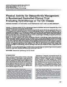
Thirty-seven subjects, 18 females and 19 males were enrolled in this study. One implant in each subject received treatment according to the randomization. Nineteen individuals were treated with the glycine powder airpolishing device (GPAP) and 18 individuals were treated using the ultrasonic device (US). One patient could not attend the 9-month examination but participated at the 12month examination. One patient dropped out due to personal reasons following the 9-months follow-up (Fig. 1). The mean age of the patients was 64.3 years. Five patients were current smokers. The baseline demographic data are presented in Table 1. Mean values of full-mouth plaque scores at baseline between the two groups were estimated to be 19.8 ! 4.1% (GPAP) and 18.5 ! 4.4% (US). Plaque scores decreased to 8.1 ! 1.4% and 8.7 ! 1.6% at 1 month. Mean full-mouth bleeding score was low 9.2 ! 2.7% (GPAP) and 9.6 ! 2.8% (US) at baseline for both groups. The difference according to full-mouth plaque score between baseline and 12 months was statistically significant (p < 0.05) for the GPAP group and according to bleeding score for the ultrasonic group (Table 2). Mean local implant plaque index at baseline was 25.5 ! 6.8% in the GPAP group and 24.1 ! 6.6% in the ultrasonic group. Local plaque index varied between the groups during the study period and was significantly reduced between baseline and 12 months in both groups to 5.6 ! 3.8% and 7.4 ! 6.4%, (p < 0.05). Mean local implant bleeding on probing was 43.9 ! 7.3% (GPAP) and 53.7 ! 7.9% (US) at baseline and was significantly reduced (p < 0.001) in both groups to 12.1 ! 3.8% (GPAP) and 18.6 ! 6.4% (US) at the end of the study (Table 3). No
© 2015 John Wiley & Sons A/S. Published by John Wiley & Sons Ltd
Treatment of peri-implant mucositis
465
Assessed for eligibility (n = 37)
Enrollment
37 Individuals
Glycine powder airpolishing
Ultrasonic
Allocated to intervention (n = 19) Received allocated intervention (n = 19)
Baseline
Allocated to intervention (n = 18) Received allocated intervention (n = 18)
Allocated to intervention (n = 19) Received allocated intervention (n = 19)
3 months
Allocated to intervention (n = 18) Received allocated intervention (n = 18)
Allocated to intervention (n = 19) Received allocated intervention (n = 19)
6 months
Allocated to intervention (n = 18) Received allocated intervention (n = 18)
Allocated to intervention (n = 19) Received allocated intervention (n = 19)
9 months
Allocated to intervention (n = 18) Received allocated intervention (n = 17)
Lost to follow-up (1) Analysis
Analyzed (n = 18)
Analyzed (n = 18)
1 year
Fig. 1. Demographic data and baseline characteristics for the GPAP and ultrasonic group.
statistically significant existed between groups.
difference
Suppuration was recorded in 1–3 sites in 10 subjects and was reduced
Table 1. Baseline examination
No. patients Male Female Age range (years) Mean age (years) Smokers Never smokers Non-smokers Packyears (years) Implant design ASTRA TECH Implant SystemTM Nobel Biocare! Implant System Straumann!Dental Implant System Referred from Department of Prosthodontics Department of Periodontology General dentist
GPAP
US
Total
19 10 9 25–85 64.4 1 13 5 4.1
18 9 9 25–86 64.3 4 8 6 8
37 19 18 25–86 64.3 5 21 11 2–45
3 12 4
2 10 6
5 22 10
6 4 9
6 3 9
12 7 18
© 2015 John Wiley & Sons A/S. Published by John Wiley & Sons Ltd
to affecting five subjects in the end of the study. Mucosal overgrowth of 1–3 mm was detected in three subjects and mucosal recession of 1–6 mm in eight subjects. No differences were found in the group or between groups at the end of the study. Periodontal pockets were grouped into sites with pockets between 0 to 3 mm and pockets ≥ 4 mm (Table 4). The percentage of periodontal pockets with ≥4 mm in probing depth at baseline was 30% in the GPAP group and 34% in the ultrasonic group. After 3 months the number of sites ≥ 4 mm were reduced to 22% and 29%, respectively. At the 12 month follow-up a reduction to 13% in the GPAP group and 20% in the ultrasonic group was observed, (p < 0.001). No
466
Riben-Grundstrom et al.
Table 2. Full-mouth plaque score and bleeding score at different time intervals (mean ! SEM). Group
Baseline 1 month 3 months 6 months 9 months 12 months
GPAP Ultrasonic GPAP Ultrasonic GPAP Ultrasonic GPAP Ultrasonic GPAP Ultrasonic GPAP Ultrasonic
N
19 18 19 18 19 18 19 18 19 17 18 18
FMPS (%)
FMBS (%)
Mean
SEM
Mean
SEM
19.8 18.5 8.1 8.7 9.5 12.4 8.2 14.4 10.7 6.3 5.7* 9.9
4.1 4.4 1.4 1.6 1.8 3.3 2.0 4.5 3.4 1.8 1.2 5.5
9.2 9.6 – – 3.8 5.1 4.8 5.1 3.3 2.0 3.4 2.2*
2.7 2.8 – – 0.9 1.9 2.4 1.8 0.9 0.6 1.1 0.8
FMPS, full-mouth plaque score; FMBS, Full-mouth bleeding score. Changes in mean full-mouth plaque and bleeding score at different time intervals. *Statistically significant difference according to full-mouth plaque score between baseline and 12 months in the GPAP group and according to bleeding score for the Ultrasonic group (p < 0.05).
Table 3. Implant plaque score and bleeding on probing at different time intervals (mean ! SEM) N
Baseline 1 month 3 months 6 months 9 months 12 months
GPAP Ultrasonic GPAP Ultrasonic GPAP Ultrasonic GPAP Ultrasonic GPAP Ultrasonic GPAP Ultrasonic
19 18 19 18 19 18 19 18 19 17 18 18
IPS (%)
IBoP (%)
Mean
SEM
Mean
SEM
25.5 24.1 7.9 16.8 6.2 13.0 13.2 14.9 13.2 4.9 5.6* 7.4*
6.8 6.6 3.5 5.5 2.6 4.2 7.3 6.0 5.8 3.1 3.8 6.4
43.9 53.7 – – 23.0 25.1 16.7 23.2 18.5 11.9 12.1* 18.6*
7.3 7.9 – – 6.1 5.6 4.6 5.4 5.7 2.4 3.8 6.4
IPS, Implant plaque score; IBS, Implant bleeding on probing. Changes in mean implant plaque score and bleeding on probing at different time intervals. *Statistically significant difference according to implant plaque score and implant bleeding on probing between baseline and 12 months for both groups (p < 0.05).
significant difference in the number of sites with 0–3 mm and ≥ 4 mm existed between the two groups. The number of diseased sites (pocket depth ≥4 mm with bleeding/ suppuration) before and after treatment are presented in Table 5. At baseline 38% of the sites in the GPAP group were diseased compared to 52% in the ultrasonic group. A reduction of diseased sites to 8% in the GPAP group and 17% in the ultrasonic group was achieved. No significant differences were recorded between the two groups at
baseline, after 12 months or according to reduction of number of diseased sites from baseline to 12 months. However, for both groups the number of diseased sites were significantly reduced between baseline and 12 months, (p < 0.01). Discussion
The non-surgical treatment in this study resulted in a significant reduction in all clinical measured variables. Gingival bleeding and bleeding on probing, the most
important findings in detecting periimplant disease were continuously reduced throughout the study. One possible reason for the positive outcome, besides the effect of therapy, could be related to the patient’s compliance in maintaining low plaque indices during the whole study period. At the 1-month examination the mean full mouth plaque score had decreased to values below 10%. Oral hygiene conditions have been suggested to be an important variable associated with peri-implant health (Zitzmann et al. 2001, Salvi & Lang 2004, Kracher & Smith 2010, Mombelli & D!ecaillet 2011). The importance of plaque levels on successful long-term supportive maintenance of dental implants diagnosed with peri-implant mucositis have been evaluated in a few studies. Costa et al. (2012) followed 80 patients diagnosed with peri-implant mucositis for a period of 5 years with and without preventive maintenance. In patients not receiving maintenance treatment 44% developed peri-implantitis compared to 18% of patients receiving maintenance treatment. Higher plaque levels were detected in the group not receiving maintenance therapy. Despite the low levels of plaque, both groups responded well to treatment. However, 8% of the sites in the GPAP group and 17% of the sites in the ultrasonic were still diseased after 12 months and highlights the difficulties in achieving complete resolution of inflammation at implants. This is in agreement with results from a short-term randomized clinical trial by (Heitz-Mayfield et al. 2011) where the authors reported a complete resolution in 38% of implants and no added benefit using adjunctive chlorhexidine gel. In a recent meta-analysis, air-abrasives had no adjunctive effect to professionally administered plaque removal (Schwarz et al. 2014). Nevertheless, it indicates that mechanical debridement in conjunction with oral hygiene is effective in reducing peri-implant soft tissue inflammation. In a recent in vitro model, 18 implants were dip-coated and placed in resin blocks with different defect morphologies. Different
© 2015 John Wiley & Sons A/S. Published by John Wiley & Sons Ltd
Treatment of peri-implant mucositis
467
Table 4. Number of sites with probing depth 0–3 mm and ≥4 mm at the different time points for implants treated with either a glycine powder air-polishing or an ultrasonic device. Baseline
GPAP US Total p-value³
3 months
Sites 0–3 mm n%
Sites ≥ 4 mm n%
Sites 0–3 mm n%
Sites ≥ 4 mm n%
48 (22) 32 (14) 80 (36)
66 (30) 76 (34) 142 (64)
66 (30) 43 (19) 109 (49)
48 (22) 65 (29) 113 (51) NS
6 months
p-value¹
0.01 NS
9 months
12 months
Sites 0–3 mm n%
Sites ≥4 mm n%
Sites 0–3 mm n%
Sites ≥ 4 mm n%
Sites 0–3 mm n%
Sites ≥4 mm n%
71 (32) 55 (25) 126 (57)
43 (19) 53 (24) 96 (43)
77 (36) 55 (25) 132 (61)
37 (17) 47 (22) 84 (39)
79 (37) 65 (30) 144 (67)
29 (13) 43 (20) 72 (33) NS
p-value²