Journal of Dental School 2013; 31(1):8-14
Original Article
Topographic Evaluation of Apex and Root Canal of Maxillary Premolars in an Iranian Population 1
Mandana Naseri 2Mojgan Momayez *3Zohreh Ahangari
1
Assistant Professor, Dept. of Endodontics, School of Dentistry, Shahid Beheshti University of Medical Sciences, Tehran, Iran. 2 General Practitioner. *3Associate Professor, Dept. of Endodontics, School of Dentistry, Shahid Beheshti University of Medical Sciences, Tehran, Iran. E-mail:
[email protected]
Abstract Objective: Knowledge about the internal anatomy of the root canal and apical foramen is a critical prerequisite for root canal therapy and is necessary for the success of endodontic treatment. This study aimed to determine the distance of apical foramen from the anatomical apex and apical constriction, evaluate the deviation of apical foramen from the anatomical apex and specify the most common canal types in maxillary premolar teeth using stereomicroscope. Methods: In this laboratory experimental study, 100 extracted first and second maxillary premolars of patients presenting to dental clinics in Tehran were selected. After the disinfection of roots and access cavity preparation, apical foramen deviation and its distance from the anatomical apex were determined by introducing a #10 hand file into the canal and observation under stereomicroscope; 2% methylene blue was then injected into the canals and demineralization and clearing process were carried out to measure the distance of apical foramen from the apical constriction and evaluate the canal type. Results: The mean distance of apical foramen from the anatomical apex and apical constriction was 0.4-0.5 and 0.5-0.7 mm, respectively in maxillary first premolars and 0.3-0.7 and 0.6-1 mm, respectively in maxillary second premolars. In less than 17% of first premolars and 37% of second premolars, the canal followed a straight path to the anatomical apex without any deviation towards the mesiodistal or buccolingual directions. The most common canal type according to Vertucci’s classification was type IV in the first and type I in the second premolars. In general, 94% (n=47) of the first premolars and 46% (n=23) of the second premolars had 2 canals while the remaining teeth had a single canal. Conclusion: This study showed that the apical foramen in premolar teeth is located at a 0.3-0.7 mm distance from the anatomical apex and 0.5-1 mm distance from the apical constriction. In more than 83% of first premolars and 63% of second premolars the apical foramen did not correspond to the anatomical apex. First and second premolars had 2 canals in 94% and 46% of cases, respectively. Key words: Anatomical apex, Apical constriction, Apical foramen, Canal deviation, Canal type, , Clearing process, Premolar. Please cite this article as follows: Naseri M, Momayez M, Ahangari Z. Topographic Evaluation of Apex and Root Canal of Maxillary Premolars in an Iranian Population. J Dent Sch 2013; 31(1): 8-14.
Received: 16.01.2013
Final Revision: 03.03.2013
Introduction: Before starting root canal therapy, the clinician needs to have sufficient knowledge about tooth morphology. Numerous in-vitro studies have evaluated root canal morphology through direct observation with the use of dye, sectioning, scanning or 3D assessment using computer
Accepted: 05.03.2013
programs. The results of these studies and use of new equipment such as dental microscope when preparing the access cavity can all decrease the odds of not finding an extra canal during endodontic therapy (1). Root anatomy at the apical region has three anatomical and histological landmarks: apical constriction, cementodentinal junction and
Naseri, et al.
9
apical foramen. Apical constriction is defined as a part of root canal with the smallest diameter. It is also considered a reference point for apical termination of root canal treatment. Cementodentinal junction is a part of root canal where cementum reaches dentin. It indicates the termination of pulp tissue and initiation of periodontal tissue. This area is clinically undetectable. Apical foramen is defined as the circumference or rounded edge, like a funnel or crater, that differentiates the termination of the cemental canal from the root surface. It represents the junction of the pulp and the periodontal ligament. During root formation, apical foramen is usually positioned at the anatomical root end. However, along with tooth development, apical foramen becomes smaller and more distant from the center (2). Apical foramen may be positioned at mesial, distal, buccal or lingual of the anatomical apex (3). Deviation of apical foramen from the apex cannot be easily detected radiographically especially when the deviation and opening are on the buccal or lingual root surface (4). Different studies have demonstrated that there is usually a 0.5 to 3 mm distance between the apical foramen and the anatomical apex and in only 17-46% of cases the two exactly correspond (5). This study aimed to assess the prevalence of apical foramen deviation from the anatomical apex at buccolingual and mesiodistal dimensions. It also measured the distances of apical foramen from the apical constriction and anatomical apex by stereomicroscope in an Iranian population.
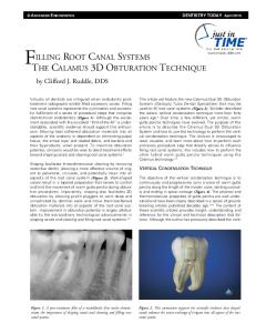
Methods: In this in-vitro experimental study, a total of 100 permanent maxillary first and second premolars (n=50 for each) with mature apex and no or minimal carious lesions were collected from several dental clinics in Tehran during a one year period. The teeth were stored in jars containing 10% formalin solution for disinfection until the experiment. Age and gender of patients and the reason of tooth extraction were not specified. The teeth were debrided from soft tissue or bony appendages and calculus by scaling and polishing. Access cavity was prepared using 008 fissure bur (Tizkavan, Iran) and high speed hand piece (Kavo, West, Germany). An endodontic explorer was used to detect canal orifices (DG 16, HuFreiday, Chicago, IL, USA). A #6 or # 8 Kfile (Dentsply Maillefer, Switzerland) was introduced into the canal until observing the file tip at the apical foramen. Specimens were then fixed on a slide with play dough (Arya, Tehran, Iran). The two variables of the distance of apical foramen (where the file tip was observed) from the anatomical apex and also the deviation direction of apical foramen relative to the anatomical apex from the proximal (to determine buccal or palatal inclination) and buccolingual aspects (to determine mesial or distal inclination of the apical foramen) were measured using stereomicroscope (SZX1LLB2, Olympus Optical, Japan) at 16X magnification and 0.01 mm readability (Figure 1).
Figure 1- Evaluation of the direction of apical foramen deviation relative to the anatomical apex in premolar teeth from the buccolingual aspect to determine mesial or distal inclination (A), from the proximal aspect to determine buccal or palatal inclination (B) and without deviation (C) using stereomicroscope at 16X magnification
Journal of Dental School 2013 10 For demineralization, specimens were immersed in 5% nitric acid solution at room temperature (20°C) for 6 days. The solution was stirred and agitated 3-4 times a day and exchanged daily. In order to ensure demineralization, tooth softening was checked by inserting a sharp point explorer into the tooth crown structure and also by radiographic comparison with the control intact tooth. After completion of the 6-day period and ensuring the demineralization process, samples were rinsed under running water for 4-6 hours. The dehydration process was accomplished by using 100° ethyl alcohol (Ararat, Tehran) for 2 hours. The dehydrated teeth were then cleared after immersion in methyl salicylate (Merck, Darmstadt, Germany) for 2 hours. All specimens were stored in this solution until the experiment. Apical foramen morphology of the teeth was evaluated under stereomicroscope (SZX-1LLB2, Olympus Optical, Japan) at 16× magnification and 0.01 mm readability. The distance of apical foramen center from the apical constriction was measured using a stereomicroscope with a micrometer. Type of canals was studied as well. Prevalence and frequency of each morphological parameter in specimens were determined and the actual values were estimated with 95% Confidence Interval. In order to achieve the descriptive objectives of the study, descriptive statistics including tables, central measures and dispersion were applied.
Results: Table 1 summarizes the prevalence and direction of apical foramen deviation from the anatomical apex in maxillary premolars. In single canal maxillary first premolars, apical foramen was deviated from the anatomical apex in 100% of cases. This rate was 89.3% and 82.9% in buccal and palatal canals of maxillary two-canal first premolars, respectively.
Prevalence of apical foramen deviation from the anatomical apex was 62.9% in maxillary singlecanal second premolars. This rate was 86.9% and 95.6% in buccal and palatal canals of twocanal maxillary second premolars, respectively. In general, in single-canal maxillary first premolars, the most prevalent deviation was towards the buccal and distal aspects; whereas, in the buccal and palatal canals of two-canal teeth the most common deviations were towards the mesiolingual and distolingual aspects, respectively (Table 1). In single-canal maxillary second premolars the apical foramen deviation from the anatomical apex was mostly towards the mesiolingual aspect while in two-canal teeth, the buccal canal was mostly deviated towards the mesiobuccal and mesiolingual and the palatal canal towards the distolingual aspect (Table 1). Based on the obtained results, the mean distance of apical foramen from the anatomical apex and apical constriction was 0.4-0.5 and 0.5-0.7 mm in the first maxillary premolars and 0.3-0.7 and 0.6-1 mm in the second maxillary premolars, respectively (Table 2). Morphological evaluation of canals revealed that maxillary first premolars had 2 canals in 94% of cases (n=47) and a single canal in 6% (n=3) of cases. These rates were 46% (n=23) and 54% (n=27) in maxillary second premolars, respectively. The most common canal types according to Vertucci’s classification (7) in maxillary first premolars were type IV (n=35) with 70% prevalence, type II (n=12) with 24% prevalence and type I (n=3) with 6% prevalence rate. In maxillary second premolars, the most frequent canal types were type I (n=27) with 54% prevalence, type II (n=9) with 18% prevalence and type IV (n=14) with 28% prevalence rate. Other canal types were not observed.
Naseri, et al.
11
Table 1- Prevalence and direction of apical foramen deviation from the anatomical apex at mesiodistal and buccolingual aspects in maxillary premolars
First premolar
Single canal (6%)
Deviation of apical foramen from the apex at the mesiodistal aspect
Buccal Lingual No deviation Mesial Distal
Deviation of apical foramen from the apex at the buccolingual aspect
Second premolar
Two canals (94%)
Two canals (46%)
25% 27.8% 47.2% 36.1% 22.2%
Buccal canal 28.6% 28.6% 42.8% 35.7% 28.6%
Palatal canal 14.3% 71.4% 14.3% 35.7% 50%
28.5%
41.7%
35.7%
14.3%
89.3%
82.9%
62.9%
86.9%
95.6%
10.6%
17%
37%
13%
4.3%
66.7% 33.3% 0% 0% 100%
Buccal canal 36.2% 38.3% 25.5% 44.7% 36.2%
Palatal canal 2.9% 71.4% 25.7% 22.9% 48.6%
No deviation
0%
19.1%
100%
0%
Deviation of apical foramen from the apex at the mesiodistal and buccolingual aspects Apical foramen without deviation from the apex at the mesiodistal and buccolingual aspects
Single canal (54%)
Table 2- The mean and SD value of the distance of apical foramen from the anatomical apex and apical constriction in maxillary premolars in mm
First premolar
Single canal (n=3)
Buccal canal
The mean distance of apical foramen from the anatomical apex
0.5 (0.4)
0.4 (0.3)
The mean distance of apical constriction from the apical foramen
0.7 (0.4)
0.5 (0.2)
The mean total distance of apical foramen from the anatomical apex The mean total distance of apical constriction from the apical foramen
Second premolar
Two canals (n=47)
Single canal (n=27)
Two canals (n=23)
Buccal canal
0.5 (0.4)
0.3 (0.2)
0.7 (0.5)
0.6 (0.3)
0.5 (0.4)
0.6 (0.2)
1 (0.4)
0.8 (0.2)
Palatal canal
0.4-0.5
0.3-0.7
0.5-0.7
0.6-1
Discussion: Internal anatomy of the root canal and apex of maxillary first and second premolars has been extensively studied by various researchers. However, their findings have been mostly controversial (7-9). This variability in results may be due to the presence of significant differences in the internal anatomy of the root
Palatal canal
canals between different individuals of various races and ethnicities. Variable classifications of root canal morphology by researchers, age and gender of understudy subjects and the methodology of the study can also play a role in this respect (10). Anatomy of the root canal system and apex can be studied using various techniques such as the laboratory methods i.e. injection of methylene blue (11), Black India ink
Journal of Dental School 2013 12 (10, 12), or Chinese ink (13), metal or plastic casting (8, 14), in-vitro endodontic access cavity preparation with the use of radiography and instrumentation (15, 16), radiopaque gel infusion and radiography (7) and in-vitro root canal therapy (17).Tooth clearing technique is an accurate method with complex laboratory phases; which was used also by Vertucci et al. in 1984 (10). This technique provides thorough 3D observation of canals and apical foramen with the use of dye injection without traumatizing the tooth structure. The apical one-third of root canals is a strategic region in terms of working length determination and interaction with the surrounding live tissue. Thus, it can directly affect the healing of periapical lesions. In this respect, the three important anatomical landmarks are the apical constriction, apical foramen and anatomical apex. Over time with advanced age and cementum formation at the apex area, apical foramen is deviated towards various directions. But, this deviation and apical foramen opening at the apex cannot be detected radiographically particularly when this opening occurs at the buccal or lingual aspects. The distance of apical constriction from the anatomical apex in maxillary premolars has reported to be highly variable from 0.052 to 2.921mm (18-20). The mean distance of apical constriction from the anatomical apex has reported to be approximately 1 mm (range 0.8-1.1) in maxillary second premolars of an Iranian population (21). In our study, apical foramen deviation was evaluated at both mesiodistal and buccolingual directions. The prevalence of apical foramen deviation from the anatomical apex was 100% and more than 83% in single canal and twocanal maxillary first premolars and 63% and more than 87% in single-canal and two-canal maxillary second premolars, respectively. These results are almost similar to those of Kuttler et al. in 1955 (22). In our study, in single-canal maxillary first premolars deviations were mostly
towards the buccal and distal directions. In twocanal teeth, buccal and palatal canals were mostly deviated towards mesiolingual and distolingual directions. In single-canal maxillary second premolars, apical foramen was mostly deviated towards the mesiolingual direction. In two-canal teeth, buccal canal in the majority of cases was deviated towards the mesiobuccal and mesiolingual while the palatal canal had distolingual deviation in most cases. These findings are different from those of Burch et al. (1972)(4). In the present study, the mean distance of apical foramen from the anatomical apex was 0.4-0.5 and 0.3-0.7mm in the first and second maxillary premolars, respectively. These rates are not in accord with the values obtained by other researchers such as Green (1960)(2 mm range) (9), Burch (1972)(0.59 mm)(4), Arora (2009)(0.052-2.91 mm)(20) and Martos (2009)(0.69 mm)(23). Differences in this respect may be attributed to different methodology of studies and ethnic differences. Another parameter evaluated in this study was the mean distance of apical constriction from the apical foramen in maxillary first and second premolars which was found to be 0.5-0.7 and 0.6-1 mm, respectively. Kuttler et al. (1955)(22) reported this distance to be 0.5 to 1.5 mm. They assessed the entire dentition in their study. Furthermore, in contrast to our study (using clearing process), they applied the tooth sectioning technique (22). Morphological assessment of root canals according to Vertucci’s classification (1984)(7) indicated that the maxillary first premolars had 2 canals in 94% of cases and type IV, type II and type I (with 70%, 24% and 6% prevalence rate, respectively) were the most prevalent canal types. In Maxillary second premolars, prevalence of two-canal teeth was 46% (the remaining were single-canal) and the most common canal types were type I, type IV and type II (with 54%, 28% and 18% prevalence
Naseri, et al.
13
rate, respectively) in decreasing order of frequency. These findings are in agreement with the results of SadrLahijani et al. (2002)(24) and Jayasimha Raj et al. (2010)(25) and slightly different from those of Partovi et al. (2005)(1) on the morphology of maxillary second premolars. This difference may be explained by the geographical location of the study area.
of type IV and type II canals in maxillary first premolars emphasizes the very high incidence of two canals in this tooth. Knowledge about these anatomical variations highlights the need for high precision during access cavity preparation to find all canal orifices and accurate preparation and obturation of canals to the proper length.
Acknowledgement Conclusion: Within the limitations of this study, we may conclude that in two-canal maxillary first and second premolars, apical foramen coincided to the anatomical apex in less than 17% and 13% of cases, respectively. In single-canal maxillary first and second premolars these rates were zero and 37%, respectively. Also, apical foramen was located within 0.3-0.7 mm distance from the anatomical apex and 0.5-1 mm distance from the apical constriction. High frequency percentage
This article was based on an undergraduate thesis by Ms. Mojgan Momayez which was successfully completed under supervision of Dr. Mandana Naseri in Dental School of Shahid Beheshti University of Medical Sciences. This study was approved and funded by the Research Deputy of Shahid Beheshti University, School of Dentistry. Conflict of Interest: “None Declared”
References: 1. 2.
3. 4. 5.
6. 7. 8. 9.
Partovi M, Mozzafari SF. Root canal Morphology of maxillary second premolars. JBUMS 2005; 7: 34-36. Asna-ashari M, Nouri M, Mozayyeni MA, Seradji F: Evaluation of apical foramen situation by anatomic apex anddiagnostic value of radiography on determination of its location (A Stereomicroscopic study). J Dent Sch 2004; 22:361-368. Yoshioka T, Kikuchi I, Fukumoto Y, Kobayashi C, Suda H. Detection of the second mesiobuccal canal in mesiobuccal roots of maxillary molar teeth ex vivo. Int Endod J 2005; 38: 124-128 . Burch JG, Hulen S. The relationship of the apical foramen to the anatomic apex of the tooth root. Oral Surg Oral Med Oral Pathol 1972;34:262-268 Vertucci FJ, Haddix JE. Tooth morphology and access cavity preparation. In: Cohen S, Hargreaves KM, eds. Pathways of the pulp.10th Ed. St. Louis: The C.V. Mosby Co. 2011; Chap 7: 138-377 Robertson D, Leeb IJ, McKee M, Brewer E. A clearing technique for the study of root canal systems. J Endod 1980; 6: 421-424. Vertucci FJ. Root canal anatomy of the human permanent teeth. Oral Surg Oral Med Oral Pathol 1984; 58: 589-599. Calişkan MK, Pehlivan Y, Sepetçioğlu F, Tűrkűn M, Tuncer SS. Root canal morphology of human permanent teeth in a Turkish population. J Endod 1995; 21: 200-204. Green D. Stereomicroscopic study of 700 root apices of maxillary and mandibular posterior teeth. Oral Surg Oral Med Oral Pathol 1960; 13: 728-733.
Journal of Dental School 2013 14 10. Tamse A, Katz A, Kablan F. Comparison of apical leakage shown by four different dyes with two
evaluating methods. Int Endod J 1998; 31: 333-337. 11. Sert S, Bayirli GS. Evaluation of the root canal configurations of the mandibular and maxillary 12. 13. 14. 15. 16. 17. 18. 19. 20. 21.
22. 23. 24.
25.
permanent teeth by gender in the Turkish population. J Endod 2004; 30: 391-398. Imura N, Hata GI, Toda T, Otani SM, Fagundes MI. Two canals in mesiobuccal roots of maxillary molars. Int Endod J 1998; 31: 410-414. Pécora JD, Woelfel FB, Sousa Neto MD, Issa EP. Morphologic study of the maxillary molars. Part II: Internal anatomy. Braz Dent J 1992; 3: 53-57. Al Shalabi RM, Omer OE, Glennon J, Fennings M, Claffey NM. Root canal anatomy ofmaxillary first and second permanent molars. Int Endod J 2000; 33: 405-414. Yang ZP, Yang SF, Lee G. The root and root canal anatomy of maxillary molars in a Chinese population. Endod Dent Traumatol 1988; 4: 215-218. Ponce EH, Vilar Fernández JA. The cemento – dentino – canal junction, the apical foramen, and the apical constriction: evaluation by optical microscopy. J Endod 2003; 29: 214-219. Peters OA. Current challenges and concepts in the preparation of root canal systems: A review. J Endod 2004; 30: 559-567. Dummer PM, McGinn JH, Rees DG. The position and topography of the apical canal constriction and apical foramen. Int Endod J 1984; 17: 192-198. Melius B, Jiang J, Zhu Q. Measurement of the distance between the minor foramen and the anatomic apex by digital and conventional radiography. J Endod 2002; 28: 125-126. Arora S, Tewari S. The morphology of the apical foramen in posterior teeth in a North Indian population. Int Endod J 2009; 42: 930-939. Nasseri M, Ahangari Z, Momayyez M. Evaluation of the distance of apical constriction from anatomic and radiographic apices in extracted maxillary second premolars using the clearing technique. J Dent Sch 2012; 30: 95-99. Kuttler Y. Microscopic investigation of root apexes. J Am Dent Assoc 1955; 50: 544-552. Martos J, Ferrer-Luque CM, Gonzalez-Rodriguez MP, Castro LA. Topographical evaluation of the major apical foramen in permanent human teeth. Int Endod J 2009; 42: 329-334. Sadr Lahijani M, Shariati M, Sadeghi M. Evaluation of root canal anatomy in mandibular anterior-teeth and mandibular and maxillary premolars in vitro. J Rafsanjan Univ Med Sci 2002; 1: 92-98. Jayasimha Raj U, Mylswamy S. Root canal morphology of maxillary second premolars in an Indian population. J Conserv Dent 2010; 13:148-151.