Volume 4, Issue 2 Editor: Allan G. Farman, BDS, PhD (odont.), DSc (odont.), Diplomate of the American Board of Oral and Maxillofacial Radiology, Professor of Radiology and Imaging Sciences, Department of Surgical and Hospital Dentistry, The University of Louisville School of Dentistry, Louisville,KY.
Featured Article: Tooth Eruptions and Dental Impactions
In The Recent Literature: Premolar Extraction Impacted Teeth Impacted Canines
Learning Objectives: Gain understanding of the role of radiography in determining the normality of tooth eruption with regard to positions and time sequence. Learn about the role of panoramic radiography in identifying and monitoring premature eruption, retarded eruption and dental impactions. Learn the proper use of panoramic radiography, its benefits and limitations, in reviewing impactions.
US $6.00
ToothEruptionandDentalImpactions By Dr. Allan G. Farman A broad range of variation exists in the normal eruption times of primary and permanent teeth in humans. However, normality is usually associated with bilateral symmetry. Furthermore, cases where eruption time is grossly beyond the extremes of normalcy may be considered to represent a pathological state [1]. Radiography plays an important part in determining the normality of tooth eruption with regard to position and time sequence. This is particularly important in patients whose teeth are undetectable by clinical means, such as those with delayed eruption or impaction. Impaction is the impeding of the eruption by tooth, bone, or pathosis, so the eruption of the tooth is rendered unlikely. Premature Eruption Occasionally, one or two primary teeth – natal teeth – are present at birth or, in the case of neonatal teeth, erupt within the first month of life [2]. Eighty-five per cent of such premature teeth are mandibular primary central incisors, 11 % are primary maxillary incisors, and 4 % are primary posterior teeth [2]. Generally such teeth are well-formed and normal in all respects. They should be retained despite nursing difficulties [3]. Although anecdotally there may be a familial occurrence, most cases defy explanation [4]. A study of 34,457 infants born in southern Finland (1997-2000) determined an incidence of 1:1000 for natal and neonatal teeth and found no association with environmental pollutants [5]. Other studies have put the incidence of natal and neonatal at between 1:700 and 1:3,500 live births with a predilection for occur-
rence in females. [6]. Natal and neonatal teeth are occasionally reported in patients having syndromes such as pachyonychia congenita, Hallerman-Streiff syndrome, and WiedemannRautenstrauch syndrome, the latter being associated with premature aging [7-14]. Radiographic inspection is not necessary. Premature eruption of a permanent tooth sporadically may follow premature loss of the overlying primary tooth. This can readily be assessed using panoramic radiographs [15]. Caries and restorations in primary teeth have also been associated with premature eruption of their successors [16]. A study of 4,468 Flemish children indicate that the emergence of the maxillary and mandibular premolars was accelerated by 2-8 months when its predecessor had been decayed or restored but had not been extracted. Very early eruption of a permanent tooth following agenesis of its primary predecessor has also been reported [17]. Cases of premature eruption involving the whole permanent dentition have been associated with Proteus syndrome [18]. Retarded Eruption Delay in dental eruption affecting the whole of one or both dentitions has been associated with various systemic conditions, including rickets, cretinism, and cleidocranial dysplasia [1]. Neonatal illness and postnatal nutrition as well as degree of premature birth have been found to affect the timing of primary tooth eruption [19]. Fibromatosis gingivae may either slow or camouflage eruption due to enlarged hyperplastic gingival tissues with dense connective tissue [20]. If the cause is local (e.g. fibromatosis gingivae, supernu-
“Wherethereisasystemiccausetodelayederuption,lackoferuptive forcecanbepermanent,andthereisnoknownremedy.Such unimpededuneruptedteetharetermedembedded.” merary tooth, or odontoma), treatment of the primary condition should promote eruption. Excessive delay in eruption should be evaluated using either panoramic or intraoral radiographs. Panoramic radiology will provide the best overview so long as the patient can cooperate for the full exposure time. Where there is a systemic cause to delayed eruption (e.g. cleidocranial dysplasia), lack of eruptive force can be permanent, and there is no known remedy [21,22]. Such unimpeded unerupted teeth are termed embedded (Fig. 1). In such cases, periodic panoramic radiography is useful to evaluate the possible development of associated pathoses, including dentigerous cysts. A variety of conditions have been associated with temporary delayed eruption – or delayed dental development. These include low birth weight [23], HIV infection in childhood [24,25], Silver-Russell syndrome [26], Kabuki syndrome [27], osteopetrosis [28], and pycnodysostosis [29]. A study of 70 HIV infected children aged 5 months to 13 years found that delay in tooth eruption is most closely linked to severity of symptoms rather than to CD4 depletion [25]. Dental Impactions Impacted teeth may be defined as those teeth prevented from eruption due to a physical barrier within the path of eruption. Any tooth can be impacted; however, teeth in the regular permanent dentition or supernumerary teeth are usually affected. A study of radiographs from 3,874 dental patients aged over 20 years determined the prevalence of impaction to be 17 % [30]; hence, this condition can be considered among the most significant affecting dental care. The most frequently affected regular teeth are the third molars (especially in the mandible)
Fig. 1: Multiple unerupted regular permanent and supernumerary teeth in a patient having cleidocranial dysplasia.
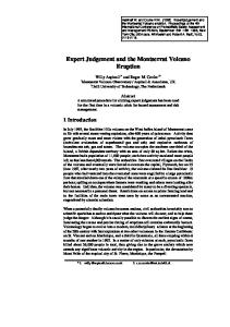
Fig. 2: Molar impaction classification according to angulation of the impacted third (and in the case of the horizontal impaction illustrated here – supernumerary/fourth) molar tooth.
Mesioangular
Vertical
Horizontal
Distoangular
2
Fig.3: Verticallyimpacted maxillary third molar tooth.
and the permanent maxillary canines. Cases can occur simply due to dental crowding, to space reduction following premature loss of primary teeth, or to an errant path of eruption. Third Molars Impacted mandibular third molars are traditionally classified according to position following the method of Winter [31] (Fig. 2). The most common type of impaction for mandibular molars is mesioangular. Mesioangularimpacted mandibular third molars lie obliquely in bone with the crown slanted in a mesial direction, generally in contact with the distal surface of the ipsilateral second permanent molar. Distoangular impacted third molars lie obliquely in bone with the crown slanted in a distal direction towards the ramus, the roots abutting the distal root of the second molar. Vertical impaction sees the third molar in normal angulation, but prevented
from eruption through impingement on the anterior ramus or distal surface of the second permanent molar tooth. With horizontal impaction, the third molar is positioned horizontally within the mandible with the crown directed towards the distal surface of the second molar. In each of these angular impactions, the third molar can be positioned at various depths within bone, and in relation to the mandibular canal. Panoramic radiographs clearly demonstrate the mesio-distal and vertical position of the impacted tooth, but do not provide details of the bucco-lingual positioning or angulation. This can be remedied in most cases by simply taking a true occlusal radiograph using a size 2 intraoral x-ray film to provide details in the third orthogonal plane. This will be sufficient if the tooth is not superimposed over the mandibular canal or intimately associated with that structure. In the few cases where this occurs and extraction is deemed necessary, the case may warrant more advanced imaging using conventional tomogra-
3
phy, computed tomography, or conebeam CT with 3-D reconstruction. Panoramic radiographs do not show bucco-lingual dimensions, so they should be supplemented because this dimension is critical for appropriate treatment planning. Damage to the contents of the mandibular canal can lead to temporary or permanent paresthesia of the ipsilateral side of the lower lip. While this complication may be unavoidable, it is less likely to occur given appropriate radiographic assessment. A prospective cohort study was performed in Sweden to measure the prevalence of disease in conjunction with mandibular third molars referred for removal [32]. Pericoronitis was found in 64 % of cases, caries in the third molar in 31 %, periodontitis in association with 8 %, caries in the second molar in 5 %, and root resorption of the second molar with 1 % of the molars having pathoses. The odds ratio for disease was highest for distoangular molars (5.8) and for impactions partially covered by soft tissue (6.7). The odds ratio for associated pathoses was between 22 and 34 times higher for molars partially covered by soft tissue than for impactions completely covered by soft or bone tissue. For distoangular molars the odds ratio for associated pathoses was 5 to 12 times higher than for molars in other positions. Impacted maxillary third molars (Fig. 3) may also be mesioangular, distoangular, vertical, or horizontal in position. In the maxilla, no structure as critical to surgical success as the mandibular canal is present; hence, pre-surgical assessment using panoramic and true occlusal radiography is almost invariably sufficient. Concerning the treatment of impacted third molars, systematic reviews have generally concluded that, in the absence of association with definite pathoses, it is best not to extract these teeth [33,34]. Fur-
ther, there is little evidence that retention of third molars has any effect on anterior crowding that might undermine orthodontic treatment [35]. Nevertheless, univariate analysis based on removal of 354 mandibular third molars identified increased patient age as a factor that predicted the surgical difficulty of third molar extractions, so delay could have a price if extraction is eventually needed [36]. Other factors ascribed to increased surgical difficulty included bony impaction, depth of tooth within bone, horizontal angulation, proximity to the inferior alveolar canal, male gender, and obesity [36]. When it is decided not to extract impacted third molars, periodic panoramic radiographs should be made to assure that no conditions develop that warrant subsequent surgical intervention. That periodic re-evaluation may be preferred to extraction of third molars in adolescence is supported by a study of 2,857 third molars assessed at age 18 years, and where 93 % were able to be clinically followed up at age 26 years [37]. Approximately 55 % of the teeth that were not considered impacted by age 18 had erupted by 26 years. Of the teeth considered impacted at age 18, 34 % had fully erupted by age 26. Of the maxillary teeth that were categorized as “impacted” at age 18 years, 36 % had fully erupted by age 26, whereas 26% of the mandibular teeth had done so (p