Disease of the Month
Thin Basement Membrane Nephropathy Karl Tryggvason and Jaakko Patrakka Division of Matrix Biology, Department of Medical Biochemistry and Biophysics, Karolinska Institutet, Stockholm, Sweden J Am Soc Nephrol 17: 813– 822, 2006. doi: 10.1681/ASN.2005070737
T
hin basement membrane nephropathy (TBMN) is the most common cause of persistent hematuria in children and adults, the other main causes being IgA nephropathy and Alport syndrome (1–3). In addition to hematuria, patients with TBMN usually have minimal proteinuria, normal renal function, and uniformly thinned glomerular basement membranes (GBM), as determined by electron microscopy. TBMN, which affects at least 1% of the population, is a lifelong nonprogressive disorder associated with family history. TBMN was first described approximately 80 yr ago as a curable form of hemorrhagic nephritis (4). Later, many cases of this microscopic, painless hematuria with good prognosis were shown to be inherited, and this fact is emphasized in the numerous names used for the disease in the literature, such as “congenital hereditary hematuria” (5), “hereditary hematuria” (6), “familial hematuric nephritis” (7), “familial benign hematuria” (8), “benign familial hematuria” (9,10), “familial benign essential hematuria” (11,12), “benign hereditary nephritis” (13), and “benign essential hematuria” (14). TBMN has also been referred to as familial hematuria (15), but that is a misnomer, as it does not distinguish it from the progressive Alport syndrome (3). Commonly used names are “thin membrane nephropathy” (16 –18), “thin basement membrane disease” (19 –25), “thin GBM nephropathy” (26), and “thin GBM syndrome” (27). The term “thin basement membrane nephropathy” is currently most widely used (1,2,28 –36), and this name is to be preferred, as it refers to a renal disorder that is associated with observable structural changes in the basement membrane (3) without necessarily being a “true disease.” It has been and still is a major clinical challenge to differentiate between TBMN characterized by nonprogressive hematuria and Alport syndrome with progressive hematuria as the main symptom. The connection between recurrent benign hematuria and thin GBM was first noted in electron microscopic analysis of kidney specimens in 1973 (11). This typical histopathologic feature of TBMN, i.e., uniform thinning of the GBM, could be found also at the early stages of Alport syndrome, which suggested that the pathomechanisms of the two diseases might overlap. This connection was verified at the
Published online ahead of print. Publication date available at www.jasn.org. Address correspondence to: Dr. Karl Tryggvason, Division of Matrix Biology, Department of Medical Biochemistry and Biophysics, Karolinska Institutet, Scheeles Va¨g 2, Stockholm S-171 77, Sweden. Phone: ⫹46-8-5248-7720; Fax: ⫹468-31-6165; E-mail:
[email protected] Copyright © 2006 by the American Society of Nephrology
gene level during the early 1990s, when the type IV collagen genes COL4A3, COL4A4, and COL4A5 were discovered and shown to be mutated in X-linked and autosomal Alport syndromes (37– 41), with subsequent demonstration of mutations in COL4A3 and COL4A4 in TBMN (10). At present, 40% of TBMN cases have been associated with the COL4A3 and COL4A4 genes (23,42), but it remains to be verified whether female carriers who are carriers for a mutation in the COL4A5 gene can develop true TBMN. In principle, DNA-based diagnosis of TBMN is possible, but such tests are not commercially available. At present, the clinical diagnosis still is made mainly on the basis of persistent hematuria with minimal proteinuria and normal renal function, combined with electron microscopy examination of biopsy showing thinned GBM; the use of immunologic examination of the type IV collagen ␣3 to ␣5 chains still is not being used extensively. However, as discussed below, these methods are insufficient; therefore, the majority of TBMN cases are still considered to be undiagnosed.
Epidemiology TBMN has been reported in all races, although most of the cases thus far have been reported in developed countries. Hematuria has been diagnosed at all ages (8,11,43), and several studies have indicated that the disease is somewhat more common in female than in male individuals, among both children and adults (8,17,18,27,42– 47), but other studies have not revealed such findings (26,48 –50). Exact prevalence of the disease is difficult to assess, as the diagnosis is made mostly on the basis of persistent hematuria combined with minimal proteinuria, whereas the number of electron microscopic analyses of renal biopsies showing thinned basement membrane have become less common (51). Thus, most of the cases remain undiagnosed. However, the prevalence may be estimated from known frequencies of persistent hematuria in the population and from the number of TBMN cases in archival series of renal biopsies, together with the knowledge of prevalence of autosomal Alport syndrome (51). Several studies have addressed the prevalence of hematuria and persistent hematuria in children and adults (52–56). Persistent hematuria is commonly defined as hematuria that is observed on at least two occasions, and in TBMN, a useful additional criterion could be that these two incidents occurred at least 2 yr apart (51). Persistence of hematuria is important from the point of view of TBMN diagnosis, as it distinguishes from other, more acute renal disorders, such as hematuria associated with streptococcal infections (51). The ISSN: 1046-6673/1703-0813
814
Journal of the American Society of Nephrology
prevalence of hematuria in children has been estimated to be 1 to 2% (52,53), but the prevalence in adults is not well known (3). According to Wang and Savige (51), persistent hematuria occurs consistently in as much as 6% of both children and adults. On the basis of both direct and indirect approaches, the overall prevalence of TBMN in the population has been estimated to be 1% (3). However, on the basis of observations of frequencies of persistent hematuria, thin basement membrane in renal biopsies, and autosomal recessive Alport syndrome, another estimate indicates a higher prevalence of ⬎1 but ⬍10% (51). When making estimations about the prevalence of TBMN by analyzing incidence of hematuria, one should keep in mind that not all patients who have TBMN have hematuria (or it is intermittent), that some patients with persistent hematuria have other signs of renal impairment (excluding the diagnosis of TBMN), and last that hematuria is not always of glomerular origin. It can be concluded, however, that TBMN is the most common inherited renal disorder.
J Am Soc Nephrol 17: 813– 822, 2006
teinuria or only minimally so, indicating that the podocyte slit diaphragm is not really affected. The proteinuria develops normally later than hematuria, and it is rarely seen in children (3). A significant proportion of adult patients, however, show mild to moderate proteinuria (3,11,17,18,48,59). Conversely, nephrotic-range proteinuria is rare even in older patients. Renal function in children with TBMN is normal (43,49,60), whereas adults have been reported to have low prevalence of renal insufficiency (16,17,26,27,46,47,61). The incidence of renal insufficiency in these patients might partially reflect the complicated differential diagnosis from autosomal or X-linked Alport syndrome or concurrent additional renal disease. Similarly, rare occurrence of hearing loss in TBMN may as well reflect the difficulties in differential diagnosis (18). Also, hypertension has been diagnosed in 11 to 31% of adults with TBMN (42,46,47,59,61) but very infrequently in pediatric patients (49). The interpretation of these results is difficult, but hypertension in patients with TBMN could be coincidental. Generally, the prognosis for the nephropathy in true TBMN is excellent.
Clinical Features The characteristic clinical manifestation of TBMN is persistent microscopic hematuria (1–3,8,11,16,17,26,27,43,44,46,57). Most patients with TBMN present only with hematuria, without additional symptoms or progression to renal impairment, and the condition is usually incidentally detected during health control. The age at diagnosis varies considerably, from as early as age 1 yr (8,43) up to age 86 yr (11). TBMN, which does not progress, has been documented for up to 30 yr (17). Microscopic analysis of the urine samples reveals red blood cells in most patients. At least a single episode of macroscopic hematuria is observed in 5 to 22% of patients, typically manifesting after exercise or during infection. However, occurrence of macroscopic hematuria seems to be more common in patients with Alport syndrome and IgA nephropathy (45,57). The urinary blood cells are dysmorphic with irregular shape and size, indicating hematuria of glomerular origin (17,18). Occasionally, the hematuria has disappeared with time (3,47,50,58). Despite hematuria, the individuals usually do not have pro-
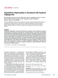
Renal Biopsy Rogers et al. (11) were the first to associate familial hematuria with thinning of the GBM. Light microscopy of renal samples in TBMN shows almost normal glomerular histology with only occasional mild mesangial cellular proliferation and matrix expansion (62,63). Slight attenuation of the GBM sometimes can be observed by Jones methenamine silver or periodic acidSchiff stains, suggesting GBM thinning. Erythrocytes may be identified in the urinary space (62). In approximately 5 to 25% of the cases, focal glomerular sclerosis and tubular fibrosis may be found with aging (26,62), but all histopathologic changes that are observed in light microscopic evaluation are nonspecific. Usually, direct immunofluorescent staining is negative for Ig and complement, but there sometimes are traces of segmental mesangial positivity for IgM or C3 and rarely IgG or IgA. Electron microscopy reveals the typical feature of TBMN, i.e., thinning of the GBM (Figure 1), but it does not distinguish between pure TBMN and thin GBM in early stages of Alport
Figure 1. Ultrastructural nature of the glomerular basement membrane in thin basement membrane nephropathy (TBMN). (A) In the normal adult male kidney, one can see a uniformly wide glomerular basement membrane (GBM) located between the fenestrated endothelial cells and the podocyte foot processes. (B) In TBMN, the GBM does not revel any structural abnormalities, but it is characteristically thinned, sometimes having only approximately half of the thickness in a normal kidney (A). Endothelial cells and podocyte foot processes maintain normal morphologic features. Bar ⫽ 500 nm. Courtesy: Kjell Hultenby, Karolinska University Hospital, Huddinge, Sweden.
J Am Soc Nephrol 17: 813– 822, 2006
syndrome. Normally, the GBM thickness varies with age and gender, and it is also influenced by the method and tissue preparations used. The GBM thinning in TBMN is uniform, which distinguishes it from patients who have focal thinning process, as is seen sometimes in normal children (64), and in patients with minimal-change nephrosis and some other forms of glomerulonephritides (65). In normally fixed kidney samples (e.g., with glutaraldehyde), the GBM appears as a trilaminar structure with a central lamina densa and an inner lamina rara interna and an outer lamina rara externa. However, in electron microscopic preparations made using quick-freezing, the GBM has a uniform appearance (66). According to a general agreement, the GBM thickness is determined from fixed samples as the distance between the outer limits of the endothelial cell and the base of podocyte foot process cell membranes, which are partially embedded in the GBM (62). As an additional rule, the thickness should be measured only in the peripheral capillaries, and multiple measurements should be made over several capillaries of multiple glomeruli (62). Normal GBM thickness has been estimated in several reports. Osawa et al. (67) compared 587 measurements from normal adult biopsies and 254 from autopsy samples and showed the average thickness to be 315 nm with a range of means from 239 to 453 nm. Haynes (68) reported the thickness in normal individuals aged between 11 and 26 yr to be 394 nm (range 372 to 632 nm). Using the complicated orthogonal intercept technique (69 –71), the mean GBM thickness in female adults was found to be 326 nm and that in male adults to be 373 nm. According to those reports, the thickness increased until a plateau at age 40 yr. According to Vogler et al. (72), the GBM thickness increases progressively in children until age 11. At ages 2 d to 1 yr, the thickness is 132 to 208 nm, at ages 1 to 6 yr 208 to 245 nm, and at age 6 to 11 yr 244 to 307 nm. As general guidelines concerning GBM thickness on the basis of several published results, the mean thickness in male adults is 370 ⫾ 50 nm and in female adults is 320 ⫾ 50 nm (71). In children, the GBM thickness is 150 nm at birth, 200 nm at age 1 yr, and approaches thickness in adults at age 11 yr (72). A central question, then, is how one should determine what constitutes thin GBM in an individual. The World Health Organization has proposed a threshold of 250 nm for adults and 180 nm for children between 2 and 11 yr of age (73). According to Vogler et al. (72), the criteria for TBMN in children vary between ⬍200 and ⬍250 nm, and in adults from ⬍200 nm (74) and ⬍250 nm (50) or up to ⬍264. This variation in recommendations is due to difficulties in standardizing the technical methods. This means that the morphologic criteria for thin GBM have to be evaluated carefully by the pathologist, and one has to take into account that the width of the GBM varies according to age and gender of the patient and also between different laboratories as a result of technical differences in sample preparation. Individual criteria should be established for each histologic laboratory. In TBMN, the cardinal findings are that the GBM is thinned in most of the glomerular capillaries and that there is absence of other significant glomerular pathology. The GBM thickness varies in individuals with TBMN (27,74 –79); at least 50% of the
Thin Basement Membrane Nephropathy
815
glomeruli have abnormally thin GBM. Only rare regions are observed with lamellation or regional thickening, a feature typically seen in Alport kidneys (1). This may pose a problem in differentiating between TBMN and Alport syndrome, as electron microscopic analysis at early stages of Alport syndrome can reveal similar uniform thinning of the GBM (see below for differential diagnosis). Immunohistochemical evaluation of the type IV collagen ␣3 to ␣5 chains in renal biopsy has become of major importance as a method to use in differentiating between TBMN and early stages of Alport syndrome with microscopic hematuria and thin GBM, as these chains usually are either absent or abnormally distributed in Alport syndrome (80,81).
Genetics of TBMN TBMN mainly manifests as an inherited disorder with dominant transmission affecting approximately one half of successive generations (11,28,48). Approximately two thirds of patients with TBMN have at least one other relative with hematuria (23). The remaining one third of patients may have de novo mutations or the nonpenetrance in other members of the family (82). Although TBMN is currently viewed as a common inherited disorder of type IV collagen, the genetic basis of this GBM disease was not understood until the late 1990s. The abnormally thin GBM could be associated with defective structure of the GBM, but this finding could not be connected with the loss of any GBM proteins, Thus, all of the GBM type IV collagen chains, ␣1 to ␣5, are present, as are all other known GBM proteins (83,84). The causes of TBMN started to unravel first through the identification of type IV collagen genes mutated in Alport syndrome. This kidney disease, that mainly affects male individuals, is characterized by early-onset, progressive hematuria that usually develops to ESRD. Type IV collagen is a specific triple-helical structural component of basement membranes that contain three ␣ chains in one individual molecule (85,86). These molecules assemble into a tightly cross-linked protein meshwork that forms the structural skeleton of the basement membranes. There are six distinct collagen IV ␣ chains, ␣1 to ␣6, that are encoded, respectively, by the specific genes COL4A1 to COL4A6 (Figure 2). The most common form of type IV collagen molecules contains ␣1 and ␣2 chains in a 2:1 ratio (85,86). In some specific basement membranes, such as the GBM, this ubiquitous form of collagen IV is replaced after birth by molecules with the chain composition ␣3:␣4:␣5 (87). This isoform of collagen IV contains more cysteines and is more cross-linked and resistant to degradation than the ubiquitous ␣1:␣1:␣2 form, and it seems to be essential for the integrity of the postnatal GBM, as well as for some other specialized basement membranes in the inner ear and lens capsule (86). The combination ␣5:␣5:␣6 is not present in the GBM, but it is present in the basement membrane of the Bowman’s capsule, as well as at some other locations in the body (88). Male individuals with progressive X chromosome–linked Alport syndrome were shown to contain mutations in the COL4A5 gene (38), whereas their mothers, carrying one normal
816
Journal of the American Society of Nephrology
J Am Soc Nephrol 17: 813– 822, 2006
Figure 2. Type IV collagen genes, ␣ chains, and GBM specific isoforms. (A) The six collagen IV genes (COL4A1 to COL4A6) located pairwise in a head-to-head manner on three different chromosomes generate six different ␣ chains that have a globular noncollagenous domain at their C-terminus (B). Three chains form triple-helical molecules that can have different combinations (C) (85,86). (D) Extracellularly, the triple-helical type IV collagen molecules form a network by associating with each other at their ends so that two molecules are cross-linked through their C-terminal globular domain (NC1) and for trimers associated with each other at the N-termini. In the embryonic GBM, the ubiquitous ␣1:␣1:␣2 trimer is the only isoform. After birth, this isoform is gradually replaced by an ␣3:␣4:␣5 isoform (87), which is more cross-linked through disulfide bonds within the collagenous regions (illustrated by black spots) and more resistant to extracellular proteolysis. Defects in the ␣3:␣4:␣5 trimers lead to TBMN or Alport syndrome, depending on the extensiveness of alleles involved (see text). Illustration by Josh Gramling—Gramling Medical Illustration.
allele in addition to the abnormal allele, most often exhibited mild hematuria. Subsequently, the autosomal recessive forms of Alport syndrome were shown to be due to mutations in the COL4A3 and COL4A4 genes that are located head to head on chromosome 2 (Figure 2) (41,89). Autosomal Alport syndrome can be caused by homozygous mutations in either of COL4A3 or COL4A4 or by combined heterozygosity for both. Lemmink et al. (10) noted that some carriers of the autosomal forms of Alport had thin GBM, and this prompted them to analyze the COL4A3 and COL4A4 genes in patients with TBMN. They first showed linkage of the disease with the COL4A3/COL4A4 locus and subsequently identified a mutation that leads to a substitution of glycine to glutamic acid in the collagenous domain of the COL4A4 gene. Thus, patients with TBMN that is linked to chromosome 2 are heterozygous for mutations in either COL4A3 or COL4A4, and they represent a carrier status for autosomal recessive Alport syndrome (Figure 3A), in a similar manner as female individuals with mutations in one COL4A5 allele are carriers for X-linked Alport syndrome in male individuals (Figure 3B). Savige et al. (23) showed that up to 36% of TBMN cases associate with the COL4A3/COL4A4 locus for
autosomal recessive Alport syndrome. More recently, several studies that aimed to identify mutations in the COL4A3 and COL4A4 genes, as well as their common promoter region, revealed that heterozygous mutations in these two genes are found in many patients with TBMN (15,23,24,34,42,90 –92). The current understanding of the mode of inheritance for autosomal TBMN and autosomal recessive Alport syndrome is depicted in Figure 3A. Thus far, 21 COL4A3 and COL4A4 gene mutations have been identified in patients with TBMN (82). Mutations in TBMN are scattered throughout the COL4A3 and COL4A4 genes without any “hot spots,” similar to what has been observed in the same genes and COL4A5 in patients with autosomal recessive and X-linked Alport syndrome, respectively. Most mutations result in single nucleotide substitutions and lead to missense or nonsense mutations. Many of the mutations lead to a replacement of a glycine in the collagenous domain but also missense mutations in nonglycine residues of the collagenous domain. Single amino acid substitutions have been identified in the noncollagenous domain, and nonsense and splice site mutations have also been identified. In addition, six insertion or deletion
J Am Soc Nephrol 17: 813– 822, 2006
Figure 3. Illustration of possible modes of inheritance for autosomal and X-linked forms of TBMN and Alport syndrome (AS). (A) TBMN and autosomal recessive AS in two intermarried families with a mutation (blue color) in COLA3 located on chromosome 2. Half-shaded symbols represent hematuria and heterozygosity state for a mutation in COL4A3 (or COL4A4), and fully shaded symbols represent homozygosity for a mutation or compound heterozygosity for mutations in COL4A3 and COL4A4 resulting in AS. First-generation heterozygotes with TBMN can be considered carriers for recessive AS. Half of the second-generation individuals can be expected to have TBMN. A third-generation offspring of a couple in which both parents have TBMN can be expected to get a child with AS, half of the children being expected to be TBMN patients. However, it has not yet been demonstrated that the offspring of TBMN-affected parents are at risk for developing AS. (B) TBMN simulates nephropathy and AS in a single family in which the mother is heterozygous for a mutation in the X chromosomal COL4A5 gene. Each child has a 25% risk for inheriting the mutant allele. Half-shaded symbols represent heterozygosity and TBMN mimicking disease in females, and fully shaded symbols represent AS (X-AS) in male proband. Illustration by Josh Gramling—Gramling Medical Illustration.
mutations have been identified. To date, no mutations have been described in the promoter region of COL4A3/COL4A4 genes in TBMN, in contrast to that observed in Alport syndrome. Heterozygous mutations that are found in patients with TBMN are very similar and, in some cases, identical to those identified in patients with autosomal recessive Alport syndrome. So far, only a few studies have attempted to correlate the type of mutation in type IV collagen to the clinical features of TBMN. It is interesting that Wang et al. (34) reported that the same mutations in different family members can result in different clinical features. Autosomal dominant Alport syndrome is also caused by heterozygous mutations in the COL4A3 or COL4A4 gene. As described above, the nephropathy in autosomal dominant Alport syndrome follows a much more deleterious course, leading to renal failure and lamellated GBM, in contrast to the symptomless hematuria observed in TBMN. It would be crucial to understand the cause of this difference in the clinical outcome of these patients with heterozygous COL4A3/COL4A4 mutations, but, so far, too few mutations have been described in autosomal dominant Alport syndrome and TBMN to distinguish the two on the basis of the genotype. The COL4A3 and COL4A4 genes are huge in size; therefore,
Thin Basement Membrane Nephropathy
817
the identification of mutations in the genes is difficult. In studies made by the Savige group, the mutation detection rate for COL4A3 and COL4A4 mutations in patients with TBMN was 11 and 17%, respectively, using single-strand conformation polymorphism (SSCP) (24,34). However, the rate increased to as high as 67% when only individuals from families in which TBMN was linked to the COL4A3/COL4A4 locus were included. The low detection rate may be due to many other reasons. First, the use of SSCP instead of direct sequencing of exons lowers the detection rate (93). Thus, sequencing of all exon regions has been shown to reveal ⬎80% of mutations in COL4A5 in X-linked Alport syndrome (93). Second, the primers used in the studies were not designed to detect possible splice site mutations. Furthermore, because of numerous polymorphisms in the genes, some pathogenic alterations in the gene sequence may be missed and interpreted as nonpathogenic polymorphisms. Practically all female carriers for X-linked Alport syndrome who have a mutation in one COL4A5 allele exhibit hematuria. In a study of 288 heterozygous female individuals from 329 families with X-linked Alport syndrome, 96% had hematuria, and a relatively large proportion (30%) had progressive disease that led to chronic renal failure and ESRD (94). This type of progression is not considered typical for TBMN (1). However, the female carriers for X-linked Alport syndrome do exhibit thinning and thickening or diffuse thinning of the GBM. Thus, such cases with thin GBM that mimics TBMN could be considered as TBMN cases. The relationship of thin GBM and Xlinked Alport syndrome is depicted in Figure 3B. TBMN is a disorder of the trimeric ␣3:␣4:␣5 isoform of type IV collagen. Therefore, it could be anticipated that a single mutation in any of the alleles for these three genes would lead to a similar phenotype, as they all affect the same trimeric protein. However, it seems to be a common view in the literature that only mutations in COL4A3 or COL4A4 lead to TBMN, as depicted in Figure 3A. It is still possible that X-linked TBMN exists with inheritance, as depicted in Figure 3B. For example, clinical studies that were performed before identification of the COL4A5 gene indicated the existence of such cases (95,96); similarly, a more recent study by Liapis et al. (97) suggested that a connection between some cases of TBMN and COL4A5 could exist. It is important to solve this controversy by more extensive sequence analyses of the COL4A5 gene in individuals with TBMN that does not show association with chromosome 2. Many families with TBMN do not show linkage to the COL4A3/COL4A4 or COL4A5 loci (98,99). This may be explained by several factors. A high rate of de novo mutations has been shown in the COL4A5 gene in X-linked Alport syndrome, and this might be the case in COL4A3 and COL4A4 genes as well (100). Incomplete penetrance of hematuria in heterozygous carriers has also been reported (101), and this can influence linkage analyses. Also, coincidental hematuria without proteinuria in family member makes it more difficult to establish linkage. Last, it is possible that some TBMN cases are due to mutations in another, as yet unknown gene. In relation to this, Rana et al. (82) recently studied the linkage of TBMN in nine families who did not show segregation to COL4A3/COL4A4
818
Journal of the American Society of Nephrology
J Am Soc Nephrol 17: 813– 822, 2006
Differential Diagnosis of TBMN and Alport Syndrome
and ruled out linkage to MYH9 (mutations in this gene result in Alport-like nephropathy) and to genes coding for GBM components laminin-5, perlecan, and fibronectin. So far, there are no data showing any genetic linkage to another loci than COL4A3/COL4A4 and COL4A5, supporting the view that TBMN is a disease of the adult form of GBM type IV collagen (␣3:␣4:␣5).
The characteristic manifestation of TBMN is persistent hematuria of glomerular origin; therefore, it is important to distinguish this benign disease from other, superimposing causes of glomerular bleeding. These include IgA nephropathy, postinfectious glomerulonephritis, mesangiocapillary glomerulonephritis, and lupus nephritis. Whereas IgA nephropathy and TBMN may be difficult to distinguish on clinical grounds alone, other forms of glomerulonephritis are frequently associated with additional clinical features that rarely are seen in TBMN, such as proteinuria, hypertension, renal impairment, and systemic symptoms. For initial diagnosis of TBMN, it is critical to be able to distinguish the condition from early stages of Alport syndrome that can yield very similar findings as TBMN. Differential diagnosis of these two is extremely important because of the dramatically different outcomes of the two disorders. X-linked Alport syndrome, which accounts for approximately 85% of Alport syndrome cases, generally occurs in 1 in 50,000 live births (104), but it can affect as many as 1 in 5000 male individuals, such as in Utah (38). Typical Alport syndrome findings, such as hearing loss, lenticonus, and severe structural changes in the GBM, develop usually first during adolescence. For early diagnosis of Alport syndrome, it very important to clarify family history. Approximately 95% of the female carriers of Alport syndrome have hematuria (105), but they cannot be distinguished from individuals with TBMN (1). However, knowledge about hearing loss,
Pathogenesis In general, development of autosomal nonprogressive TBMN involves heterozygous mutations in either COL4A3 or COL4A4, whereas homozygosity or combined heterozygosity of mutations in these genes results in autosomal recessive Alport syndrome characterized by deterioration of the GBM (3,92). Similarly, a TBMN-like phenotype may be caused in a female individual with heterozygosity for a mutation in the X-chromosomal COL4A5 gene. This could be explained by a “dose effect” whereby absence of one normal allele leads to lower production of the ␣3:␣4:␣5 trimer and thin GBM, whereas the loss of two alleles results in lack of the ␣3:␣4:␣5 trimer and Alport syndrome (35). However, there are reports that a single heterozygous mutation in COL4A3 or COL4A4 can cause Alport syndrome in adult life (15,102,103). This could mean that some mutations are more serious than others, but it still cannot be excluded yet that in these cases, there is another unknown mutation somewhere else in the other allele or gene that could lead to absence of the chain. To date, too few mutations have been identified in TBMN and autosomal dominant Alport for making conclusions about the differences in pathogenesis.
Table 1. Expression of collagen IV ␣3, ␣4 and ␣5 chains in normal, TBMN, and AS kidneysa X-Linked AS Normal
GBM ␣3(IV) ␣4(IV) ␣5(IV) TBM (distal and collecting) ␣3(IV) ␣4(IV) ␣5(IV) Bowman’s capsule ␣3(IV) ␣4(IV) ␣5(IV) Epidermal BM ␣5(IV)
TBMN
b
c
Male
Female
Autosomal Recessive ASd
⫹ ⫹ ⫹
⫹ ⫹ ⫹
⫺ ⫺ ⫺
Mosaic Mosaic Mosaic
⫺ ⫺ ⫺
⫹ ⫹ ⫹
⫹ ⫹ ⫹
⫺ ⫺ ⫺
Mosaic Mosaic Mosaic
⫺ ⫺ ⫹
⫹ ⫹ ⫹
⫹ ⫹ ⫹
⫺ ⫺ ⫺
Mosaic Mosaic Mosaic
⫺ ⫺ ⫹
⫹
⫹
⫺
Mosaic
⫹
a Modified from reference (81). AS, Alport syndrome; GBM, glomerular basement membrane; TBMN, thin basement membrane nephropathy; b Approximately 20% of male individuals with X-linked AS show positive renal basement membrane staining for ␣3, ␣4, and ␣5 and for ␣5 in epidermal basement membranes. c Approximately 30% of female carriers for X-linked AS exhibit positive uninterrupted staining for renal basement membranes for the ␣3, ␣4, and ␣5 chains. d Some patients with autosomal recessive AS show positive staining for ␣3, ␣4, and ␣5 in renal basement membranes.
J Am Soc Nephrol 17: 813– 822, 2006
lenticonus, or retinopathy in other relatives can give a hint about X-linked Alport syndrome. If the existence of X-linked Alport syndrome is not clear, then renal biopsy should be performed. The characteristic uniform thinning of the GBM that is observed in electron microscopy often confirms the diagnosis of TBMN. However, the importance of this finding must not be overemphasized, as abnormally thin GBM can also be observed at the early stages of Alport syndrome. Therefore, differential diagnosis from X-linked or autosomal recessive Alport syndrome should include immunohistochemical analysis of the type IV collagen ␣3 to ␣5 chains (2,81). In both forms of Alport syndrome, the expression of all three chains is usually significantly reduced or absent, whereas in TBMN, the expression is comparable to normal levels (Table 1). Such analysis can be performed in many cases on skin biopsies, such as in X-linked Alport syndrome, in which the ␣5 chain may be absent from the epidermal basement membrane, whereas it is not absent in TBMN (2,81). However, immunohistochemical analysis can yield in some cases false-negative results, because it has been shown that approximately 20% of autosomal Alport syndrome cases and 30% of cases of female carriers for X-linked Alport syndrome express low or even normal levels (80,102). Genetic analyses can verify the diagnosis, but DNA analysis unfortunately is not generally available. Segregation of the disease with the COL4A5 locus and sequencing of all of the exon regions can reveal ⬎80% of the mutations in X-linked Alport syndrome (93), whereas more easily performed methods, such as SSCP, yield only between 37 and 50% of the mutations (106 –108). Unfortunately, an assay for screening COL4A5 mutations is currently not commercially available. Similar sequencing of the COL4A3 and COL4A4 genes is also possible in specialized laboratories. It would be important to be able to provide DNA sequencing of the COL4A3, COL4A4, and COL4A5 genes for making diagnosis of TBMN and Alport syndrome that together form a very large group of renal disorders. With today’s technologies, such analysis methods should not have to be very costly.
Management of TBMN Currently, no clear evidence-based treatment protocols are available for TBMN. Although the condition generally has an excellent outcome and in many cases cannot really be considered a disease, it must be noted that it may not always be benign, and the worsening of the clinical picture has been reported (61,109). It is still a dilemma for the physician to know whether such cases are in fact true TBMN cases or misdiagnosed Alport syndrome. In any case, patients who receive a diagnosis of TBMN should be monitored for the appearance of hypertension, proteinuria, or renal insufficiency, and patients who develop these complications should be treated accordingly.
Conclusion TBMN is one of the most common disorders of the kidney, affecting at least 1% of the population. It seems to be a disease of the adult GBM type IV collagen trimer ␣3:␣4:␣5. Genetic
Thin Basement Membrane Nephropathy
819
evidence indicates that autosomal TBMN is caused by heterozygous mutations in either COL4A3 or COL4A4, whereas homozygous or combined heterozygous mutations in the same genes lead to autosomal recessive Alport syndrome. Thus, individuals with autosomal TBMN are carriers for autosomal Alport syndrome. Heterozygosity for mutations in COL4A5 in female carriers for X-linked Alport syndrome can also mimic a TBMN condition, although some of those individuals will later develop progressive hematuria (94). It is not clear whether the individuals with TBMN and female carriers who simulate TBMN and develop progressive disease could have a second mutation in some other of the six alleles that encode the ␣3: ␣4:␣5 collagen form. The main clinical problem is to differentiate between TBMN and X-linked or autosomal recessive Alport syndrome, as Alport syndrome has severe outcome. Although family history and electron microscopy of renal biopsies are helpful for diagnosis, immunohistological examination of expression of the type IV collagen ␣3 to ␣5 chains is currently the most informative method. It would be of utmost importance to make sequencing analyses of the COL4A3, COL4A4, and COL4A5 genes available to the clinic as a routine diagnostic method of TBMN and Alport syndrome.
Acknowledgments We are grateful to Dr. Martin Gregory for critical comments on this manuscript.
References 1. Savige J, Rana K, Tonna S, Buzza M, Dagher H, Wang YY: Thin basement membrane nephropathy. Kidney Int 64: 1169 –1178, 2003 2. Kashtan CE: Familial hematuria due to type IV collagen mutations: Alport syndrome and thin basement membrane nephropathy. Curr Opin Pediatr 16: 177–181, 2004 3. Gregory MC: The clinical features of thin basement membrane nephropathy. Semin Nephrol 25: 140 –145, 2005 4. Baehr G: Benign and curable form of hemorrhagic nephritis. JAMA 86: 1001–1004, 1926 5. Reyersbach GC, Butler AM: Congenital hereditary hematuria. N Engl J Med 251: 377–380, 1954 6. Russell E, Smith N: Hereditary hematuria. Am J Dis Child 98: 353–358, 1959 7. Rome L, Cuppage F, Vertes V: Familial hematuric nephritis. Pediatrics 38: 808 – 818, 1966 8. McConville JM, McAdams AJ: Familial and nonfamilial benign hematuria. J Pediatr 69: 207–214, 1966 9. Tina L, Jenis E, Jose P, Medani C, Papadopoulou Z, Calcagno P: The glomerular basement membrane in benign familial hematuria. Clin Nephrol 17: 1– 4, 1982 10. Lemmink HH, Nillesen WN, Mochizuki T, Schroder CH, Brunner HG, van Oost BA, Monnens LA, Smeets HJ: Benign familial hematuria due to mutation of the type IV collagen alpha4 gene. J Clin Invest 98: 1114 –1118, 1996 11. Rogers PW, Kurtzman NA, Bunn SM Jr, White MG: Familial benign essential hematuria. Arch Intern Med 131: 257– 262, 1973 12. Gubler MC, Levy M, Naizot C, Habib R: Glomerular basement membrane changes in hereditary glomerular diseases. Ren Physiol 3: 405– 413, 1980
820
Journal of the American Society of Nephrology
13. Peterson AS, Schubert JJ: Benign hereditary nephritis. J Fam Pract 4: 437– 441, 1977 14. Mihatsch MJ, Zollinger HU: Kidney disease. Pathol Res Pract 167: 88 –117, 1980 15. Longo I, Porcedda P, Mari F, Giachino D, Meloni I, Deplano C, Brusco A, Bosio M, Massella L, Lavoratti G, Roccatello D, Frasca G, Mazzucco G, Muda AO, Conti M, Fasciolo F, Arrondel C, Heidet L, Renieri A, De Marchi M: COL4A3/COL4A4 mutations: From familial hematuria to autosomal-dominant or recessive Alport syndrome. Kidney Int 61: 1947–1956, 2002 16. Dische FE, Anderson VE, Keane SJ, Taube D, Bewick M, Parsons V: Incidence of thin membrane nephropathy: Morphometric investigation of a population sample. J Clin Pathol 43: 457– 460, 1990 17. Perry GJ, George CR, Field MJ, Collett PV, Kalowski S, Wyndham RN, Newland RC, Lin BP, Kneale KL, Lawrence JR: Thin-membrane nephropathy—A common cause of glomerular haematuria. Med J Aust 151: 638 – 642, 1989 18. Aarons I, Smith PS, Davies RA, Woodroffe AJ, Clarkson AR: Thin membrane nephropathy: A clinico-pathological study. Clin Nephrol 32: 151–158, 1989 19. Takemura T, Yanagida H, Yagi K, Moriwaki K, Okada M: Alport syndrome and benign familial hematuria (thin basement membrane disease) in two brothers of a family with hematuria. Clin Nephrol 60: 195–200, 2003 20. Basta-Jovanovic G, Venkataseshan VS, Gil J, Kim DU, Dikman SH, Churg J: Morphometric analysis of glomerular basement membranes (GBM) in thin basement membrane disease (TBMD). Clin Nephrol 33: 110 –114, 1990 21. Colville D, Savige J, Branley P, Wilson D: Ocular abnormalities in thin basement membrane disease. Br J Ophthalmol 81: 373–377, 1997 22. Sakai K, Muramatsu M, Ogiwara H, Kawamura T, Arai K, Aikawa A, Ohara T, Mizuiri S, Joh K, Naito I, Hasegawa A: Living related kidney transplantation in a patient with autosomal-recessive Alport syndrome. Clin Transplant 17[Suppl 10]: 4 – 8, 2003 23. Buzza M, Wilson D, Savige J: Segregation of hematuria in thin basement membrane disease with haplotypes at the loci for Alport syndrome. Kidney Int 59: 1670 –1676, 2001 24. Buzza M, Dagher H, Wang YY, Wilson D, Babon JJ, Cotton RG, Savige J: Mutations in the COL4A4 gene in thin basement membrane disease. Kidney Int 63: 447– 453, 2003 25. Ueda T, Nakajima M, Akazawa H, Maruhashi Y, Shimoyama H, Sakagami Y, Takagawa K, Kamitsuji H, Naito I, Sado Y, Yoshioka A: Quantitative analysis of glomerular type IV collagen alpha3–5 chain expression in children with thin basement membrane disease. Nephron 92: 271– 278, 2002 26. Nieuwhof CM, de Heer F, de Leeuw P, van Breda Vriesman PJ: Thin GBM nephropathy: Premature glomerular obsolescence is associated with hypertension and late onset renal failure. Kidney Int 51: 1596 –1601, 1997 27. Abe S, Amagasaki Y, Iyori S, Konishi K, Kato E, Sakaguchi H, Shimoyama K: Thin basement membrane syndrome in adults. J Clin Pathol 40: 318 –322, 1987 28. Gauthier B, Trachtman H, Frank R, Valderrama E: Familial thin basement membrane nephropathy in children with asymptomatic microhematuria. Nephron 51: 502–508, 1989 29. Lang S, Stevenson B, Risdon RA: Thin basement mem-
J Am Soc Nephrol 17: 813– 822, 2006
brane nephropathy as a cause of recurrent haematuria in childhood. Histopathology 16: 331–337, 1990 30. Makino H, Ichiyasu A, Ota Z: Two cases of aortitis syndrome associated with thin basement membrane nephropathy. Clin Nephrol 37: 106 –107, 1992 31. Roy-Chaudhury P, Simpson JG, Edward N: Profuse haematuria and clot colic in thin basement membrane nephropathy. Nephrol Dial Transplant 7: 1139 –1141, 1992 32. Saxena S, Davis DJ: Glomerular alterations in idiopathic haematuria—Ultrastructural and morphometric analysis. Indian J Pathol Microbiol 35: 326 –332, 1992 33. Collar JE, Ladva S, Cairns TD, Cattell V: Red cell traverse through thin glomerular basement membranes. Kidney Int 59: 2069 –2072, 2001 34. Wang YY, Rana K, Tonna S, Lin T, Sin L, Savige J: COL4A3 mutations and their clinical consequences in thin basement membrane nephropathy (TBMN). Kidney Int 65: 786–790, 2004 35. Gregory MC: Alport syndrome and thin basement membrane nephropathy: Unraveling the tangled strands of type IV collagen. Kidney Int 65: 1109 –1110, 2004 36. Saxena U, Auerbach BJ, Ferguson E, Wolle J, Marcel YL, Weisgraber KH, Hegele RA, Bisgaier CL: Apolipoprotein B and E basic amino acid clusters influence low-density lipoprotein association with lipoprotein lipase anchored to the subendothelial matrix. Arterioscler Thromb Vasc Biol 15: 1240 –1247, 1995 37. Hostikka SL, Eddy RL, Byers MG, Hoyhtya M, Shows TB, Tryggvason K: Identification of a distinct type IV collagen alpha chain with restricted kidney distribution and assignment of its gene to the locus of X chromosome-linked Alport syndrome. Proc Natl Acad Sci U S A 87: 1606 –1610, 1990 38. Barker DF, Hostikka SL, Zhou J, Chow LT, Oliphant AR, Gerken SC, Gregory MC, Skolnick MH, Atkin CL, Tryggvason K: Identification of mutations in the COL4A5 collagen gene in Alport syndrome. Science 248: 1224 –1227, 1990 39. Mariyama M, Leinonen A, Mochizuki T, Tryggvason K, Reeders ST: Complete primary structure of the human alpha 3(IV) collagen chain. Coexpression of the alpha 3(IV) and alpha 4(IV) collagen chains in human tissues. J Biol Chem 269: 23013–23017, 1994 40. Leinonen A, Mariyama M, Mochizuki T, Tryggvason K, Reeders ST: Complete primary structure of the human type IV collagen alpha 4(IV) chain. Comparison with structure and expression of the other alpha (IV) chains. J Biol Chem 269: 26172–26177, 1994 41. Mochizuki T, Lemmink HH, Mariyama M, Antignac C, Gubler MC, Pirson Y, Verellen-Dumoulin C, Chan B, Schroder CH, Smeets HJ, Reeders ST: Identification of mutations in the alpha 3(IV) and alpha 4(IV) collagen genes in autosomal recessive Alport syndrome. Nat Genet 8: 77– 81, 1994 42. Badenas C, Praga M, Tazon B, Heidet L, Arrondel C, Armengol A, Andres A, Morales E, Camacho JA, Lens X, Davila S, Mila M, Antignac C, Darnell A, Torra R: Mutations in the COL4A4 and COL4A3 genes cause familial benign hematuria. J Am Soc Nephrol 13: 1248 –1254, 2002 43. Yoshikawa N, Matsuyama S, Iijima K, Maehara K, Okada S, Matsuo T: Benign familial hematuria. Arch Pathol Lab Med 112: 794 –797, 1988 44. Marks MI, McLaine PN, Drummond KN: Proteinuria in children with febrile illnesses. Arch Intern Med 45: 250 –253, 1970 45. Piqueras AI, White RH, Raafat F, Moghal N, Milford DV:
J Am Soc Nephrol 17: 813– 822, 2006
Renal biopsy diagnosis in children presenting with haematuria. Pediatr Nephrol 12: 386 –391, 1998 46. Auwardt R, Savige J, Wilson D: A comparison of the clinical and laboratory features of thin basement membrane disease (TBMD) and IgA glomerulonephritis (IgA GN). Clin Nephrol 52: 1– 4, 1999 47. Goel S, Davenport A, Goode NP, Shires M, Hall CL, Harrison PR, Maciver AG: Clinical features and outcome of patients with thin and ultrathin glomerular membranes. QJM 88: 785–793, 1995 48. Blumenthal SS, Fritsche C, Lemann J Jr: Establishing the diagnosis of benign familial hematuria. The importance of examining the urine sediment of family members. JAMA 259: 2263–2266, 1988 49. Roth KS, Amaker BH, Chan JC: Pediatric hematuria and thin basement membrane nephropathy: What is it and what does it mean? Clin Pediatr (Phila) 40: 607– 613, 2001 50. Tiebosch AT, Frederik PM, van Breda Vriesman PJ, Mooy JM, van Rie H, van de Wiel TW, Wolters J, Zeppenfeldt E: Thin-basement-membrane nephropathy in adults with persistent hematuria. N Engl J Med 320: 14 –18, 1989 51. Wang YY, Savige J: The epidemiology of thin basement membrane nephropathy. Semin Nephrol 25: 136 –139, 2005 52. Dodge WF, West EF, Smith EH, Bruce H 3rd: Proteinuria and hematuria in schoolchildren: Epidemiology and early natural history. J Pediatr 88: 327–347, 1976 53. Vehaskari VM, Rapola J, Koskimies O, Savilahti E, Vilska J, Hallman N: Microscopic hematuria in school children: Epidemiology and clinicopathologic evaluation. J Pediatr 95: 676 – 684, 1979 54. Ritchie CD, Bevan EA, Collier SJ: Importance of occult haematuria found at screening. BMJ (Clin Res Ed) 292: 681– 683, 1986 55. Hogg RJ, Harris S, Lawrence DM, Henning PH, Wigg N, Jureidini KF: Renal tract abnormalities detected in Australian preschool children. J Paediatr Child Health 34: 420 – 424, 1998 56. Chadban SJ, Briganti EM, Kerr PG, Dunstan DW, Welborn TA, Zimmet PZ, Atkins RC: Prevalence of kidney damage in Australian adults: The AusDiab kidney study. J Am Soc Nephrol 14[Suppl 2]: S131–S138, 2003 57. Trachtman H, Weiss RA, Bennett B, Greifer I: Isolated hematuria in children: Indications for a renal biopsy. Kidney Int 25: 94 –99, 1984 58. Packham DK, Perkovic V, Savige J, Broome MR: Hematuria in thin basement membrane nephropathy. Semin Nephrol 25: 146 –148, 2005 59. Dische FE, Weston MJ, Parsons V: Abnormally thin glomerular basement membranes associated with hematuria, proteinuria or renal failure in adults. Am J Nephrol 5: 103– 109, 1985 60. Schroder CH, Bontemps CM, Assmann KJ, Schuurmans Stekhoven JH, Foidart JM, Monnens LA, Veerkamp JH: Renal biopsy and family studies in 65 children with isolated hematuria. Acta Paediatr Scand 79:630 – 636, 1990 61. van Paassen P, van Breda Vriesman PJ, van Rie H, Tervaert JW: Signs and symptoms of thin basement membrane nephropathy: A prospective regional study on primary glomerular disease—The Limburg Renal Registry. Kidney Int 66: 909 –913, 2004 62. Foster K, Markowitz GS, D’Agati VD: Pathology of thin basement membrane nephropathy. Semin Nephrol 25: 149 – 158, 2005
Thin Basement Membrane Nephropathy
821
63. Kincaid-Smith P: Thin basement membrane disease. In: Textbook of Nephrology, Baltimore, Williams & Wilkins, 1995, pp 760 –764 64. Bloom P, Hartmann J, Vernier R: An electron microscopic evaluation of the width of normal basement membrane in children with hematuria. J Pathol 142: 263–267, 1984 65. Hill G, Jenis E, Goodloe S: The nonspecificity of the ultrastructural alterations in hereditary nephritis with additional observations in benign familial hematuria. Lab Invest 31: 516 –532, 1974 66. Inoue S: Ultrastructural architecture of basement membranes. Contrib Nephrol 107: 21–28, 1994 67. Osawa G, Kimmelstiel P, Seiling V: Thickness of glomerular basement membranes. Am J Clin Pathol 45: 7–20, 1966 68. Haynes WD: The normal human renal glomerulus. Virchows Arch B Cell Pathol Incl Mol Pathol 35: 133–158, 1981 69. Gundersen HJ, Jensen TB, Osterby R: Distribution of membrane thickness determined by lineal analysis. J Microsc 113: 27– 43, 1978 70. Jensen EB, Gundersen HJ, Osterby R: Determination of membrane thickness distribution from orthogonal intercepts. J Microsc 115: 19 –33, 1979 71. Steffes MW, Barbosa J, Basgen JM, Sutherland DE, Najarian JS, Mauer SM: Quantitative glomerular morphology of the normal human kidney. Lab Invest 49: 82– 86, 1983 72. Vogler C, McAdams AJ, Homan SM: Glomerular basement membrane and lamina densa in infants and children: an ultrastructural evaluation. Pediatr Pathol 7: 527–534, 1987 73. Churg J, Bernstein J, Glassock R: Renal Disease: Classification and Atlas of Glomerular Diseases, 2nd Ed., New York, IgakuShoin, 1995 74. Cosio FG, Falkenhain ME, Sedmak DD: Association of thin glomerular basement membrane with other glomerulopathies. Kidney Int 46: 471– 474, 1994 75. Shindo S, Yoshimoto M, Kuriya N, Bernstein J: Glomerular basement membrane thickness in recurrent and persistent hematuria and nephrotic syndrome: Correlation with sex and age. Pediatr Nephrol 2: 196 –199, 1988 76. Saxena S, Davies DJ, Kirsner RL: Thin basement membranes in minimally abnormal glomeruli. J Clin Pathol 43: 32–38, 1990 77. Marquez B, Stavrou F, Zouvani I, Anastasiades E, Patsias C, Pierides A, Kyriacou K: Thin glomerular basement membranes in patients with hematuria and minimal change disease. Ultrastruct Pathol 23: 149 –156, 1999 78. Nogueira M, Cartwright J Jr, Horn K, Doe N, Shappell S, Barrios R, Coroneos E, Truong LD: Thin basement membrane disease with heavy proteinuria or nephrotic syndrome at presentation. Am J Kidney Dis 35: E15, 2000 79. Dische FE: Measurement of glomerular basement membrane thickness and its application to the diagnosis of thin-membrane nephropathy. Arch Pathol Lab Med 116: 43– 49, 1992 80. Gubler MC, Knebelmann B, Beziau A, Broyer M, Pirson Y, Haddoum F, Kleppel MM, Antignac C: Autosomal recessive Alport syndrome: Immunohistochemical study of type IV collagen chain distribution. Kidney Int 47: 1142– 1147, 1995 81. Kashtan CE: The nongenetic diagnosis of thin basement membrane nephropathy. Semin Nephrol 25: 159 –162, 2005 82. Rana K, Wang YY, Buzza M, Tonna S, Zhang KW, Lin T,
822
Journal of the American Society of Nephrology
Sin L, Padavarat S, Savige J: The genetics of thin basement membrane nephropathy. Semin Nephrol 25: 163–170, 2005 83. Lajoie G: Approach to the diagnosis of thin basement membrane nephropathy in females with the use of antibodies to type IV collagen. Arch Pathol Lab Med 125: 631– 636, 2001 84. Pettersson E, Tornroth T, Wieslander J: Abnormally thin glomerular basement membrane and the Goodpasture epitope. Clin Nephrol 33: 105–109, 1990 85. Hudson BG, Reeders ST, Tryggvason K: Type IV collagen: Structure, gene organization, and role in human diseases. Molecular basis of Goodpasture and Alport syndromes and diffuse leiomyomatosis. J Biol Chem 268: 26033–26036, 1993 86. Hudson BG, Tryggvason K, Sundaramoorthy M, Neilson EG: Alport’s syndrome, Goodpasture’s syndrome, and type IV collagen. N Engl J Med 348: 2543–2556, 2003 87. Miner JH, Sanes JR: Collagen IV alpha 3, alpha 4, and alpha 5 chains in rodent basal laminae: Sequence, distribution, association with laminins, and developmental switches. J Cell Biol 127: 879 – 891, 1994 88. Saito K, Naito I, Seki T, Oohashi T, Kimura E, Momota R, Kishiro Y, Sado Y, Yoshioka H, Ninomiya Y: Differential expression of mouse alpha5(IV) and alpha6(IV) collagen genes in epithelial basement membranes. J Biochem (Tokyo) 128: 427– 434, 2000 89. Lemmink HH, Mochizuki T, van den Heuvel LP, Schroder CH, Barrientos A, Monnens LA, van Oost BA, Brunner HG, Reeders ST, Smeets HJ: Mutations in the type IV collagen alpha 3 (COL4A3) gene in autosomal recessive Alport syndrome. Hum Mol Genet 3: 1269 –1273, 1994 90. Ozen S, Ertoy D, Heidet L, Cohen-Solal L, Ozen H, Besbas N, Bakkaoglu A, Antignac C: Benign familial hematuria associated with a novel COL4A4 mutation. Pediatr Nephrol 16: 874 – 877, 2001 91. Gross O, Netzer KO, Lambrecht R, Seibold S, Weber M: Novel COL4A4 splice defect and in-frame deletion in a large consanguine family as a genetic link between benign familial haematuria and autosomal Alport syndrome. Nephrol Dial Transplant 18: 1122–1127, 2003 92. Tazon Vega B, Badenas C, Ars E, Lens X, Mila M, Darnell A, Torra R: Autosomal recessive Alport’s syndrome and benign familial hematuria are collagen type IV diseases. Am J Kidney Dis 42: 952–959, 2003 93. Martin P, Heiskari N, Zhou J, Leinonen A, Tumelius T, Hertz JM, Barker D, Gregory M, Atkin C, Styrkarsdottir U, Neumann H, Springate J, Shows T, Pettersson E, Tryggvason K: High mutation detection rate in the COL4A5 collagen gene in suspected Alport syndrome using PCR and direct DNA sequencing. J Am Soc Nephrol 9: 2291–2301, 1998 94. Jais JP, Knebelmann B, Giatras I, De Marchi M, Rizzoni G, Renieri A, Weber M, Gross O, Netzer KO, Flinter F, Pirson Y, Dahan K, Wieslander J, Persson U, Tryggvason K, Martin P, Hertz JM, Schroder C, Sanak M, Carvalho MF, Saus J, Antignac C, Smeets H, Gubler MC: X-linked Alport syndrome: Natural history and genotype-phenotype correlations in girls and women belonging to 195 families: A “European Community Alport Syndrome Concerted Action” study. J Am Soc Nephrol 14: 2603–2610, 2003 95. Rumpelt HJ, Langer KH, Scharer K, Straub E, Thoenes W: Split and extremely thin glomerular basement membranes
J Am Soc Nephrol 17: 813– 822, 2006
in hereditary nephropathy (Alport’s syndrome). Virchows Arch A Pathol Anat Histol 364: 225–233, 1974 96. Habib R, Gubler MC, Hinglais N, Noel LH, Droz D, Levy M, Mahieu P, Foidart JM, Perrin D, Bois E, Grunfeld JP: Alport’s syndrome: Experience at Hopital Necker. Kidney Int Suppl 11: S20 –S28, 1982 97. Liapis H, Gokden N, Hmiel P, Miner JH: Histopathology, ultrastructure, and clinical phenotypes in thin glomerular basement membrane disease variants. Hum Pathol 33: 836 – 845, 2002 98. Yamazaki H, Nakagawa Y, Saito A: No linkage to COL4A3 gene locus in Japanese thin basement membrane disease families. Nephrology 1: 315–321, 1995 99. Piccini M, Casari G, Zhou J, Bruttini M, Volti SL, Ballabio A, Renieri A: Evidence for genetic heterogeneity in benign familial hematuria. Am J Nephrol 19: 464 – 467, 1999 100. Lemmink HH, Schroder CH, Monnens LA, Smeets HJ: The clinical spectrum of type IV collagen mutations. Hum Mutat 9: 477– 499, 1997 101. Dagher H, Yan Wang Y, Fassett R, Savige J: Three novel COL4A4 mutations resulting in stop codons and their clinical effects in autosomal recessive Alport syndrome. Hum Mutat 20: 321–322, 2002 102. van der Loop FT, Heidet L, Timmer ED, van den Bosch BJ, Leinonen A, Antignac C, Jefferson JA, Maxwell AP, Monnens LA, Schroder CH, Smeets HJ: Autosomal dominant Alport syndrome caused by a COL4A3 splice site mutation. Kidney Int 58: 1870 –1875, 2000 103. Ciccarese M, Casu D, Ki Wong F, Faedda R, Arvidsson S, Tonolo G, Luthman H, Satta A: Identification of a new mutation in the alpha4(IV) collagen gene in a family with autosomal dominant Alport syndrome and hypercholesterolaemia. Nephrol Dial Transplant 16: 2008 –2012, 2001 104. Levy M, Feingold J: Estimating prevalence in single-gene kidney diseases progressing to renal failure. Kidney Int 58: 925–943, 2000 105. Dagher H, Buzza M, Colville D, Jones C, Powell H, Fassett R, Wilson D, Agar J, Savige J: A comparison of the clinical, histopathologic, and ultrastructural phenotypes in carriers of X-linked and autosomal recessive Alport’s syndrome. Am J Kidney Dis 38: 1217–1228, 2001 106. Knebelmann B, Breillat C, Forestier L, Arrondel C, Jacassier D, Giatras I, Drouot L, Deschenes G, Grunfeld JP, Broyer M, Gubler MC, Antignac C: Spectrum of mutations in the COL4A5 collagen gene in X-linked Alport syndrome. Am J Hum Genet 59: 1221–1232, 1996 107. Renieri A, Bruttini M, Galli L, Zanelli P, Neri T, Rossetti S, Turco A, Heiskari N, Zhou J, Gusmano R, Massella L, Banfi G, Scolari F, Sessa A, Rizzoni G, Tryggvason K, Pignatti PF, Savi M, Ballabio A, De Marchi M: X-linked Alport syndrome: An SSCP-based mutation survey over all 51 exons of the COL4A5 gene. Am J Hum Genet 58: 1192–1204, 1996 108. Kawai S, Nomura S, Harano T, Harano K, Fukushima T, Osawa G: The COL4A5 gene in Japanese Alport syndrome patients: Spectrum of mutations of all exons. The Japanese Alport Network. Kidney Int 49: 814 – 822, 1996 109. Frasca GM, Soverini L, Gharavi AG, Lifton RP, Canova C, Preda P, Vangelista A, Stefoni S: Thin basement membrane disease in patients with familial IgA nephropathy. J Nephrol 17: 778 –785, 2004