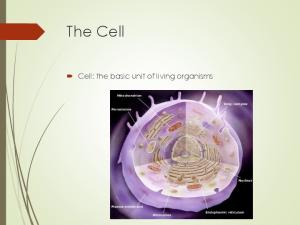
The Tissue Level of Organisms • Tissues are groups of similar cells • Common Embryonic Origin • Common Function
• Histology: the study of tissue
Epithelial Tissue › Covers surfaces because cells are contact › Lines hollow organs, cavities and ducts › Forms glands when cells sink under the
surface
Connective Tissue › Material found between cells
› Supports and binds structures together › Stores energy › Provides immunity to disease
Muscle Tissue › Cells shorten in length producing movement
Nervous Tissue › Cells that conduct electrical impulses › Detects changes inside and outside the
body › Responds with nerve impulses
Generally thought of as the protective layer. Closely packed cells forming a continuous sheet Cells rest on a basement membrane Have an upper free (apical) surface Avascular- without a blood supply › Nutrients diffuse from underlying connective tissue
Good nerve supply Rapid cell division 2 types › Covering/lining › Glandular types
Is like double sided sticky tape b/c it holds epithelium to connective tissues
Guide for cellular migration during development
Covering and Lining Epithelium › Epidermis of skin › Lining of blood vessels and ducts › Lining respiratory, reproductive, urinary, & GI tract
Glandular Epithelium › Secreting portion of glands
› Thyroid, adrenal, and sweat glands
1. Protect underlying structures › Ex. Oral cavity epithelium protects underlying glands from abrasions. 2. Acts as a barrier › Ex. Reduces water loss from body 3. Permits the passage of substances › Ex. O2 & CO2 exchange through lungs 4. Secretes Substances › Ex. Mucous or sweat 5. Absorbs substances › Ex. Epithelial of the intestines absorb molecules of digested food.
Classified by arrangement of cells into layers › Simple – one layer thick › Stratified – many cell layers thick › Pseudostratified – single layer of cells where all cells
don’t reach apical surface
Nuclei are found at different layers so it looks stratified
Classified by shape of surface cells › Squamous – flat
› Cuboidal – cube shaped › Columnar – tall column › Transitional – shape varies with tissue stretching
Single layer of flat cells › Lines blood vessels (endothelium), body cavities
(mesothelium) › Very thin- controls diffusion, osmosis, & filtration › Nuclei centrally located
Cells in direct contact with each other
Single layer of cube shaped cells viewed from the side Nuclei round and centrally located Absorption or secretion
Sectional view of kidney tubules
Single layer rectangular cells Unicellular glands – goblet cells secrete mucus › Lubricate GI, respiratory, reproductive and urinary systems Microvilli – fingerlike projections › For absorption in GI tract (stomach – anus)
Function & location determine type!!!
Single layer cell All cells attached to basement membrane, but not all cells reach apical (free surface) Nuclei are at varying depths Respiratory system, male urethra, and epididymis
Free surface Nonkeratinized stratified squamous cells Nuclei Basement membrane
Several cell layers thick Surface cells flat Keratinized- surface cells dead and filled with keratin › Skin (epidermis) Nonkeratinized- no keratin in moist living cells at surface › Mouth, vagina
Multilayered Surface cells cuboidal
› rare (only found in
sweat gland ducts & male urethra)
Absorption, secretion, & protection
Multilayered Surface cells columnar Rare (mammary glands, male urethra, & larynx) Secretion, protection, & some absorption
4-21
Multilayered Surface cells varying in shape from round to flat if stretched Lines hollow organs that expand from within (urinary bladder)
4-22
Glandular Epithelia
Derived from epithelial cells that sank below the surface during development
Exocrine glands › cells that secrete---sweat,
ear wax, saliva, digestive enzymes onto free surface of epithelial layer › connected to the surface by tubes (ducts) › unicellular glands or multicellular glands
Endocrine glands secrete hormones into the bloodstream › hormones help maintain homeostasis ›
Exocrine- sebacious glands of skin
• Figure 4.4
Endocrine- no ducts sm intestines
Cells rarely touch due to extracellular matrix Matrix(fibers & ground substance secreted by cells Consistency varies from liquid, gel to solid Does not occur on free surface Good nerve & blood supply except cartilage & tendons
425
Collagen (25% of protein in your › tough, resistant to pull, yet pliable › formed from the protein collagen
body)
Elastin
(lungs, blood vessels, ear cartilage) › smaller diameter fibers formed from protein elastin surrounded by glycoprotein (fibrillin) › can stretch up to 150% of relaxed length and return to original shape
Reticular (spleen and lymph nodes) › thin, branched fibers that form framework of
organs 4-26 › formed from protein collagen
Loose connective tissue Dense connective tissue Cartilage Bone Blood Lymph (not in this lecture)
4-27
Loosely woven fibers throughout tissues Types of loose connective tissue
› areolar tissue
› adipose tissue › reticular tissue
4-28
Cell types = fibroblasts, plasma cells, macrophages, mast cells and a few white blood cells All 3 types of fibers present Gelatinous ground substance 4-29
Black = elastic fibers, Pink = collagen fibers Nuclei are mostly fibroblasts
4-30
Peripheral nuclei due to large fat storage droplet Deeper layer of skin, organ padding, yellow marrow Reduces heat loss, energy storage, protection Brown fat found in infants has more blood vessels and mitochondria and responsible for heat 4generation 31
Network of fibers & cells that produce framework of organ Holds organ together (liver, spleen, lymph nodes, 4bone marrow) 32
More fibers present but fewer cells Types of dense connective tissue
› dense regular connective tissue › dense irregular connective tissue › elastic connective tissue
4-33
Collagen fibers in parallel bundles with fibroblasts between bundles of collagen fibers White, tough and pliable when unstained (forms tendons) 4 Also known as white fibrous connective tissue 34
Collagen fibers are irregularly arranged (interwoven) Tissue can resist tension from any direction Very tough tissue -- white of eyeball, dermis 435 of skin
Branching elastic fibers and fibroblasts Can stretch & still return to original shape Lung tissue, vocal cords, ligament between 436 vertebrae
Network of fibers in rubbery ground substance Resilient and can endure more stress than loose or dense connective tissue Types of cartilage
› hyaline cartilage › fibrocartilage › elastic cartilage
4-37
Bluish-shiny white rubbery substance Chondrocytes sit in spaces called lacunae No blood vessels or nerves so repair is very slow Reduces friction at joints as articular cartilage
4-38
Many more collagen fibers causes rigidity & stiffness Strongest type of cartilage (intervertebral 4discs) 39
Elastic fibers help maintain shape after deformations 4 Ear, nose, vocal cartilages
40
Spongy bone › sponge-like with spaces and trabeculae › trabeculae = struts of bone surrounded by
red bone marrow › no osteons (cellular organization)
Compact bone › solid, dense bone › basic unit of structure is osteon (haversian
system- canal containing blood supply)
Protects, provides for movement, stores minerals, site of blood cell formation 4-41
Osteon = lamellae (rings) of mineralized matrix › calcium & phosphate---give it its hardness
› interwoven collagen fibers provide strength
Osteocytes in spaces (lacunae) in between lamellae 442 Canaliculi (tiny canals) connect cell to cell
Connective tissue with a liquid matrix = the plasma Cell types = red blood cells (erythrocytes), white blood cells (leukocytes) and cell fragments called platelets Provide clotting, immune functions, carry O2 443 and CO2
Cells that shorten Provide us with motion, posture and heat Types of muscle
› skeletal muscle › cardiac muscle › smooth muscle
4-44
Cells are long cylinders with many peripheral nuclei Visible light and dark banding (looks striated) Voluntary or conscious control 445
Cells are branched cylinders with one central nuclei Involuntary and striated Attached to and communicate with each other 446 by intercalated discs
Spindle shaped cells with a single central nuclei Walls of hollow organs (blood vessels, GI tract, bladder) Involuntary and nonstriated 4
47
Cell types -- nerve cells and neuroglial (supporting) cells Nerve cell structure
› nucleus & long cell processes conduct nerve signals dendrite --- signal travels towards the cell body axon ---- signal travels away from cell body
448