Gene 238 (1999) 291–300 www.elsevier.com/locate/gene
Review
The Sp-family of transcription factors G. Suske * Institut fu¨r Molekularbiologie und Tumorforschung, Philipps-Universita¨t Marburg, Emil-Mannkopff-Straße. 2, D-35037 Marburg, Germany Received 14 June 1999; accepted 6 August 1999; Received by A.J. van Wijnen
Abstract GC-boxes and related motifs are frequently occurring DNA-elements present in many promoters and enhancers. In contrast to other elements it was generally thought that the transcription factor Sp1 is the only factor acting through these motifs. The cloning of paralogous genes of the Sp1 factor uncovered the existence of a small protein family consisting of Sp1, Sp2, Sp3 and Sp4. All four proteins exhibit very similar structural features. They contain a highly conserved DNA-binding domain composed of three zinc fingers close the C-terminus and serine/threonine- and glutamine-rich domains in their N-terminal regions. The high degree of structural conservation between these four proteins suggested that they do exert similar functions. Molecular, genetic and biochemical analyses, however, demonstrated that Sp2, Sp3 and Sp4 are not simply functional equivalents of Sp1. Here, I will summarize and discuss recent advances which have been made towards understanding the mode of action and biological function of individual family members. © 1999 Elsevier Science B.V. All rights reserved. Keywords: Review; Sp1; Sp2; Sp3; Sp4; Zinc finger
1. Introduction Transcriptional regulation is exerted by the combinatorial action of proteins binding to distinct promoter and enhancer elements. Usually a limited number of cisacting DNA elements is recognized not only by a single transcription factor but by a set of different proteins which are often structurally related (Latchman, 1995). Important and widely distributed promoter elements are G-rich elements such as the GC-box (GGGGCGGGG) and the related GT/CACCC-box (GGTGTGGGG). These elements are required for the appropriate expression of many ubiquitous, tissue-specific and viral genes. In addition, they occur frequently in the regulatory region of genes which are under a specific mode of control such as cell cycle regulation, hormonal activation and developmental patterning. For some time it has been known that the general transcription factor Sp1 (Specificity protein 1) can bind to and act through the GC-boxes and it was generally accepted that this protein is an extremely versatile protein involved in the expression of many different genes docu* Tel.: +49-6421-2866697. fax: +49-6421-2865398. E-mail address:
[email protected] (G. Suske)
mented by more than 2600 citations. More recently, however, it became clear that Sp1 is not the only protein acting through ‘Sp1-binding sites’ but simply represents the first identified and cloned protein of a small protein family. Currently this family consists of four proteins designated Sp1, Sp2, Sp3 and Sp4. Accordingly our view on Sp1 and its function has changed significantly. Here, I will summarize and discuss advances which have been directed towards understanding the properties and function of the individual Sp-proteins.
2. Molecular cloning of Sp transcription factors Sp1 was originally identified as the transcription factor which binds to and activates transcription from multiple GC-boxes in the simian virus 40 (SV40) early promoter (Dynan and Tjian, 1983; Gidoni et al., 1984) and the thymidine kinase ( TK ) promoter (Jones et al., 1985). Molecular cloning of a partial human Sp1 cDNA from HeLa cells was described in 1987 ( Kadonaga et al., 1987). Since that time, the cDNA sequence published and deposited in databases is still incomplete. It encodes for the 696 C-terminal amino acids of human Sp1. The entire human Sp1 protein consists of 778 amino acids.
0378-1119/99/$ – see front matter © 1999 Elsevier Science B.V. All rights reserved. PII: S0 3 7 8 -1 1 1 9 ( 9 9 ) 0 0 35 7 - 1
292
G. Suske / Gene 238 (1999) 291–300
Table 1 Function and properties of Sp-factors Factor
Accession numbers
Chromosomal localisation
Distribution
Transcriptional properties
Knockout phenotype
Features/ miscellaneous
Sp1
Human: J03133; Mouse: AF062566, AF022363; Rat: D12768
Human: 12q13 (Gaynor et al., 1993; Matera and Ward, 1993); Mouse: 15 (Saffer et al., 1990); Rat: 7q36 (Scohy et al., 1998)
Ubiquitous, developmental variations (Saffer et al., 1991)
Activator (Courey and Tjian, 1988) Synergistic activation (Courey et al., 1989) ; Superactivation (Pascal and Tjian, 1991)
Lethal at embryonic day 10 (Marin et al., 1997)
Sp2
Human: M97190, D28588
Various cell lines; tissues unknown ( Kingsley and Winoto, 1992)
Unknown
Unknown
Sp3
Human: X68560, S52144; Mouse: AF062567
Human: 17q21.3-q22 (Scohy et al., 1998); Rat: 10q31-q32.1 (Scohy et al., 1998); Human: 2q31 ( Kalff-Suske et al., 1996); Rat: 3q24-q31 (Scohy et al., 1998)
Ubiquitous (Hagen et al., 1992) and G. Suske, unpublished
Repressor of Sp1-mediated transcription (Hagen et al., 1994) Activator ( Udvadia et al., 1995; Dennig et al., 1996)
Unknown
Sp4
Human: X68561, S50516; Mouse: U62522; Rat: U07610
Human: 7p15.3-p21 ( Kalff-Suske et al., 1995); Mouse: 12 (Supp et al., 1996); Rat: 6q33 (Scohy et al., 1998)
Predominantly in neuronal cells; also in certain epithelia (Hagen et al., 1992; Supp et al., 1996) and G. Suske, unpublished
Activator (Hagen et al., 1994, 1995)
Growth retardation; males do not breed (Supp et al., 1996)
Two glutamine-rich activation domains (Courey and Tjian, 1988; Gill et al., 1994); phosphorylated (Jackson et al., 1990); glycosylated (Jackson and Tjian, 1988) Original Sp2 clone incomplete; complete sequence in data bank D28588 Two glutamine-rich activation domains; three isoforms (Hagen et al., 1994; Kennett et al., 1997); inhibitory domain (Dennig et al., 1996); translational start site of full length protein unknown Two glutamine-rich activation domains (Hagen et al., 1995); entire human genomic sequence in data bank: Accession No. AC004595
Its sequence could be obtained from the Tjian laboratory upon request. More recently, highly conserved full length Sp1 cDNAs from rat and mouse have been cloned (Imataka et al., 1992; Yajima et al., 1998) ( Table 1). The Sp1-related transcription factors Sp3 and Sp4 (originally also designated SPR-2 and SPR-1, respectively) were cloned by recognition site screening using the GT-box motif of the uteroglobin promoter as a probe ( Hagen et al., 1992). Independently, Sp3 was obtained along with Sp2 from a human T-cell library by low stringency screening with the zinc finger-encoding region of human Sp1 as a probe ( Kingsley and Winoto, 1992). The originally published human Sp2 cDNA sequence assigned as complete coding sequence turned out to be incomplete. The complete coding sequence of Sp2 is deposited in the data bank as human mRNA for the KIAA0048 gene (Accession number D28588). The cDNAs coding for the murine homologues of Sp3 and Sp4 have been published recently (Supp et al., 1996; Yajima et al., 1998).
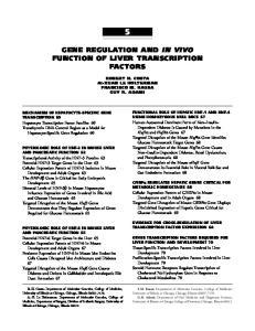
3. Structural features of Sp-family members All four human Sp-family members have similar domain structures (Fig. 1A). They contain three zinc fingers close to the C-terminus and glutamine-rich domains adjacent to serine/threonine stretches in their N-terminal region. The 81 amino acids C2H2-type zinc finger region which represents the DNA-binding domain is the most highly conserved part of the proteins. Alignment of that region shows that Sp1, Sp3 and Sp4 are more closely related to each other than to Sp2 ( Fig. 1B). According to structural studies on zinc finger proteins bound to DNA (Pavletich and Pabo, 1991; Fairall et al., 1993), one could predict that the amino acids KHA within the first, RER within the second and RHK within the third zinc finger contact specific bases. Especially these critical amino acids are all conserved in Sp1, Sp3 and Sp4 but not in Sp2. Consistently, Sp1, Sp3 and Sp4 recognize the classical Sp1-binding site with identical affinity (Hagen et al., 1992, 1994). In Sp2,
G. Suske / Gene 238 (1999) 291–300
293
Fig. 1. Structural features of Sp-proteins. (A) Schematic representation of the four human Sp-family members Sp1, Sp2, Sp3 and Sp4. Their length in amino acids is indicated on the right. The length of Sp3 refers to the full length isoform according to ( Kingsley and Winoto, 1992). Colored boxes indicate regions of the proteins which are rich in glutamine (red ) and serine/threonine (yellow) residues. The region preceding the first zinc finger (+/−) is rich in charged amino acids. The black boxes represent the zinc fingers. Lines above the draw of the Sp1 protein indicate the extent of four regions (A, B, C and D) which contribute to the transcriptional properties of Sp1 as defined by Tjian and coworkers (Courey and Tjian, 1988). Known activation (AD) and inhibitory domains (ID) are indicated. (B) Protein sequence alignment of the zinc finger domains. Stars below the sequence indicate sequence identity. Cysteine and histidine residues which coordinate zinc ions are underlayed in green and protein regions which contact the DNA in blue. Arrows point to the amino acids which determine the recognition specificity by contacting specific bases of the DNA (adapted from Pavletich and Pabo, 1991; Fairall et al., 1993). The black lines and the zig-zag lines indicate a-helical and b-sheet regions respectively.
the important histidine residue within the first zinc finger is replaced by a leucine residue ( Fig. 1B). In accordance with this structural difference it was shown that Sp2 does not bind to the GC-box but to a GT-rich element within the T-cell receptor gene 5∞-flanking region ( Kingsley and Winoto, 1992).
4. Evolutionary relationship of the four Sp genes The structural similarity of the four Sp-proteins suggests that they are evolutionally closely related. This is indeed the case and documented by their chromosomal localization in the human genome. All four Sp genes are found on paralogous chromosomal regions on human chromosomes 12q13 (Sp1), 17q21.3-q22 (Sp2), 2q31 (Sp3) and 7q21.3-q22 (Sp4) (Matera and Ward, 1993; Kalff-Suske et al., 1995, 1996; Scohy et al., 1998). The human Sp genes are linked to the homeobox gene cluster (O’Brien et al., 1993) on the corresponding chromosome (Sp1/HOX C, Sp2/HOX B, Sp3/HOX D and Sp4/HOX A). This chromosome configuration is maintained in rodents with the exception of Sp4 which dissociated from the Hox a genes in the mouse and in the rat (Saffer et al., 1990; Supp et al., 1996; Scohy et al., 1998). Except for the human Sp4 gene, the sequence and
the exon–intron structures of the individual chromosomal Sp genes are only partially known ( Fig. 2). The entire sequence of the human Sp4 gene is contained within the BAC clone RG023M10 (Accession No. AC004595). Cloning and mapping of the entire murine Sp4 gene (G. Suske, unpublished data) revealed that the exon–intron structure of the Sp4 gene is conserved between mouse and man. Partial gene structure information of the mouse Sp1 (Chestier and Charnay, 1992; Marin et al., 1997) and Sp3 genes (G. Suske, unpublished data) are also known ( Fig. 2). The available data show that the exon–intron structures are conserved among individual Sp-family members supporting the conclusion that the four genes arise from a single ancestor gene. Little is known about Sp genes in non-mammalian vertebrates and invertebrates. In vitro binding studies and biochemical characterizations have demonstrated the existence of GC-box binding proteins in fish which share immunological properties with Sp1 and Sp3 (Baudler et al., 1997). In Drosophila melanogaster, the head-specific protein buttonhead (btd) encodes a zinc finger type transcription factor with significant sequence and functional similarity to the Sp-proteins ( Wimmer et al., 1993). However, the arrangement of the zinc finger region relative to the other domains is slighly different in btd. The region C-terminal to the zinc finger
294
G. Suske / Gene 238 (1999) 291–300
Fig. 2. Known genomic structures of Sp1, Sp3 and Sp4 genes substantiate their close evolutional relationship. The central drawing shows the structure of the Sp-proteins schematically. The glutamine-rich activation domains A and B and the zinc fingers are indicated. Known exons (numbered 1 to 6 in the case of the Sp4 genes) of the mouse Sp1, Sp3 and Sp4 genes are connected with the corresponding protein regions. Both glutamine-rich activation domains are encoded on a single large exon. The human Sp4 gene contains a CpG island in its 5∞-flanking region.
region is extended and contains a serine/threonine-rich domain. Btd is required for development of the antennal, intercalary and mandibular segments of the fly head. A human Sp1 transgene rescued the btd null phenotype partially. It supported development of the mandibular segment in the head of btd mutant embryos when expressed in the spatial pattern of btd ( Wimmer et al., 1993). It would be interesting to know whether any of the other three Sp-proteins would have the capacity to rescue fully the btd null phenotype in flies. Since Sp4 is expressed predominantly in the brain, it could be a good candidate gene.
5. Functional properties of individual Sp-family members 5.1. Sp1: The prototype of the family Molecular cloning of Sp1 and its subsequent dissection revealed the functional domains of the protein. Both glutamine-rich regions (designated A and B) (Fig. 1A) can act as strong activation domains (Courey and Tjian, 1988). Mapping of the activation domain revealed that interspersed bulky hydrophobic amino acids are essential for transcriptional activation and not, per se, the glutamine residues (Gill et al., 1994). More recently, an inhibitory domain has been mapped to the N-terminus (Murata et al., 1994). Synergistic activation of promoters by Sp1 through multiple GC-boxes requires in addition the short C-terminal domain D (Pascal and Tjian, 1991). Sp1 is known to be phosphorylated (Jackson et al., 1990) and glycosylated (Jackson and Tjian, 1988), and it is capable of forming homotypic interactions leading to multimeric complexes (Mastrangelo et al., 1991; Pascal and Tjian, 1991; Su
et al., 1991). In addition, many heterotypic interactions with different classes of nuclear proteins have been reported. These include factors belonging to the general transcription machinery, such as the TATA-box binding protein TBP ( Emili et al., 1994) and the TBP-associated factors dTAFII110/hTAFII130 (Hoey et al., 1993; Tanese et al., 1996), and hTAFII55 (Chiang and Roeder, 1995). Other proteins which have been shown to interact with Sp1 are cell cycle regulators such as the retinoblastoma-related protein p107 (Datta et al., 1995) and transcription factors such as YY1 (Lee et al., 1993; Seto et al., 1993) or E2F ( Karlseder et al., 1996; Lin et al., 1996). Sp1 can bind to its target sequence in assembled nucleosomes (Li et al., 1994), and in this regard it is interesting to note the interaction with a large coactivator complex called CRSP (cofactor required for Sp1 activation) which stimulates Sp1-mediated transcription in vitro ( Ryu et al., 1999). 5.2. Sp2 There exists no functional analysis of Sp2 and correspondingly little is known about this Sp-family member. The presence of a potential glutamine-rich activation domain suggests that it may act as an activator. The T-cell antigen receptor a (TCRa) gene might be a target for Sp2 since it binds in vitro to a GT-box promoter element within the TCRa promoter ( Kingsley and Winoto, 1992). 5.3. Sp3: Activator versus repressor Unraveling the transcriptional role of Sp3 was complicated by the fact that three Sp3 isoforms exist, a 110-115 kDa Sp3 protein and two approximately
G. Suske / Gene 238 (1999) 291–300
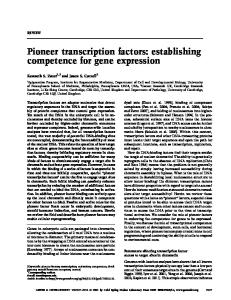
60-70 kDa Sp3 species. Very likely, the full length Sp3 protein is derived by translational initiation at a nonAUG which has been arbitrarily assigned to an AUU codon ( Kingsley and Winoto, 1992). However, other potential non-AUG start codons are also present in that region and thus it remains to determine the N-terminus of Sp3. It has also been suggested that the published Sp3 cDNA lacks 5∞-untranslated sequences and the extreme N-terminus of the Sp3 protein. I consider this possibility unlikely since in vitro translation of the full length isoform of Sp3 from the published mRNA has been described ( Kingsley and Winoto, 1992; Kennett et al., 1997). The two smaller Sp3 species arise from the first two internal AUG codons (G. Suske, unpublished observation, and Kennett et al., 1997). Consistently, antibodies raised against the N-terminus of Sp3 recognize in EMSAs the slow migrating complex which contains the full length Sp3 isoform but not the two fast migrating complexes which contain the two N-terminally truncated isoforms ( Fig. 3). Reports on the transcriptional properties of Sp3 appear, at first sight, contradictory. Sp3 has been shown to act as a transcriptional activator similar to Sp1 ( Udvadia et al., 1995; Liang et al., 1996; Ihn and Trojanowska, 1997; Zhao and Chang, 1997, and many others). In other experiments, Sp3 remained inactive or acted only as a very weak activator ( Hagen et al., 1994; Majello et al., 1994; Dennig et al., 1995; Kumar and Butler, 1997 and others). Most of these reports are based on co-transfection experiments into the insect cell line SL2. Usually, a distinct promoter fragment containing appropriate Sp-binding sites fused to a reporter gene was co-transfected along with Sp1 and Sp3 or both together. If Sp3 is expressed to the same extent as Sp1 but does not act as a strong activator, it will compete for the same binding site and thus lower Sp1-mediated activation. There are only a few reports in which additional approaches have been chosen to determine the potential role of Sp3 for the activity of a certain promoter (Noti, 1997; Hata et al., 1998). An antisense strategy which knocked out endogenous Sp3 expression in the myelomonocytic cell line HL60 revealed that Sp3 participates in the activation of the CD11c and CD11b promoters (Noti, 1997). The experimental conditions which are needed for Sp3 to act as a strong activator or a transcriptional inactive molecule which represses Sp1-mediated activation are not completely understood. The structure and the arrangement of the recognition sites appear to determine whether Sp3 is transcriptionally inactive and can repress Sp1-mediated activation or whether it acts as a strong activator. Promoters containing a single binding site are activated, whereas promoters containing multiple binding sites often are not activated or respond weakly to Sp3 (Birnbaum et al., 1995; Dennig et al., 1996). Whether Sp3 acts as an activator or as a repressor
295
Fig. 3. Sp1 and the three isoforms of Sp3 are visible in an electrophoretic mobility shift assay ( EMSA). A schematic representation of Sp3 is shown at the top. The activation domains A and B, the inhibitory domain (ID), the charged region (+/−) preceding the DNA-binding domain and the three zinc fingers (black boxes) are depicted. Asterisks indicate the N-termini of the three isoforms. The black lines indicate the segments of Sp3 which have been used for generation of the antibodies aSp3N and aSp3. The EMSA was performed with a 32P-labeled GC-box oligonucleotide and nuclear extract from CV-1 cells. Antisera against Sp1 (aSp1, lane 3), the N-terminal part of Sp3 (aSp3N, lane 5), the C-terminal part of Sp3 (aSp3, lane 2) or the corresponding preimmune sera (Pre, lanes 1, 4 and 6), respectively, were included in the binding reactions as indicated. Complexes containing Sp1 or Sp3 are indicated at the left. The antiserum directed to the C-terminus shifted all three Sp3-containing complexes ( lane 2) whereas the antiserum directed to the N-terminus shifted only the slow migrating complex ( lane 5).
of Sp1-mediated activation might also depend of the cellular context. Transfected Sp3 stimulated transcription from the HERV-H long-terminal repeat in the teratocarcinoma cell line NTera2-D1 but acted as a repressor in HeLa and insect cells (Sjottem et al., 1996). It has been suggested that the two small Sp3 isoforms might act as repressor molecules whereas the full length Sp3 isoform does act as an activator ( Kennett et al., 1997). Although attractive and simple, this model does not seem to hold true. Exclusive expression of full length Sp3 triggered by an artificial leader sequence can also repress Sp1-mediated activation (Dennig et al., 1996).
296
G. Suske / Gene 238 (1999) 291–300
It is clear that both N-terminal glutamine-rich regions can act as strong activation domains on their own in both insect and in mammalian cells (Dennig et al., 1996; Majello et al., 1997). The molecular basis for the inactivity of Sp3 under certain conditions has been mapped to an inhibitory domain located between the second glutamine-rich activation domain and the first zinc finger. The amino acid triplet KEE within this domain is absolutely essential for repressor function (Dennig et al., 1996). Mutation of these amino acids to alanine residues converted almost inactive Sp3 to a strong activator. The inhibitory domain of Sp3 acts as an independent modul in cis. It can be transferred to other activation domains which in turn lose their activation properties (Dennig et al., 1996). Yet, we do not know how this domain functions mechanistically. Purified recombinant Sp3 expressed in SL2 cells (Braun and Suske, 1999) act in an in vitro system as strong activator similar to Sp1 (H. Braun and G. Suske, unpublished ). From this, and other observations, it can be concluded that additional proteins which act as co-repressors are involved in the inhibitory function of this domain. Our recent cloning of a protein designated SIF-1 (Sp3-interacting protein 1) which specifically interacted with the wild type inhibitory domain but not with the mutated form support this idea (A. Doll and G. Suske, unpublished ). 5.4. Sp4: The tissue-specific Sp-family member Sp4 was cloned along with Sp3 by virtue of its binding to the GT-box of the uteroglobin promoter (Hagen et al., 1992). In contrast to Sp1 and Sp3 which are ubiquitous transcription factors, Sp4 expression appears to be restricted to a few tissues. High levels of Sp4 are predominantly found in the brain (Hagen et al., 1992; Supp et al., 1996). Consistent with its high expression in the central nervous system, targeted disruption of the mouse Sp4 gene by homologous recombination led to behavioral defects (see Section 7). Sp4 exhibits specific functional properties distinct from Sp1 and Sp3. The transactivation function of Sp4 resides, like that of Sp1, in the N-terminal glutaminerich regions. In contrast to Sp1, Sp4 is not able to act synergistically through adjacent binding sites although Sp4 can function as a target for the Sp1 activation domains in a superactivation assay suggesting that the activation domains of Sp1 and Sp4 are functionally related (Hagen et al., 1995).
6. Regulation by the ratio of Sp1 and Sp3 It is clear that in a given cell type, co-expression of Sp1 and Sp3 occurs and we have to assume that these two proteins compete for the same binding sites in vivo. The initial characterization of Sp1 and Sp3 shows that
they differ in their capacity to activate or repress transcription. Independently of whether Sp3 acts as an activator or as a repressor of Sp1-mediated activation, the relative abundance of Sp1 and Sp3 should allow regulation of gene activities. The abundance of Sp1 and Sp3 varies among different cell types. In endothelial cells which contain high levels of both Sp1 and Sp3, the Sp1/Sp3 ratio is higher than in non-endothelial cells ( Hata et al., 1998). The endothelial-specific activity of the KDR/flk-1 promoter was mapped to an Sp1/Sp3-binding site, and Sp3 attenuated Sp1-mediated KDR/flk-1 promoter activation. Thus, it was suggested that endothelial selective KDR/flk-1 expression may be partially mediated by the high Sp1/Sp3 ratio in these cells (Hata et al., 1998). Alterations in the relative abundance of Sp1 and Sp3 upon different conditions have been reported in several cases. In primary keratinocytes, Sp3 levels exceed those of Sp1. The Sp3/Sp1 ratio beomes inverted if these cells differentiate in vitro on treatment with high calcium suggesting that cell type differential transcription of several genes might be regulated by Sp1 and Sp3 (Apt et al., 1996). Worth mentioning in this context is that in differentiated keratinocytes only Sp3, but not Sp1, overexpression enhanced the induction of the p21 promoter (Prowse et al., 1997). A change in the Sp1/Sp3 ratio occurred also when C2C12 myocytes were cultivated under hypoxic conditions. Hypoxia caused a progressive depletion of Sp3 whereas the Sp1 protein level remained unchanged (Discher et al., 1998). Like other glycolytic enzymes the muscle-specific pyruvate kinase-M and b-enolase are upregulated under low oxygen pressure. The hypoxia responsive promoter elements in these two genes have been mapped to GC-rich elements bound by Sp1 and Sp3. Thus, it was concluded that hypoxia activates these glycolytic enzyme gene promoters by down-regulating Sp3, thereby removing the associated transcriptional repression (Discher et al., 1998). In another report, it was shown that downregulation of a2-integrin gene expression in mammary epithelial cells by Erb-B2 and v-Hras is mediated by two adjacent Sp1/Sp3 binding sites in the a2-integrin promoter. In that case, however, the Sp3 DNA-binding activity remained unaltered whereas the Sp1 DNA-binding activity was reduced ( Ye et al., 1996). Although not shown so far, one could imagine that alterations of the transcriptional capacity of Sp1 or Sp3 could be obtained by other mechanisms. Especially, Sp3 could be a target of signal transduction pathways since it has both activation and repression functions. Modifications in the protein might alter the potency of the protein in one or the other direction. Altogether, further unraveling of the molecular structure and modifications of the protein should give insights into the specific role of Sp3.
G. Suske / Gene 238 (1999) 291–300
7. Physiological function of Sp-proteins In the past, a large variety of biological functions have been assigned to Sp1-binding sites and to Sp1. However, the identification of the three paralogous proteins Sp2, Sp3 and Sp4 raises the question as to which tasks are performed by which protein. This question is particularly interesting for Sp1 and Sp3 because both proteins are present in the same cell and are indistinguishable in their DNA-binding specificity. Gene disruption in mice is a powerful tool for obtaining information on specific functions of individual Sp-proteins. Given that Sp1 is implicated in the activation of a very large number of genes, such as housekeeping, tissue-specific and cell cycle-regulated genes, and is required to prevent methylation of CpG islands (Brandeis et al., 1994; Macleod et al., 1994), one would expect that cells lacking Sp1 would not survive. Surprisingly, this is not the case. S. Philipsen and his coworkers have disrupted the mouse Sp1 gene and found that Sp1-deficient embryonic stem cells ( ES cells) are viable, have normal growth characteristics and can be induced to differentiate and form embryoid bodies as efficiently as wild type ES cells (Marin et al., 1997). Nevertheless, Sp1 is essential for normal mouse embryogenesis. The Sp1-knockout embryos are severely retarded in development and they all died around day 11 of gestation. They displayed a marked heterogeneity in phenotype indicating that Sp1 has indeed a general function in many cell types. Interestingly, the defects in Sp1-/- mice are caused by a cell autonomous mechanism. Sp1-/- ES cells injected into blastocysts contributed efficiently to chimaeric embryos at early stages but after day E 11 they rapidly declined with no contribution to newborn mice. Thus, Sp1 appears to be a transcription factor whose function is essential for differentiated cells after day10 of development. Independently of the severe developmental defects of the Sp1 null mice, the embryos express many putative Sp1 target genes at normal levels, including housekeeping and cell-cycle regulated genes. In addition, CpGislands remained methylation free. So far, the only genes which were found to be expressed at a lower level in Sp1-/- mice are the thymidine kinase and the methylCpG binding protein 2 (MeCP2) genes (Marin et al., 1997). Since there are striking similarities between the phenotypes of the MeCP2 ( Tate et al., 1996) and the Sp1 knockouts, it was suggested that the MeCP2 gene might be a key target of Sp1. However, whether Sp1 acts as a direct regulator of MeCP2 expression by binding to promoter, enhancer or local control region elements in the MeCP2 gene, or whether additional proteins mediate downregulation of MeCP2 remains to be established. The Sp1-knockout demonstrated that Sp1 is not
297
essential for the expression of many genes previously shown be activated in cell culture transfection experiments. One could speculate that other Sp-family members compensate at early embryonic stages, at least in part for the loss of Sp1 activity. Sp3 would be a good candidate because it is also ubiquitously expressed, has the potential to activate transcription and its DNAbinding activity is indistinguishable from Sp1. In that regard, the phenotype of Sp3 knockout mice will be of great interest. Sp4, the tissue restricted member of the Sp-family, is predominantly expressed in the brain but also detectable in epithelial tissues, testis and developing teeth (Hagen et al., 1992; Supp et al., 1996). Disruption of the mouse Sp4 gene revealed that it is important for early postnatal survival (Supp et al., 1996). Approximately two thirds of the Sp4-/- mice die within a few days of birth. The cause of the early death remains unknown. Those mice which survive are significantly smaller than their wild type littermates. The reduced body weight appears to result from an unknown, but growth hormone independent, mechanism (Supp et al., 1996). Interestingly, surviving mice exhibit a striking sex-specific abnormality. While fertility of the female mutants appears normal, males do not breed although their reproductive organs are fully developed and apparently normal. It appears that male Sp4-/- mice are unable to copulate. The most likely cause of this abnormal behavior is a neurological defect. The hypothalamus and the vomeronasal organ are known to play important roles in reproductive physiology and behavior. However, both structures are histologically normal in Sp4-/- mice. Thus, we await further investigation to understand the role of Sp4 and to identify its target genes. In that context, it should be noted that for both the Sp1 and the Sp4 knockouts, the zinc finger regions have been chosen to inactivate the genes. In both cases the N-terminal part encoding the transactivation domains are still expressed (Supp et al., 1996; Marin et al., 1997). One could speculate that the activation domains on their own might act as a gain-of-function or might interfere with other Sp-family members. In the case of the Sp4 knockout such a scenario does not seem to be the case. Knockout Sp4-/- mice which do not express the N-terminal part of the protein manifest the same phenotype (G. Suske, unpublished observations).
8. Yet more and more GC/GT-box binding proteins In the past, essential GC-boxes in promoters were often equated with ‘‘Sp1-binding sites’ thereby overlooking the fact that Sp1 is not the only protein which recognizes this important element. In addition to Sp3 and Sp4 there exist at least three other proteins, BTEB1 (basic transcription element binding protein 1) (Imataka
298
G. Suske / Gene 238 (1999) 291–300
et al., 1992), TIEG1 and TIEG2 ( TGFb-inducible early protein genes 1 and 2) (Subramaniam et al., 1995; Cook et al., 1998; Fautsch et al., 1998) which have a binding specificity very similar, if not identical, to Sp1 (Sogawa et al., 1993b; Cook et al., 1998). In BTEB1, TIEG1 and TIEG2 the DNA-binding domain is found also close to the C-terminus and consists of three C2H2-type zinc fingers. Most significantly, all amino acids which are believed to specifically contact the DNA are conserved between these three proteins and Sp1, Sp3 and Sp4. However, the N-terminal domains of these proteins do not share any similarity with the Sp-proteins. BTEB1 contains acidic and hydrophobic amino acid stretches, and TIEG1 as well as the closely related protein TIEG2 are rich in proline residues. In addition, another subgroup of zinc finger proteins, including the so called Kru¨ppel-like factors, has to be considered when essential ‘Sp1-binding sites’ are analyzed in promoters. These proteins also contain three zinc fingers close to their C-terminus and bind preferentially to so called GT-or CACCC-boxes. Sp-family members also recognize these elements. Thus, Sp-proteins and Kru¨ppel-like factors have an overlapping binding specificity. Currently this subfamily of zinc finger proteins consists of at least 10 different members, the so called Kru¨ppel like factors EKLF (Miller and Bieker, 1993), BKLF/TEF-2 (Crossley et al., 1994), GKLF (Shields et al., 1996), IKLF (Conkright et al., 1999), LKLF (Anderson et al., 1995), UKLF (Matsumoto et al., 1998) and FKLF (Asano et al., 1999) and ZNF741 (only sequence in data base), AP-2rep (Imhof et al., 1999) and ZF9 (Ratziu et al., 1998). Another factor called BTEB2 (Sogawa et al., 1993a) is very likely an N-terminally truncated human ortholog of murine IKLF. Understanding the biological function and the mode of action of all these GC/GT-box binding proteins will be a scientific challenge.
9. Conclusions The cloning and initial characterization of a family of Sp-proteins as well as other GC/GT-box binding proteins provides much information about the potential functions and activities of these proteins. The most obvious question, however, concerns the specificity of the individual family members especially of Sp1 and Sp3. Neither binding site preferences nor differential expression patterns seem to confer specificity of these two proteins. However, activation of a given promoter requires the binding of multiple transcription factors which might bind cooperatively to their sites or might act synergistically by other mechanisms. Thus far, little is known how Sp1 and Sp3 act on natural promoters in combination with other transcription factors in vivo. Specificity could be obtained also by the interaction
with co-activators or co-repressors. In that regard it might be interesting to know whether the co-activator complex CRSP (Ryu et al., 1999) is specific for Sp1 or whether it can also cooperate with other Sp-family members. The most interesting question at this stage concerns the key target genes for Sp1 and Sp3. The Sp1-knockout suggests that there are only a few genes which are regulated by Sp1. However, only a few genes have actually been tested. New techniques such as DNA microchip arrays could be helpful in identifying additional genes whose expression is dependent on Sp1. On the other hand, knocking out a single family member may not reflect the whole truth because overlapping functions with other Sp-family members might conceal important in vivo functions. In addition, the Sp1-knockout mice do not provide information on Sp1 in differentiated tissues in the adult animal. For that we have to await for the conditional disruption of the Sp1 gene in specific tissues and at specific stages during development.
Acknowledgements I thank H. Braun and H. Go¨llner for discussions and M. Kalff-Suske for critically reading the manuscript. The Deutsche Forschungsgemeinschaft and the Stiftung P.E. Kempkes financially support research in my group.
References Anderson, K.P., Kern, C.B., Crable, S.C., Lingrel, J.B., 1995. Isolation of a gene encoding a functional zinc finger protein homologous to erythroid Kru¨ppel-like factor: identification of a new multigene family. Mol. Cell. Biol. 15, 5957–5965. Apt, D., Watts, R.M., Suske, G., Bernard, H.U., 1996. High Sp1/Sp3 ratios in epithelial cells during epithelial differentiation and cellular transformation correlate with the activation of the HPV-16 promoter. Virology 224, 281–291. Asano, H., Li, X.S., Stamatoyannopoulos, G., 1999. FKLF, a novel Kru¨ppel-like factor that activates human embryonic and fetal betalike globin genes. Mol. Cell. Biol. 19, 3571–3579. Baudler, M., Duschl, J., Winkler, C., Schartl, M., Altschmied, J., 1997. Activation of transcription of the melanoma inducing Xmrk oncogene by a GC box element. J. Biol. Chem. 272, 131–137. Birnbaum, M.J., van Wijnen, A.J., Odgren, P.R., Last, T.J., Suske, G., Stein, G.S., Stein, J.L., 1995. Sp1 trans-activation of cell cycle regulated promoters is selectively repressed by Sp3. Biochemistry 34, 16 503–16 508. Brandeis, M., Frank, D., Keshet, I., Siegfried, Z., Mendelsohn, M., Nemes, A., Temper, V., Razin, A., Cedar, H., 1994. Sp1 elements protect a CpG island from de novo methylation. Nature 371, 435–438. Braun, H., Suske, G., 1999. Vectors for inducible expression of dual epitope-tagged proteins in insect cells. BioTechniques. in press Chestier, A., Charnay, P., 1992. Difference in the genomic organizations of the related transcription factors Sp1 and Krox-20, possible evolutionary significance. DNA Sequence 2, 325–327.
G. Suske / Gene 238 (1999) 291–300 Chiang, C.M., Roeder, R.G., 1995. Cloning of an intrinsic human TFIID subunit that interacts with multiple transcriptional activators. Science 267, 531–536. Conkright, M.D., Wani, M.A., Anderson, K.P., Lingrel, J.B., 1999. A gene encoding an intestinal-enriched member of the Kru¨ppel-like factor family expressed in intestinal epithelial cells. Nucleic Acids Res. 27, 1263–1270. Cook, T., Gebelein, B., Mesa, K., Mladek, A., Urrutia, R., 1998. Molecular cloning and characterization of TIEG2 reveals a new subfamily of transforming growth factor-ß-inducible Sp1-like zinc finger-encoding genes involved in the regulation of cell growth. J. Biol. Chem. 273, 25 929–25 936. Courey, A.J., Holtzman, D.A., Jackson, S.P., Tjian, R., 1989. Synergistic activation by the glutamine-rich domains of human transcription factor Sp1. Cell 59, 827–836. Courey, A.J., Tjian, R., 1988. Analysis of Sp1 in vivo reveals multiple transcriptional domains, including a novel glutamine-rich activation motif. Cell 55, 887–898. Crossley, M., Tsang, A.P., Bieker, J.J., Orkin, S.H., 1994. Regulation of the erythroid Kru¨ppel-like factor ( EKLF ) gene promoter by the erythroid transcription factor GATA-1. J. Biol. Chem. 269, 15 440–15 444. Datta, P.K., Raychaudhuri, P., Bagchi, S., 1995. Association of p107 with Sp1: genetically separable regions of p107 are involved in regulation of E2F- and Sp1-dependent transcription. Mol. Cell. Biol. 15, 5444–5452. Dennig, J., Beato, M., Suske, G., 1996. An inhibitor domain in Sp3 regulates its glutamine-rich activation domains. EMBO J. 15, 5659–5667. Dennig, J., Hagen, G., Beato, M., Suske, G., 1995. Members of the Sp transcription factor family control transcription from the uteroglobin promoter. J. Biol. Chem. 270, 12 737–12 744. Discher, D.J., Bishopric, N.H., Wu, X., Peterson, C.A., Webster, K.A., 1998. Hypoxia regulates beta-enolase and pyruvate kinase-M promoters by modulating Sp1/Sp3 binding to a conserved GC element. J. Biol. Chem. 273, 26 087–26 093. Dynan, W.S., Tjian, R., 1983. The promoter-specific transcription factor Sp1 binds to upstream sequences in the SV40 early promoter. Cell 35, 79–87. Emili, A., Greenblatt, J., Ingles, C.J., 1994. Species-specific interaction of the glutamine-rich activation domains od Sp1 with the TATA box-binding protein. Mol. Cell. Biol. 14, 1582–1593. Fairall, L., Schwabe, J.W., Chapman, L., Finch, J.T., Rhodes, D., 1993. The crystal structure of a two zinc-finger peptide reveals an extension to the rules for zinc-finger/DNA recognition. Nature 366, 483–487. Fautsch, M.P., Vrabel, A., Subramaniam, M., Hefferen, T.E., Spelsberg, T.C., Wieben, E.D., 1998. TGFb-inducible early gene ( TIEG) also codes for early growth response alpha ( EGRa): evidence of multiple transcripts from alternate promoters. Genomics 51, 408–416. Gaynor, R.B., Shieh, B.H., Klisak, I., Sparkes, R.S., Lusis, A.J., 1993. Localization of the transcription factor SP1 gene to human chromosome 12q12-->q13.2. Cytogenet. Cell. Genet. 64, 210–212. Gidoni, D., Dynan, W.S., Tjian, R., 1984. Multiple specific contacts between a mammalian transcription factor and its cognate promoters. Nature 312, 409–413. Gill, G., Pascal, E., Tseng, Z.H., Tjian, R, 1994. A glutamine-rich hydrophobic patch in transcription factor Sp1 contacts the dTAF 110 component of the Drosophila TFIID complex and II mediates transcriptional activation. Proc. Natl. Acad. Sci. USA 91, 192–196. Hagen, G., Dennig, J., Preiss, A., Beato, M., Suske, G., 1995. Functional analyses of the transcription factor Sp4 reveal properties distinct from Sp1 and Sp3. J. Biol. Chem. 270, 24 989–24 994. Hagen, G., Mu¨ller, S., Beato, M., Suske, G., 1992. Cloning by recogni-
299
tion site screening of two novel GT box binding proteins: a family of Sp1 related genes. Nucleic Acids Res. 20, 5519–5525. Hagen, G., Mu¨ller, S., Beato, M., Suske, G., 1994. Sp1-mediated transcriptional activation is repressed by Sp3.. EMBO J. 13, 3843–3851. Hata, Y., Duh, E., Zhang, K., Robinson, G.S., Aiello, L.P., 1998. Transcription factors Sp1 and Sp3 alter vascular endothelial growth factor receptor expression through a novel recognition sequence. J. Biol. Chem. 273, 19 294–19 303. Hoey, T., Weinzierl, R.O.J., Gill, G., Chen, J.-L., Dynlacht, B.D., Tjian, R., 1993. Molecular cloning and functional analysis of Drosophila TAF110 reveal properties expected of coactivators. Cell 72, 247–260. Ihn, H., Trojanowska, M., 1997. Sp3 is a transcriptional activator of the human alpha2(I ) collagen gene.. Nucleic Acids Res. 25, 3712–3717. Imataka, H., Sogawa, K., Yasumoto, K., Kikuchi, Y., Sasano, K., Kobayashi, A., Hayami, M., Fujii-Kuriyama, Y., 1992. Two regulatory proteins that bind to the basic transcription element (BTE), a GC box sequence in the promoter region of the rat P-4501A1 gene. EMBO J. 11, 3663–3671. Imhof, A., Schuierer, M., Werner, O., Moser, M., Roth, C., Bauer, R., Buettner, R., 1999. Transcriptional regulation of the AP-2a promoter by BTEB-1 and AP- 2rep, a novel wt-1/egr-related zinc finger repressor. Mol. Cell. Biol. 19, 194–204. Jackson, S.P., MacDonald, J.J., Lees-Miller, S., Tjian, R., 1990. GC box binding induces phosphorylation of Sp1 by a DNA-dependent protein kinase. Cell 63, 155–165. Jackson, S.P., Tjian, R., 1988. O-Glycosylation of eukaryotic transcription factors: implications for mechanisms of transcriptional regulation. Cell 55, 125–133. Jones, K.A., Yamamoto, K.R., Tjian, R., 1985. Two distinct transcription factors bind to the HSV thymidine kinase promoter in vitro. Cell 42, 559–572. Kadonaga, J.T., Carner, K.R., Masiarz, F.R., Tjian, R., 1987. Isolation of cDNA encoding transcription factor Sp1 and functional analysis of the DNA binding domain. Cell 51, 1079–1090. Kalff-Suske, M., Kunz, J., Grzeschik, K.-H., Suske, G., 1995. Human Sp4 transcription factor gene (SP4) maps to chromosome 7p15. Genomics 26, 631–633. Kalff-Suske, M., Kunz, J., Grzeschik, K.-H., Suske, G., 1996. Human Sp3 transcriptional regulator gene (SP3) maps to chromosome 2q31. Genomics 37, 410–412. Karlseder, J., Rotheneder, H., Wintersberger, E., 1996. Interaction of Sp1 with the growth- and cell cycle-regulated transcription factor E2F. Mol. Cell. Biol. 16, 1659–1667. Kennett, S.B., Udvadia, A.J., Horowitz, J.M., 1997. Sp3 encodes multiple proteins that differ in their capacity to stimulate or repress transcription. Nucleic Acids Res. 25, 3110–3117. Kingsley, C., Winoto, A., 1992. Cloning of GT box-binding proteins: a novel Sp1 multigene family regulating T-cell receptor gene expression. Mol. Cell. Biol. 12, 4251–4261. Kumar, A.P., Butler, A.P., 1997. Transcription factor Sp3 antagonizes activation of the ornithine decarboxylase promoter by Sp1. Nucleic Acids Res. 25, 2012–2019. Latchman, D.S., 1995. in: Eukaryotic Transcription Factors, 2nd Ed, Academic Press, London. Lee, J.S., Galvin, K.M., Shi, Y., 1993. Evidence for physical interaction between the zinc-finger transcription factors YY1 and Sp1. Proc. Natl. Acad. Sci. USA 90, 6145–6149. Li, B., Adams, C.C., Workman, J.L., 1994. Nucleosome binding by the constitutive transcription factor Sp1. J. Biol. Chem. 269, 7756–7763. Liang, Y., Robinson, D.F., Dennig, J., Suske, G., Fahl, W.E., 1996. Transcriptional regulation of the SIS/PDGF-B gene in human osteosarcoma cells by the Sp family of transcription factors. J. Biol. Chem. 271, 11 792–11 797. Lin, S.-Y., Black, A.R., Kostic, D., Pajovic, S., Hoover, C.N., Azizk-
300
G. Suske / Gene 238 (1999) 291–300
han, J.C., 1996. Cell cycle-regulated association of E2F and Sp1 is related to their functional interaction. Mol. Cell. Biol. 16, 1668–1675. Macleod, D., Charlton, J., Mullins, J., Bird, A.P., 1994. Sp1 sites in the mouse aprt gene promoter are required to prevent methylation of the CpG island. Genes Dev. 8, 2282–2292. Majello, B., De Luca, P., Hagen, G., Suske, G., Lania, L., 1994. Different members of the Sp1 multigene family exert opposite transcriptional regulation of the long terminal repeat of HIV-1. Nucleic Acids Res. 22, 4914–4921. Majello, B., De Luca, P., Lania, L., 1997. Sp3 is a bifunctional transcription regulator with modular independent activation and repression domains. J. Biol. Chem. 272, 4021–4026. Marin, M., Karis, A., Visser, P., Grosveld, F., Philipsen, S., 1997. Transcription factor Sp1 is essential for early development but dispensable for cell growth and differentiation. Cell 89, 619–628. Mastrangelo, I.A., Courey, A.J., Wall, J.S., Jacsdon, S.P., Hough, P.V.C, 1991. DNA looping and Sp1 multimer links: A mechanism for transcriptional synergism and enhancment. Proc. Natl. Acad. Sci. USA 88, 5670–5674. Matera, A.G., Ward, D.C., 1993. Localization of the human Sp1 transcription factor gene to 12q13 by fluorescence in situ hybridization. Genomics 17, 793–794. Matsumoto, N., Laub, F., Aldabe, R., Zhang, W., Ramirez, F., Yoshida, T., Terada, M., 1998. Cloning the cDNA for a new human zinc finger protein defines a group of closely related Kru¨ppel-like transcription factors. J. Biol. Chem. 273, 28 229–28 237. Miller, I.J., Bieker, J.J., 1993. A novel, erythroid cell-specific murine transcription factor that binds to the CACCC element and is related to the Kru¨ppel family of nuclear proteins. Mol. Cell. Biol. 13, 2776–2786. Murata, Y.H.G.K., Rogers, K.T., Udvadia, A.J., Horowitz, J.M., 1994. Negative regulation of Sp1 trans-activation is correlated with the binding of cellular proteins to the amino terminus of the Sp1 trans-activation domain. J. Biol. Chem. 269, 20 674–20 681. Noti, J.D., 1997. Sp3 mediates transcriptional activation of the leukocyte integrin genes CD11C and CD11B and cooperates with c-Jun to activate CD11C. J. Biol. Chem. 272, 24 038–24 045. O’Brien, S.J., Womack, J.E., Lyons, L.A., Moore, K.J., Jenkins, N.A., Copeland, N.G., 1993. Anchored reference loci for comparative genome mapping in mammals. Nat. Genet. 3, 103–112. Pascal, E., Tjian, R., 1991. Different activation domains of Sp1 govern formation of multimers and mediate transcriptional synergism. Genes Dev. 5, 1646–1656. Pavletich, N.P., Pabo, C.O., 1991. Zinc finger-DNA recognition: crys˚ . Science 252, tal structure of a Zif268-DNA complex at 2.1 A 809–817. Prowse, D.M., Bolgan, L., Molnar, A., Dotto, G.P., 1997. Involvement of the Sp3 transcription factor in induction of p21Cip1/WAF1 in keratinocyte differentiation. J. Biol. Chem. 272, 1308–1314. Ratziu, V., Lalazar, A., Wong, L., Dang, Q., Collins, C., Shaulian, E., Jensen, S., Friedman, S.L., 1998. Zf9, a Kru¨ppel-like transcription factor up-regulated in vivo during early hepatic fibrosis. Proc. Natl. Acad. Sci. USA 95, 9500–9505. Ryu, S., Zhou, S., Ladurner, A.G., Tjian, R., 1999. The transcriptional cofactor complex CRSP is required for activity of the enhancerbinding protein Sp1. Nature 397, 446–450. Saffer, J.D., Jackson, S.P., Annarella, M.B., 1991. Developmental expression of Sp1 in the mouse. Mol. Cell Biol. 11, 2189–2199. Saffer, J.D., Thurston, S.J., Annarella, M.B., Compton, J.G., 1990.
Localization of the gene for the trans-acting transcription factor SP1 to the distal end of mouse chromosome 15. Genomics 8, 571–574. Scohy, S., Van Vooren, P., Szpirer, C., Szpirer, J., 1998. Assignment of Sp genes to rat chromosome bands 7q36 (Sp1), 10q31-- >q32.1 (Sp2), 3q24-->q31 (Sp3) and 6q33 (Sp4) and of the SP2 gene to human chromosome bands 17q21.3-->q22 by in situ hybridization. Cytogenet. Cell. Genet. 81, 273–274. Seto, E., Lewis, B., Shenk, T., 1993. Interaction between transcription factors Sp1 and YY1. Nature 365, 462–464. Shields, J.M., Christy, R.J., Yang, V.W., 1996. Identification and characterization of a gene encoding a gut-enriched Kru¨ppel-like factor expressed during growth arrest. J. Biol. Chem. 271, 20 009–20 017. Sjottem, E., Anderssen, S., Johansen, T., 1996. The promoter activity of long terminal repeats of the HERV-H family of human retrovirus-like elements is critically dependent on Sp1 family proteins interacting with a GC/GT box located immediately 3∞ to the TATA box. J. Virol. 70, 188–198. Sogawa, K., Imataka, H., Yamasaki, Y., Kusume, H., Abe, H., FujiiKuriyama, Y., 1993a. cDNA cloning and transcriptional properties of a novel GC box-binding protein, BTEB2. Nucleic Acids Res. 21, 1527–1532. Sogawa, K., Kikuchi, Y., Imataka, H., Fujii-Kuriyama, Y., 1993b. Comparison of DNA-binding properties between BTEB and Sp1. J. Biochem. Tokyo 114, 605–609. Su, W., Jackson, S., Tjian, R., Echols, H., 1991. DNA looping between sites for transcriptional activation: self-association of DNA-bound SP1. Genes Dev. 5, 820–826. Subramaniam, M., Harris, S.A., Oursler, M.J., Rasmussen, K., Riggs, B.L., Spelsberg, T.C., 1995. Identification of a novel TGFb-regulated gene encoding a putative zinc finger protein in human osteoblasts. Nucleic Acids Res. 23, 4907–4912. Supp, D.M., Witte, D.P., Branford, W.W., Smith, E.P., Potter, S.S., 1996. Sp4, a member of the Sp1-family of zinc finger transcription factors, is required for normal murine growth, viability, and male fertility. Dev. Biol. 176, 284–299. Tanese, N., Saluja, D., Vassallo, M.F., Chen, J.L., Admon, A., 1996. Molecular cloning and analysis of two subunits of the human TFIID complex: hTAFII130 and hTAFII100. Proc. Natl. Acad. Sci. USA 93, 13 611–13 616. Tate, P., Skarnes, W., Bird, A., 1996. The methyl-CpG binding protein MeCP2 is essential for embryonic development in the mouse. Nat. Genet. 12, 205–208. Udvadia, A.J., Templeton, D.J., Horowitz, J.M., 1995. Functional interactions between the retinoblastoma (Rb) protein and Sp-family members: superactivation by Rb requires amino acids necessary for growth suppression. Proc. Natl. Acad. Sci. USA 92, 3953–3957. Wimmer, E.A.Ja¨ckle, Wimmer, E.A., Ja¨ckle, H., Pfeifle, C., Cohen, S.M., 1993. A Drosophila homologue of human Sp1 is a headspecific segmentation gene. Nature 366, 690–694. Yajima, S., Lee, S.H., Minowa, T., Mouradian, M.M., 1998. Sp family transcription factors regulate expression of rat D2 dopamine receptor gene. DNA Cell. Biol. 17, 471–479. Ye, J., Xu, R.H., Taylor-Papadimitriou, J., Pitha, P.M., 1996. Sp1 binding plays a critical role in Erb-B2- and v-ras-mediated downregulation of alpha2-integrin expression in human mammary epithelial cells. Mol. Cell. Biol. 16, 6178–6189. Zhao, L., Chang, L.S., 1997. The human POLD1 gene. Identification of an upstream activator sequence, activation by Sp1 and Sp3, and cell cycle regulation. J. Biol. Chem. 272, 4869–4882.