IJCRI 201 2;3(5):11 –1 5.
Çiçek et al.
www.ijcasereportsandimages.com
CASE SERIES
11
OPEN ACCESS
The root canal treatment in maxillary and mandibular molars with five root canals: Two case reports with two years follow up Ersan Çiçek, Ebru Özsezer Demiryürek, Semih Özsevik
ABSTRACT
Introduction: One of the most important steps in successful root canal treatment process is to understand the morphology of the root canal. Therefore, the clinicians should consider and release the anatomic variations in diagnosis and treatment of the mandibular and maxillary molars. Case Series: The aim of this case series is to present the successful root canal treatments completed in lower right first molar and in upper left first molar. In the first case; in right lower first molar five root canals were found, one root canal was in the mesibuccal root, one root canal was in the mesiolingual root and three root canals were in the distal root. This root canal treatment was completed in one session. In the second case, five root canals were found, two of them were in the mesibuccal root, in upper left first molar, two of them were in the distobuccal root and the fifth was in the palatinal root. The root canal treatment process was completed in three sessions. The clinical follow up performed after two years revealed that no symptoms were observed in both cases and the teeth were radiographically healthy. Conclusion: Successful endodontic treatment
Ersan Çiçek1 , Ebru Özsezer Demiryürek1 , Semih Özsevik2 Affiliations: 1 Ondokuz Mayis University, Faculty of Dentistry, Department of Endodontics, Samsun-Turkey; 2 Ondokuz Mayis University, Faculty of Dentistry, Department of Restorative Dentistry, Samsun-Turkey. Corresponding Author: Ersan Çiçek, PhD. Ondokuz Mayis University, Faculty of Dentistry, Department of Endodontics 551 39, Samsun-Turkey; Ph: +90 362 31 2 1 9 1 9-3002; Email:
[email protected] Received: 24 August 2011 Accepted: 1 4 November 2011 Published: 31 May 201 2
starts with proper clinical and radiographic examinations. It is important for clinicians to be aware of all possible anatomic variations for a good endodontic practice.
Keywords: Anatomic variations, Maxillary and mandibular molar teeth, Root canal treatment
*********
Çiçek E, Demiryürek EÖ, Özsevik S. The root canal treatment in maxillary and mandibular molars with five root canals: Two case reports with two years follow up. International Journal of Case Reports and Images 2012;3(5):11–15.
*********
doi:10.5348/ijcri201205117CS2
INTRODUCTION
One of the most important steps of a successful root canal treatment process is to understand the morphology of the root canal. Therefore, the clinicians should consider and release the anatomic variations in the process of diagnosis and treatment of the maxillary and mandibular molars. Ingle et al. [1] stated that one of the main reasons of endodontic failure is the incomplete obturation of the root canal system. Hence, the correct location, biomechanic instrumentation and hermetic obturation of all canals are essential procedures. Martinez–Berna et al. investigated the anatomical configuration and the number of root canals of the mandibular molars in several in vitro and in vivo studies [2]. They reported 29 teeth with five root canals in a sample of 2362 mandibular permanent molars. Fabra–Campos [3] studied 145 mandibular first molars and found that 2.75% of the teeth had five canals. A
IJCRI – International Journal of Case Reports and Images, Vol. 3 No. 5, May 201 2. ISSN – [0976-31 98]
IJCRI 201 2;3(5):11 –1 5.
www.ijcasereportsandimages.com
radiographic study performed on extracted teeth reported mandibular first molars had three mesial canals in 13.3% of specimens, four mesial canals in 3.3% of specimens, and three distal canals in 1.7% of specimens [2]. Clinical evaluations have shown a small but significant number of mandibular molars with five canals [2, 5]. Some authors [6–8] reported that the incidens of a mesiobuccal (MB) root with two canals varies between 64% and 96 %. However, the incidence of two canals in the distobuccal (DB) root is unusual. Sert et al. reported that the incidence of two distobuccal canals was 9.5% [9]. Quite less frequent is the occurrence of five canals in maxillary first molars. Gray et al. reported five canals in 2.4% of two mesiobuccal, two distobuccal and one palatal canal [10]. The aim of this case report is to present two cases with successful root canal treatments completed in lower right first molar and upper left first molar. In the first case five root canals were found, one root canal was in the mesibuccal root of lower right first molar, the other was in the mesiolingual root and three root canals were in the distal root. This root canal treatment was completed in one session. In the second case, five root canals were found, two of them were in the mesibuccal root of upper left fist molar, two of them were in the distobuccal root and the last one was in the palatinal root. The root canal treatment process was completed in three sessions.
Çiçek et al.
Brazil). Next, the root canals were filled with AH plus (Dentsply, De Trey, Konstanz, Germany) and gutta percha (Dentsply, Maillefer, Brazil and Dia–Dent, Maillefer, Korea) by using the cold lateral compaction technique (Figure 3). Upon completion of the root canal therapy, the tooth was restorated with composite resin materials (Clearfil APX; Kuraray Medical Inc, Tokyo, Japan). An 18month postobturation xray confirmed the success of endodontic therapy (Figure 4). Case 2: A 22yearsold male patient presented to Ondokuz Mayis University, Faculty of Dentistry, Department of Endodontics with short and discontinous pain in left upper first molar. He gave a history of pulp capping treatment and amalgam filling in the left upper first molar tooth approximately one year back (Figure 5). When the patient presented to our clinic approximately one year later, the patient reported spontaneous pain in the tooth, especially during the night. The patient was diagnosed with irreversible pulpitis.
CASE SERIES
Case 1: Dental history was taken from 47yearsold male patient who presented to Ondokuz Mayis University, Faculty of Dentistry, Department of Endodontics, and he informed that he had complaint in the right lower first molar. The patient had no significant medical history. No caries and no restoration were detected on clinical and radiographic examinations. Late response of the tooth to electrical pulp test was detected. It was concluded that the tooth could be partially nonvital. Also, there was a periodontal inflamation causedly an angler bone defect between rigth first molar and second molar teeth. The patient was referred Department of Periodontology. He was advised root canal treatment before periodontal flap and bone grefting operation. After a local anesthetic, ultracaine DS fort (4% articaine with epinephrine 1/100000, HoechstMarion Roussel, Frankfurt, Germany) was administered by mandibular anesthesia, a rubberdam was placed and access cavity was opened. When the access cavity preparation was complete and pulp tissue was removed, the canal orifices were localized easily (Figure 1). Five root canals were detected in total, three root canals in the distal root and one each in the mesiobuccal and mesiolingual root. The root canal treatment was completed in one session. Working length was defined with periapical radiography (Figure 2). The root canals were enlarged up to F3 with ProTaper rotary NiTi system (Dentsply,
12
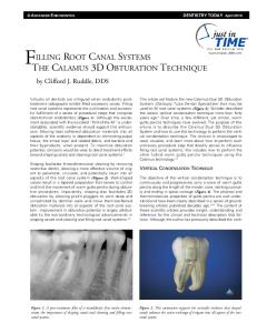
Figure 1: Working lenght radiography (Case 1).
Figure 2: Access cavity preparation (Case 1).
IJCRI – International Journal of Case Reports and Images, Vol. 3 No. 5, May 201 2. ISSN – [0976-31 98]
IJCRI 201 2;3(5):11 –1 5.
Çiçek et al.
www.ijcasereportsandimages.com
13
percha (Dentsply, Brazil and Dia–Dent, Maillefer, Korea) using the cold lateral compaction technique and the tooth was restorated with amalgam (Figure 8). Two years postobturation Xray confirmed the success of the root canal treatment (Figure 9).
DISCUSSION
Figure 3: Root canal obturation (Case 1).
Anatomical variations are more common in the molar teeth. So the variations play an important role in root canal treatment. Several studies reported the anatomy of root canal systems and the anatomical variations found in the different types of teeth. Studies on the anatomy of root canals carried out by Vande Voorde et al. [11] Badanelli et al. [12] and Fabracampos et al. [3] reinforced the importance of an accurate clinical evaluation of the possible fourth and fifth root canal to ensure success of the endodontic treatment.
Figure 4: Periapical radiography after 24 months (Case 1).
After ultracaine DS fort (4% articain with epinephrine 1/100000, HoechstMarion Roussel, Frankfurt, Germany) local anesthetic was administered by periapical infiltration, a rubberdam was placed and access cavity was opened. When the access cavity preparation was completed and the mesiobuccal, distobuccal, and palatal root canals were easily detected. After removing the pulp tissue, one root canal was found in distobuccal root. The working length was defined with periapical radiography. The root canals were enlarged up to F3 with ProTaper rotary NiTi system (Dentsply, Brazil). One week later the filling was removed and root canal treatment was renewed because of progressing pain. The root canal filling was removed and an extra canal in mesiobuccal region (MB2, Figure 7) was found next to first mesiobuccal canal (MB1, Figure 7), (Figure 6, 7). The canals were filled with medicament that contains calcium hydroxide paste (Kalsin, Aktu Tic., İzmir, Türkiye) and left for two weeks. After recovery of symptoms, the root canals were filled with AH plus (Dentsply, De Trey, Konstanz, Germany) and gutta
Figure 5: Preoparative radiography (Case 2).
Figure 6: Working lenght radiography (Case 2).
IJCRI – International Journal of Case Reports and Images, Vol. 3 No. 5, May 201 2. ISSN – [0976-31 98]
IJCRI 201 2;3(5):11 –1 5.
Çiçek et al.
www.ijcasereportsandimages.com
Figure 7: Access cavity preparation (Case 2).
14
Martinez–Berna et al. [2] remarked the importance of investigating the existence of a fourth and even a fifth root canal. Moreover, few studies [4, 14–17] found a third distal canal was in mandibular first molar. A mandibular first molar with three distal canals was first reported by Berthiaume et al. [13]; however, the three distal canals ended in two apical foramina. Examples of mandibular first molars with three distal canals, all of which ended in separate apical foramina, have also been described [14–16]. In addition, Quackenbush et al. [17] reported the existence of three separate distal canals in two extracted mandibular first molars. In the present study, the distal root which had three separate canals ended in one apical foramina. The maxillary first molar most commonly has three or four root canals, with one canal in both palatal and distobuccal roots and one or two in the mesiobuccal root. Çalışkan et al. [6] stated that a second canal is found in 65% of mesiobuccal roots of maxillary first molars. Bond et al. [18] reported a case of a maxillary first molar with six canals: two canals in the mesiobuccal root, two canals in the distobuccal root and two canals in the palatal root. Hulsmann et al. [19] presented a maxillary first molar with two canals in the distobuccal root. MartinezBerna et al. [20] reported three cases of maxillary first molars with six canals, three canals in the mesiobuccal root, two canals in the distobuccal root and one canal in the palatal root. One palatal root canal ended in one apical foramina, two separate mesiobuccal canals ended in one apical foramina and two separate distobuccal canals ended in two separate apical foramina in the present study.
CONCLUSION
Figure 8: Root canal obturation (Case 2).
Successful endodontic treatment starts with proper clinical and radiographic examinations. It is important for clinicians to be aware of all possible anatomic variations for a good endodontic practice.
*********
Author Contribution
Ersan Çiçek – Substantial contributions to conception and design, acquisition of data, Drafting the article, revising it critically for important intellectual content, Final approval of the version to be published Ebru Özsezer Demiryürek – Substantial contributions to conception and design, analysis and interpretation of data, Drafting the article, revising it critically for important intellectual content, Final approval of the version to be published Semih Özsevik – Substantial contributions to conception and design, Drafting the article, Final approval of the version to be published
Guarantor Figure 9: Periapical radiography after 24 months (Case 2).
The corresponding submission.
author
is
the
guarantor
IJCRI – International Journal of Case Reports and Images, Vol. 3 No. 5, May 201 2. ISSN – [0976-31 98]
of
IJCRI 201 2;3(5):11 –1 5.
www.ijcasereportsandimages.com
Conflict of Interest
Authors declare no conflict of interest.
Copyright
© Ersan Çiçek et al. 2012; This article is distributed under the terms of Creative Commons attribution 3.0 License which permits unrestricted use, distribution and reproduction in any means provided the original authors and original publisher are properly credited. (Please see www.ijcasereportsandimages.com /copyrightpolicy.php for more information.)
REFERENCES 1.
2. 3. 4. 5. 6. 7. 8. 9. 10. 11. 12. 13. 14. 15.
Çiçek et al.
15
16. Friedman S, Moshonov J, Stabholz A. Five root canals in a mandibular first molar. Endodontics and Dental Traumatolog 1986;2:226–8. 17. Quackenbush LE. Mandibular molar with three distal root canals. Endodontics and Dental Traumatology 1986;2:48–9. 18. Bond JL, Harwell G, Portell FR. Maxillary first molar with six canals. Journal of Endodontics 1988;14:258–60. 19. Hulsmann M. A maxillary first molar with two distobuccal root canals. Journal of Endodontics 1997;23:707–8. 20. MartinezBerna A, RuizBadanelli P. Maxillary first molars with six canals. Journal of Endodontics 1983;9:375–1.
Ingle JI. Endodontics, 3rd edt. Philadelphia, PA: Saunders 1985. MartinezBerna A, Badanelli P. investigación clinica de molares inferiores con cinco conductos. Boletín de information Dental 1983;43:27–41. FabraCampos CH. Unusual root anatomy of mandibular first molars. Journal of Endodontics 1985;11:568–2. Goel NK, Gill KS, Taneja JR. Study of root canals configuration in mandibular first permanent molar. Journal of Indian Society of Pedodontics and Preventive Dentistry 1991;8:12–4. Bueno CE, Cunha RS, Dotto SR, Ferreira R. Um molar inferior com cinco canaiscaso reportado. Revista da Faculdade de Odontologia da Universida de Passo Fundo 2002;7:51–3. Caliskan MK, Pehlivan Y, Sepetcioglu F, Türkün M, Tuncer SS. Root canal morphology oh human permanent teeth in a Turkish population. Journal of Endodontics 1995;21:200–4. Stropto JJ. Canal morphology of maxillary molars: Clinical observations of canal configuraitons. Journal of Endodontics 1999;25:446–50. Kulild JC, Peters DD. Incidence and configuraiton of canal systems in the mesiobuccal root of maxillary first and second molars. Journal of Endodontics 1990;16:311–7. Sert S, Bayirli GS. Evaluation of the root canal configuraitons of the mandibular and maxillary permanent teeth by gender in the Turkish population. Journal of Endodontics 2004;30:391–8. Gray R. The maxillary First molar. In Bjorndal A, Skidmore E, eds. Anatomy and morphology of the human teeth. Lowa City: University of lowa College of Dentistry 1983;31–40. Vande Voorde HE, Odendahl D, Davis J. Molar 4th canals: frequent cause of endodontic failure. III. Dental Journal 1975;44:779–6. Badanelli P, MartinezBerna A. Obturation de un molar inferior con cinco conductos. In: Lasala A, ed. Endodonticia. Barcelona: Salvat S A 1979:407. Berthiaume JT. Five canals in a lower first molar. The Journal of the Michigan Dent Association 1983;65:213–4. Stroner WF, Remeikis NA, Carr GB. Mandibular first molar with three distal canals. Oral Surgery 1984;57:554–7. Beatty RG, Interian CM. A mandibular first molar with five canals: report of case. The Journal of the American Dental Association 1985;111:769–1.
IJCRI – International Journal of Case Reports and Images, Vol. 3 No. 5, May 201 2. ISSN – [0976-31 98]