The Role of the Fallopian Tube in Ovarian Cancer Alicia A. Tone, PhD, Shannon Salvador, MD, Sarah J. Finlayson, MD, Anna V. Tinker, MD, Janice S. Kwon, MD, MPH, Cheng-Han Lee, MD, PhD, Trevor Cohen, MD, Tom Ehlen, MD, Marette Lee, MD, Mark S. Carey, MD, Mark Heywood, MD, Judith Pike, MD, Paul J. Hoskins, MD, Gavin C. Stuart, MD, Kenneth D. Swenerton, MD, David G. Huntsman, MD, C. Blake Gilks, MD, Dianne M. Miller, MD, and Jessica N. McAlpine, MD
Dr. Tone is a post-doctoral research fellow at the BC Cancer Agency and the Department of Pathology and Laboratory Medicine at UBC, and Dr. Huntsman is Medical Director at the Center for Translational and Applied Genomics, BC Cancer Agency, in Vancouver, Canada. Dr. McAlpine is a gynecologic oncologist and translational researcher at UBC and the BC Cancer Agency in Vancouver, Canada. Drs. Finlayson, Miller, Stuart, Ehlen, Heywood, Kwon, Carey, and Lee are gynecologic oncologists at UBC and the BC Cancer Agency. Dr. Hoskins, Swenerton, and Tinker are medical oncologists in the Division of Medical Oncology, BC Cancer Agency. Dr. Pike is a longstanding general practice oncologist with the BC Cancer Agency. Drs. Salvador and Cohen are Fellows in Gynecologic Oncology at UBC. Dr. Lee and Dr. Gilks are pathologists in the Department of Pathology and Laboratory Medicine, University of British Columbia, in Vancouver, Canada. All authors are part of British Columbia’s Ovarian Cancer Research Group (OvCaRe), co-founded by Drs. Huntsman and Miller, and comprising the cohesive voice behind the salpingectomy campaign of 2010. Address correspondence to: Jessica N. McAlpine, MD Assistant Professor University of British Columbia Department of Gynecology and Obstetrics Division of Gynecologic Oncology 2775 Laurel St., 6th Floor Vancouver, British Columbia Phone: 604-875-5608 Fax: 604-875-4869 E-mail:
[email protected] Keywords Ovarian cancer, fallopian tubes, high-grade serous carcinoma, serous tubal intraepithelial carcinomas
Abstract: High-grade serous carcinoma (HGSC) is the most common and lethal subtype of ovarian cancer. Research over the past decade has strongly suggested that “ovarian” HGSC arises in the epithelium of the distal fallopian tube, with serous tubal intraepithelial carcinomas (STICs) being detected in 5–10% of BRCA1/2 mutation carriers undergoing risk-reducing surgery and up to 60% of unselected women with pelvic HGSC. The natural history, clinical significance, and prevalence of STICs in the general population (ie, women without cancer and not at an increased genetic risk) are incompletely understood, but anecdotal evidence suggests that these lesions have the ability to shed cells with metastatic potential into the peritoneal cavity very early on. Removal of the fallopian tube (salpingectomy) in both the average and high-risk populations could therefore prevent HGSC, by eliminating the site of initiation and interrupting spread of potentially cancerous cells to the ovarian/peritoneal surfaces. Salpingectomy may also reduce the incidence of the 2 next most common subtypes, endometrioid and clear cell carcinoma, by blocking the passageway linking the lower genital tract to the peritoneal cavity that enables ascension of endometrium and factors that induce local inflammation. The implementation of salpingectomy therefore promises to significantly impact ovarian cancer incidence and outcomes.
Introduction Ovarian cancer is the leading cause of death due to gynecologic malignancies, and the fifth most common cause of cancer deaths in women. Historically, all ovarian cancers have been grouped together as one disease. Morphologic classification of epithelial ovarian carcinoma (EOC) has delineated distinct histologic subtypes: high-grade serous carcinoma (HGSC), clear cell carcinoma (CCC), endometrioid carcinoma (EC), mucinous carcinoma, and low-grade serous carcinoma, which differ vastly with respect to presentation, propensity to metastasize, response to therapy, prognosis, and site of origin.1-4 Seventy percent of EOCs are HGSC histology.3 Almost all HGSCs have TP53 mutations, which seem to occur as an early event in disease progression.2,5 Approximately half of HGSCs have BRCA dysfunction, through a combination of germline and somatic mutations or epigenetic silencing (Kasmintan A. Schrader, MD,
296 Clinical Advances in Hematology & Oncology Volume 10, Issue 5 May 2012
T h e R o l e o f th e Fa l l o p ia n T u b e i n O varia n C a n c e r
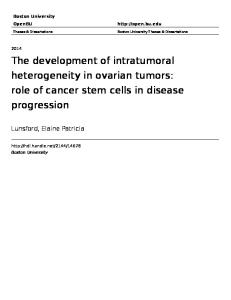
Figure 1. Surgically removed specimen highlighting the fallopian tube origin of “ovarian” high-grade serous carcinoma. The right side of the photo (corresponding to the left side of the patient) shows the distal end of the fallopian tube that is grossly distorted with disease and adherent to the rectosigmoid colon, a portion of which had to be removed. In contrast, both ovaries and the uterus appear normal.
personal communication).6-10 TP53 mutations and BRCA deficiency lead to incompetence in DNA repair, making HGSC highly responsive to chemotherapy, often repeatedly. However, the majority of patients recur, develop disease resistance, and ultimately succumb to their disease. Historically, tumors of the fallopian tube were described as rare. The fallopian tube has a unique and delicate role in capturing the egg released from the ovary in its fimbriated end and providing a conduit for transport of egg and sperm to meet for fertilization. It may also serve as a passageway for endometrial cells or infectious pathogens to ascend from the lower genital tract into the peritoneal cavity. The focus of attention on the fallopian tube has been primarily in younger women, who can experience infection leading to symptoms (eg, salpingitis, pelvic inflammatory disease), blockage of the fallopian tube(s) impacting fertility, and the potentially life-threatening situation of ectopic pregnancy. In women’s postreproductive years, the fallopian tube was believed to be of little importance. It was usually not considered to be an independent structure in surgical decision-making, and its anatomic proximity to the ovary meant that surgical removal or preservation depended on the surgical plan of the ovary. In women in whom widespread mullerian car-
cinoma was found, the pathology of the ovary (size and distortion) usually far overshadowed the fallopian tube, drawing the attention of pathologists and resulting in a diagnosis of ovarian primary carcinoma. We now appreciate the key role the fallopian tube plays in the development of ovarian cancer (Figure 1). Herein, we will outline the evidence suggesting that the distal fallopian tube is the site of origin of the most common type of ovarian cancer, HGSC, and describe its crucial role in the development of nonserous ovarian cancers. We will also share how this knowledge is being harnessed in designing intervention strategies that will impact ovarian cancer incidence and mortality over the next decade. Evidence That the Fallopian Tube Is the Site of Origin of the Majority of HGSCs Lessons Learned From Patients With Familial Risk While the lifetime risk for developing ovarian cancer is low (1.6%) in the general population, this risk increases to up to 60% and up to 30% for BRCA1 or BRCA2 mutation carriers, respectively.6-9 A retrospective study of 493 BRCA1/2 mutation carriers (245 of whom underwent surgery) observed a 98% reduction in risk for ovarian,
Clinical Advances in Hematology & Oncology Volume 10, Issue 5 May 2012 297
T O NE E T A L
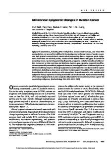
Figure 2. Detection of serous tubal intraepithelial carcinomas (STICs). (A) The SEE-FIM (Sectioning and Extensively Examining the FIMbriated end) protocol from Medeiros and colleagues19 examines salpingectomy–oophorectomy specimens in toto with length-wise (sagittal) sectioning of the fimbriated end to maximize exposure of the tubal epithelium. (B) This technique has previously been used in our center and others (eg, Salvador and coworkers69) to detect STIC lesions (inset) in BRCA1/2 mutation carriers undergoing risk-reducing salpingo-oophorectomy.
A
B fallopian tube, and primary peritoneal cancer with prophylactic (risk-reducing) bilateral salpingo-oophorectomy (RRSO).9 As mutation carriers are genetically predisposed to developing HGSC, a proportion of these specimens would be expected to harbor premalignant histologic or molecular changes involved in serous carcinogenesis. Importantly, intensive study of ovaries removed during RRSO in BRCA1/2 mutation carriers has failed to reveal reproducible premalignant epithelial changes.10-14 In stark contrast, serous tubal intraepithelial carcinomas (STICs) have been found in the fallopian tubes in 5–10% of these women.15-23 These lesions are predominantly found in the fimbriated end of the fallopian tube adjacent to the ovary, frequently in the absence of ovarian pathology. They also appear to occur at a higher incidence in BRCA1 compared to BRCA2 mutation carriers,18,23 possibly reflecting the decreased penetrance of BRCA2 and an older average age of onset.7,9,24 STICs are characterized by frequent
mutations in TP53, increased cellular proliferation and a secretory cell phenotype similar to invasive HGSC, and show evidence of DNA damage.19,21,25,26 Similar gene copy number changes have been reported in STICs and corresponding invasive HGSC from the same case discovered by RRSO, suggesting a clonal relationship.27 These unexpected findings in women at an increased risk for “ovarian” cancer led to the development of specialized protocols designed to maximize the detection of precursors/early fallopian tube cancers,19,28 most notably the Sectioning and Extensively Examining the FIMbria (SEE-FIM) protocol (Figure 2).19 Use of SEE-FIM subsequently revealed tubal involvement in 70%, and the presence of (primarily fimbrial) STICs in 40–60%, of unselected women diagnosed with ovarian27,29,30 or primary peritoneal HGSC.29-32 Importantly, identical mutations of TP53 have been observed in both ovarian/peritoneal HGSC and co-existing STICs, suggesting that these enti-
298 Clinical Advances in Hematology & Oncology Volume 10, Issue 5 May 2012
T h e R o l e o f th e Fa l l o p ia n T u b e i n O varia n C a n c e r
ties are causally related. In addition, the proportion of cases reported to contain an STIC increased with more complete examination of the fallopian tube.31,32 These findings clearly highlight the important role of the fallopian tube in pelvic HGSC irrespective of BRCA1/2 mutation status or family history, and provide a likely explanation for the observed residual risk (up to 11%) of peritoneal HGSC following prophylactic oophorectomy when the tubes are left in place.33-36 Importantly, STICs were not observed in women with ovarian carcinomas of nonserous histology or in nongynecologic or benign conditions in a recent study.30 In addition to STICs, putative precursor lesions have been described in up to 50% of RRSO specimens.21,26,37-39 Most notably, Lee and colleagues26 were the first to describe foci of strong p53 immunopositivity (termed “p53 signatures”) in benign-appearing fallopian tube epithelium. Similar to STICs, they are characterized by a predominantly fimbrial location, frequent TP53 mutations, secretory cell phenotype, and evidence of DNA damage. Unlike STICs, p53 signatures do not exhibit marked nuclear atypia or proliferative activity. Although p53 signatures were equally common in nonmalignant fallopian tube epithelium from BRCA mutation carriers and controls in the initial study26 and 2 subsequent studies,21,39 they were more frequently observed in fallopian tubes also containing STICs.26 In addition, a shared TP53 mutation was detected in 1 case with a contiguous p53 signature and STIC lesion. Importantly, Folkins and coworkers reported the presence of p53 signatures in 38% of BRCA1/2mutated fallopian tubes, while no lesions were detected in ovarian cortical inclusion cysts in the same patients.39 A New Model of Serous Carcinogenesis Emerges In light of these unexpected findings over the past decade, a new model for pelvic HGSC has emerged. As articulated by Crum and associates,40 the first step entails genotoxic injury to the secretory epithelial cells of the distal fallopian tube, which may lead to unrepaired DNA damage, cell cycle arrest, and TP53 mutations in a subset of cells. Clonal expansion of a TP53-mutated cell would then result in focal accumulation of p53, detectable by immunohistochemistry (p53 signature). The similar frequency of p53 signatures in BRCA1/2 mutation carriers and control patients (who are at a much lower risk for ovarian cancer), suggests that inheritance of a BRCA1/2 mutation may increase the risk of malignant transformation rather than formation of the precursor itself.26,40 The second step involves re-initiation of cell proliferation in a subset of these p53 signatures, leading to development of an STIC. Highly aggressive subclones of STICs may locally expand and invade the underlying tubal stroma, presenting as primary fallopian tube carcinoma. Alternatively, an STIC lesion may exfoliate onto the closely associated ovarian surface/peritoneal cavity and present as primary ovarian/peritoneal carcinoma.40
The discovery of p53 signatures and STICs predominantly in the fimbria, rather than randomly throughout the fallopian tube, has several potential explanations. Firstly, the abundance of surface area in this region may mathematically increase the risk that a neoplasm will arise. Secondly, there may be differences in the mucosa of the fimbria that make them inherently more susceptible to carcinogenesis compared to proximal segments of the fallopian tube. Thirdly, the proximity of the fimbria to the peritoneal cavity and ovarian surface may be important.19,26,40 As discussed in later sections, this microenvironment appears to be uniquely proinflammatory and protumorigenic. A recently described ex vivo primary human secretory/ciliated fallopian tube epithelial cell coculture system demonstrated a distinct response of secretory cells to ionizing radiation, including a limited ability to resolve the resulting damage over time.41 This suggests that secretory cells may be more sensitive to DNA-damaging agents and a proinflammatory microenvironment. The same group also demonstrated the ability of transformed secretory epithelial cells to give rise to HGSC tumors, including the characteristic chromosomal instability and unique immunophenotype, in xenograft models.42 Despite these recent advances in our understanding of serous carcinogenesis, some aspects are still incompletely understood. Firstly, the prevalence of STICs in women without cancer in the general population (ie, those women not at an increased genetic risk) is controversial. One study reported the presence of STICs in 3% of women at average risk for ovarian cancer,21 however, this finding was based on a small sample size. In contrast, in our preliminary study of 685 salpingectomy specimens from unselected women older than 35 years, we observed STICs only in women with a confirmed mutation in BRCA1/2 or HGSC (Blake Gilks, MD, personal communication). No intraepithelial carcinomas were observed in the ovaries removed from the same patients. Secondly, it is unclear how long STICs are present in their noninvasive state, what proportion of STICs develop into advanced-stage HGSC, and how best to manage these women. Models developed by Brown and Palmer43 estimated that the clinically occult period for HGSC (including intraepithelial [stage 0], stage I, and stage II tumors) could last up to 4 years. Reports in the literature31 also suggest that, when found in isolation, STICs have a very low risk of recurrence. However, 2 women cared for by our gynecologic oncology team, including 1 patient with an isolated STIC less than 1 mm and 1 patient with a lesion less than 1 mm showing some but not all the features of STIC (“atypia of uncertain clinical significance”), went on to develop widespread peritoneal cancer 2 years following surgery. Neither of these women received chemotherapy as per standard protocol. These
Clinical Advances in Hematology & Oncology Volume 10, Issue 5 May 2012 299
T O NE E T A L
examples underscore the ability of STICs to shed cells with metastatic potential into the peritoneal cavity very early on, and hence show the importance of removing at-risk fallopian tube tissue to reduce the risk of ovarian cancer. They also suggest that the morphologic spectrum of clinically significant in situ neoplasia in the fallopian tube may be broader than is currently appreciated. Another question is how to account for those 30% of pelvic HGSCs that do not show any evidence of tubal involvement. Kurman and Shih1 and others have suggested that, as a function of their close association, fallopian epithelial cells may become entrapped in the ovary during disruption of the ovarian surface at ovulation. HGSC may then develop from included tubal cells following exposure to the unique hormone- and cytokinerich ovarian stromal environment. This is in contrast to the previous long-standing theory, prior to the discoveries in the fallopian tube, that cortical inclusion cysts lined by ovarian surface epithelial (OSE) cells undergo metaplasia and eventual malignant transformation.44 Other recent theories have included involvement of the peritonealtubal junction45 and OSE-tubal junction.43,44 Despite these current gaps in knowledge, data over the past decade strongly suggest a tubal source for a majority of pelvic HGSC, and underscore the fallopian tube (STICs) as an attractive target for early detection and prevention. Microenvironment of the Fallopian Tube, and the Fallopian Tube as a Conduit In addition to highly penetrant mutations in BRCA1/2, epidemiologic studies have determined additional (lowpenetrance) modifiers of ovarian cancer risk associated with a woman’s reproductive history, most notably oral contraceptive (OCP) use, parity, breastfeeding, tubal ligation, and pelvic inflammatory disease (PID). All of these factors can impact the fallopian tube microenvironment, with ovulation, menstruation, and inflammation playing central protumorigenic roles. “Incessant ovulation” has long been proposed to promote ovarian carcinogenesis,46,47 as epidemiologic studies consistently observe a positive association between lifetime number of ovulatory cycles and ovarian cancer risk, either directly48 or indirectly. Consistent with their suppressive effects on ovulation, multiple studies have reported a substantial protective effect for OCP use49-51 and parity/breastfeeding49,50,52,53 in both BRCA1/2 mutation carriers and women at baseline risk. Greater risk reduction is observed with increasing duration of OCP use, an increased number of full-term pregnancies, and a longer duration of total lactation time. Two recent studies also reported an inverse correlation between the presence of an STIC/p53 signature and duration of OCP use54 or parity55 in BRCA1/2 mutation carriers.
Importantly, an acute proinflammatory environment is created following ovulation within the distal tube, the location of the overwhelming majority of occult carcinomas in RRSO specimens. Each ovulatory cycle involves infiltration by leukocytes and production of inflammatory mediators,56,57 and nonsteroidal anti-inflammatory drugs (NSAIDs) have been shown to inhibit ovulation.58-60 Ovulation results in the release of an oocyte with its adherent cumulus granulosa cells into the adjacent fallopian tube, bathing the ovarian surface and fimbria with follicular fluid rich in inflammatory cytokines and reactive oxygen species (ROS),61 and pro-inflammatory cytokines secreted by released cumulus cells.62 BRCA1-mutated nonmalignant fallopian tube epithelium obtained during the postovulatory luteal phase show global gene expression profiles more closely resembling that of HGSC than fallopian tube epithelium collected during the postmenstrual follicular phase or from control patients,63 and further analysis strongly suggests a role for ovulation-associated inflammatory signaling in predisposition to HGSC (Alicia A. Tone, PhD, manuscript in submission). Similarly, Karst64 and others26,65 have proposed that exposure of the fallopian tube to the ovulatory microenvironment contributes to development of histologic HGSC precursors. As previously discussed, secretory tubal epithelial cells, the likely cell of origin of HGSC, show a distinct response to genotoxic stress.41 The repetitive genotoxic stress associated with the ovulationassociated inflammatory environment could therefore lead to DNA damage and TP53 mutation; progression to an STIC, and, eventually, invasive HGSC would occur upon acquisition of further aberrations. In contrast to ovulation, several studies have reported an inverse association of sterilization by tubal ligation and ovarian cancer risk in the general population49,56,66-69 and BRCA1 mutation carriers.66 Of particular note, the Nurse’s Health Study (>100,000 women) reported a substantial reduced risk for women who had undergone tubal ligation after 1267 and 2868 years of follow-up. Studies suggest that tubal ligation does not significantly impact ovarian function (ie, ovarian hormone levels/ovulation),70-77 hence some authors have suggested that the protective effect lies in reducing the potential for local inflammation. Tubal ligation would block the transport of genital tract irritants (such as talc)56 from the lower genital tract, thereby limiting the exposure to initiators of inflammation. Regular exposure of the fallopian tube to sexually transmitted infections and menstrual cytokines through retrograde flow from the endometrium may also promote carcinogenesis.56,69 A history of PID has been linked to an increased risk of ovarian cancer,78 and chronic salpingitis was present in 53% of examined ovarian carcinomas in one study.79 A recent review proposed that the distal fallopian tube would have uniquely prolonged exposure to bloody peritoneal
300 Clinical Advances in Hematology & Oncology Volume 10, Issue 5 May 2012
T h e R o l e o f th e Fa l l o p ia n T u b e i n O varia n C a n c e r
fluid and catalytic iron that has collected in the Douglas pouch as a result of retrograde menstruation; tumorigenesis would then be promoted by iron-induced oxidative stress.80 The relevance of inflammation in ovarian cancer is clearly highlighted by the decreased risk associated with NSAID use. Importantly, one study reported a protective effect of NSAIDs in women who had never used OCPs, but not women who had used OCPs. Similarly, NSAIDs reduced risk among nulliparous but not parous women, suggesting that NSAIDs are particularly beneficial to women in higher risk groups.81 The Fallopian Tube and Nonserous Ovarian Cancer Histologies Although the 2 next most common subtypes of EOC do not originate in the fallopian tubes, they are believed to play a permissive role in their development. CCC and EC, each accounting for 10% of EOC, are thought to arise from endometriotic lesions on or around the ovary.82,83 Endometriosis, defined as the presence of “ectopic” endometrium outside the uterine cavity, affects 10–15% of women of reproductive age and likely develops through reflux of endometrial tissue through the fallopian tubes into the pelvis.84-86 Endometriosis is a chronic inflammatory disease, characterized by altered function of immune cells, continuous production of proinflammatory cytokines, chemokines, and prostaglandins, and high concentrations of pro-oxidant factors (free iron and heme) as a result of incessant retrograde menstruation or ovarian hemorrhage.87-90 Although endometriosis is itself benign, it increases ovarian cancer risk91-93 and frequently coexists with EC and CCC.82,94-97 In 60% of endometriosis-associated ovarian cancers (EAOC), the carcinoma is adjacent to, or in direct continuity with, atypical endometriosis.98,99 Shared mutations in ARID1A100 and PIK3CA101 have also been observed in tumor specimens and adjacent endometriotic epithelium. The importance of the fallopian tube as a conduit is highlighted by the extent of protection provided by tubal ligation for EAOC. Recent data obtained from the Ovarian Cancer Association Consortium (OCAC) suggest that tubal ligation is associated with a 38% and 52% reduction in risk for EC and CCC, respectively, compared to a 19% drop in HGSC (Shannon Salvador, MD, personal communication). This is similar to the greater protective effect observed for EC (80%) compared to all histotypes combined (30%) by Tung and coworkers.102 These data likely reflect the fact that tubal ligation would interrupt the passage of endometrial tissue to the ovary/peritoneal cavity through the fallopian tube (and subsequent development of endometriosis), while distal epithelial cells would still be able to shed to the ovarian/peritoneal surfaces unless a fimbriectomy was performed.
Strategies in Ovarian Cancer Reduction Screening/Early Detection When confined to the ovary (stage I), nonserous ovarian cancer has a 5-year survival rate of greater than 95%. In contrast, 5-year survival rates for advanced stage disease with spread beyond the ovary (stage III/IV) range between 15–30%. Unfortunately, the majority of women with ovarian cancer, including those with the most common subtype HGSC, are diagnosed at an advanced stage. In fact, HGSCs account for 90% of advanced stage disease, and less than 1.5% of comprehensively staged serous carcinomas are in International Federation of Gynecology and Obstetrics (FIGO) stage I.32 In theory, therefore, if detection of HGSCs could be shifted from a late to an early stage through the use of screening modalities, such as transvaginal ultrasound (TVUS) and serum CA-125, the survival rates for ovarian cancer would be impacted greatly. However, despite tremendous international efforts,103-106 screening studies to date have not only resulted in no difference in cancer-specific mortality but have also been associated with a considerable rate of complications and unnecessary surgery. Of particular note, 3 large randomized controlled trials, namely the Prostate, Lung, Colorectal, Ovarian (PLCO) Cancer Trial in the United States, the United Kingdom Collaborative Trial of Ovarian Cancer Screening (UKCTOS), and Japan’s Shizuoka Cohort Study of Ovarian Cancer Screening Trial, have recently been performed. These trials involved a total of 363,341 women (range 78,216–202,638 women), and each unsuccessfully attempted to improve the early detection of ovarian cancer by annual screening of asymptomatic, postmenopausal women in the general population using serum CA-125 or a combination of CA-125 and TVUS. Even attempts at screening in high-risk women (ie, BRCA1/2 mutation carriers) have proven to be ineffective at diagnosing early-stage disease,107-110 despite the greatly increased incidence of disease in this subpopulation. Of particular note, despite following 888 BRCA1/2 carriers for 13 years with annual screening, Hermsen and associates109 reported an equal number of interval and screen-detected cancers, with no difference in stage distribution. Despite the failure of screening in asymptomatic women, the recent prospective DOvE (Diagnosing Ovarian Cancer Early) study from Montreal, Quebec, Canada, aimed to improve early detection by fast-tracking assessment of symptomatic women.111 This is based on the fact that most women with ovarian cancer experience symptoms, including but not limited to, loss of appetite, bloating, unplanned weight loss, pelvic or abdominal pain, and increased urinary urgency or frequency, prior to their diagnosis.112 However, a recent (retrospective) study by Lim and associates has demonstrated that there
Clinical Advances in Hematology & Oncology Volume 10, Issue 5 May 2012 301
T O NE E T A L
is a marked acceleration in the onset of symptoms in the 3 months just prior to diagnosis, and that there is little difference in the symptoms experienced by those women ultimately diagnosed with early-stage versus late-stage disease.113 Not surprisingly, therefore, the DOvE trial failed to shift the diagnosis of ovarian cancer to an earlier stage, with similar or higher proportions of stage III–IV cancers diagnosed than those observed in asymptomatic women from the general or high-risk population. The failure of screening to improve ovarian cancer outcomes has several plausible explanations. For instance, the use of TVUS is based on the assumption that an enlarged ovary is an early manifestation of disease, when this is clearly not the case for HGSC. Secondly, only 25–50% of stage I tumors overall have elevated CA-125,114 whereas increased levels are seen in many conditions other than ovarian cancer, including but not limited to pregnancy, endometriosis, menstruation, and liver disease.115 Several groups have also demonstrated that HGSC is inherently aggressive and distinct from ovarian cancers typically presenting at an early stage.1,116 Although Brown and Palmer43 proposed that there is a “window of opportunity” during which time an early HGSC could be detected, they estimate that to achieve 50% sensitivity, an annual screening test would need to detect tumors more than 200 times smaller than clinically apparent serous cancers. Much work therefore needs to be done in developing more sensitive, cancer-specific biomarkers before screening can impact ovarian cancer outcomes in a meaningful way. Treatment Despite the great attention and resources dedicated to advancing surgical technique, conventional chemotherapy drug choices, routes of delivery, and schedules (eg, dose intensity and proposed maintenance therapies), survival rates in ovarian cancers have changed minimally since the introduction of taxanes in the mid-1980s. In British Columbia, ovarian cancer survival rates at 1, 3, and 5 years have changed a total of 1%, 5%, and 4% over the past 10 years. We have gained a better understanding of the morphologic/histologic subtypes of EOC and have identified disease subgroups that have excellent prognosis (eg, >90–95% cure rates with surgery alone), sparing some women from unnecessary or ineffective therapies. With a histology-specific approach to ovarian cancers and with deeper genomic interrogation of these tumors, we can envision the emergence of targeted therapies specific to an individual’s tumor, however, we do not have sufficient evidence to suggest that any of the current molecular-targeted therapies lead to a clinically relevant survival advantage over current standard of care.117,118 At present, non-specific chemotoxic agents are the norm for all women with EOC. It is important to emphasize that
advances in treatment, no matter how novel and exciting, will largely be for the treatment of disseminated disease. These may lead to prolonged survival, but will infrequently achieve cure. True impact on ovarian cancer, therefore, lies in its prevention. Prevention The frequent finding of intraepithelial carcinoma in the fallopian tubes of women undergoing RRSO or with pelvic HGSC suggests that removal of the fallopian tube (salpingectomy) could prevent this type of ovarian cancer, by interrupting the spread of cells to the ovarian or peritoneal surfaces (Figure 3). As already discussed, RRSO reduces ovarian, fallopian tube, and peritoneal cancer by 98% in BRCA1/2 mutation carriers.9 This practice may therefore also be effective in women at “average” risk, who ultimately make up the bulk of ovarian cancers diagnosed. In addition, the protective effect of tubal ligation has been observed in both high-risk (BRCA1/2 mutationassociated) and sporadic ovarian cancers. We hypothesize that salpingectomy should be of even greater benefit than tubal ligation in reducing ovarian cancer risk in both of these populations, and that the impact should be seen in both serous and nonserous histologies. Interestingly, up to 20% of ovarian cancer patients from British Columbia had previously had a hysterectomy, and 10–15% had previously undergone tubal ligation (CBOCOU database BCCA and Ken Swenerton, MD, personal communication). Hysterectomy and tubal ligation are 2 common gynecologic procedures in which the fallopian tubes (fimbria) are surgically accessible, but in premenopausal women they are usually left in place/in situ. Removal of the fallopian tubes during these procedures may have prevented the development of ovarian cancer (often many years postsurgery) in these women. In addition, as 20–25% of women diagnosed with HGSC have inherited mutations in BRCA1/2,119,120 which are associated with an up to 50% lifetime cancer risk, identification of these women should prompt consideration of risk-reducing surgeries in their family members. This offers an additional opportunity to reduce ovarian cancer through the improved identification of women with genetic susceptibility. In light of this information, the Ovarian Cancer Research Program (OvCaRe) of British Columbia launched a province-wide educational campaign in September 2010 directed at all gynecologists. We summarized the evidence for the tubal origin of HGSC, and asked them to consider full removal of the fallopian tube at the time of hysterectomy and removal of the fallopian tubes in women seeking permanent irreversible contraception (eg, in place of tubal ligation). A brief instructional surgical film was included in their information packet demonstrating laparoscopic salpingectomy. Furthermore,
302 Clinical Advances in Hematology & Oncology Volume 10, Issue 5 May 2012
T h e R o l e o f th e Fa l l o p ia n T u b e i n O varia n C a n c e r
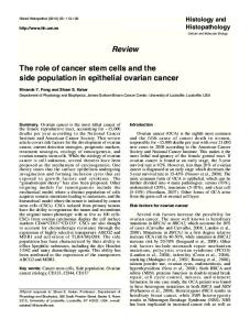
Figure 3. The role of the fallopian tube in ovarian carcinoma and the protective effect of tubal surgery. This cartoon highlights the role of the fallopian tube as the source of “ovarian” high-grade serous carcinoma (HGSC) and as a conduit for the development of endometrioid (EC) and clear cell carcinoma (CCC). Contributing factors include retrograde menstruation (leading to local inflammation and establishment of endometriotic lesions with the potential to develop into EC or CCC), ascending infection and irritants, and the inflammatory microenvironment of the distal fallopian tube as a result of ovulation, thought to play a key role in HGSC initiation. Consistently, oral contraceptives and pregnancy (which effectively prevent ovulation, decrease tubal epithelial cell motility, and increase cervical mucus thickness) and nonsteroidal anti-inflammatory drugs have been shown to decrease ovarian cancer risk. Using this knowledge, EC and CCC could be reduced (or possibly prevented if intervention is early enough) by blocking the passageway for causative factors via salpingectomy, consistent with multiple studies reporting a substantial protective effect of tubal ligation for these histotypes. The most common and lethal type of ovarian cancer, HGSC, could also be prevented by removing the tissue at risk for malignant transformation, namely the fimbriated end of the fallopian tube. The implementation of salpingectomy therefore promises to significantly impact ovarian cancer incidence and outcomes.
we recommended the referral of all women with HGSC of the ovary/fallopian tube/peritoneum for genetic counseling and BRCA1/2 mutation testing (Kasmintan A. Schrader, MD, personal communication).121,122 We believe that these simple changes in clinical and surgical practice will have an important impact on the number of HGSC cases over the next 2 decades, with an estimated 10% reduction through the increased identification of mutation carriers prompting risk-reduction surgeries, and a further 30% reduction from women undergoing salpingectomy at the time of otherwise indicated gynecologic surgery. We also anticipate a reduction in CCC and EC, as the usual conduit for retrograde menstruation and distribution of endometriosis to the ovarian surface and peritoneal cavity is interrupted with salpingectomy. Careful analysis of the incidence of ovarian cancer and the distribution of histologic subtypes of these diagnoses over the next several decades is essential. In addition to our provincial educational campaign, we surveyed all gynecologists of Canada to assess the baseline knowledge and interest in the implementation of a national strategy for the prevention of ovarian/fallopian tube cancer. We received 192 completed surveys, with
representation from the Society of Gynecologic Oncology of Canada (GOC), the Society of Obstetricians and Gynaecologists of Canada (SOGC), and all provinces. Ninety percent of respondents had previously heard about our recommendations, and 53% agreed that there would be a population-based benefit for recommending risk-reducing salpingectomy at the time of hysterectomy in the general population. Of note, 45% of respondents already routinely performed a bilateral salpingectomy at the time of hysterectomy (without bilateral oophorectomy); fallopian tubes were removed for the purpose of cancer prevention in 90% of these cases. In contrast, only 7% of respondents routinely removed the entire fallopian tube at the time of tubal ligation. Major (perceived) barriers associated with implementing salpingectomy for ovarian cancer prevention included increased complications, increased operating times, and irreversibility, with a greater number of respondents expressing concerns with performing salpingectomy in place of tubal ligation. Consistently, a much lower proportion of respondents reported that they had changed or intended to change the way they perform tubal ligations (28%) versus hysterectomy (68%).
Clinical Advances in Hematology & Oncology Volume 10, Issue 5 May 2012 303
T O NE E T A L
In September 2011, the GOC endorsed our approach, recommending that “physicians discuss the risks and benefits of bilateral salpingectomy with patients undergoing hysterectomy or requesting permanent, irreversible contraception,” and that an “ovarian cancer prevention study focused on fallopian tube removal is a GOC priority” (http://www.g-o-c.org). Local and international interest for this campaign has been substantial. Change in practice has been primarily on an individual surgeon and/or practice group level, but we are aware of numerous pathologists and surgical colleagues who advocate for salpingectomy outside of Canada.1 In addition to cancer prevention, they are often motivated by the many other potential adverse effects of retained fallopian tubes, including hydrosalpinx (often confused with ovarian pathology), tubal prolapse, torsion, PID, and tubo-ovarian abscess.123 To definitively establish whether this initiative will reduce the incidence of ovarian cancer in British Columbia, and to address concerns raised through our national survey, we are currently embarking on a long-term multisector study. If our cancer risk reduction program is found to be effective, we will establish a national campaign and dramatically shift our focus in ovarian cancer from one of treatment to prevention. Briefly, we plan to launch further (both physician- and patient-directed) education campaigns and determine whether our collective knowledge translation efforts result in an increased number of BRCA1/2 mutation referrals and preventative surgeries being performed on a provincial level. We will objectively assess the costs and complications associated with risk-reducing salpingectomy to ensure that these changes in surgical practice are both cost-effective and safe to women. In addition, we will monitor gradual changes in the distribution of tumor histologies and patient history at diagnosis (eg, BRCA status, whether patients have a history of tubal ligation or hysterectomy) as a result of this initiative, and eventually determine if we have decreased the number of ovarian cancers diagnosed per year, specifically HGSC and those types thought to pass through the fallopian tube during their development (eg, EC and CCC arising from endometriosis). Finally, as we are in a unique position of being able to evaluate a large number of fallopian tubes from the general population, we will assess the frequency of, and model the risk associated with, STICs in both women at high risk and baseline risk for ovarian cancer. Summary We believe that the evidence supporting the fallopian tube as the site of origin of the most common type of ovarian cancer is indisputable. The fallopian tube also plays a key role in
ovarian cancer development by acting as a conduit, linking the lower genital tract to the peritoneal cavity and enabling the ascension of endometrium and factors that induce local inflammation. Screening and treatment have not advanced significantly in the last 2 decades. We have an opportunity to capitalize on this increased appreciation of the role of the fallopian tube and embrace a new surgical paradigm for ovarian cancer prevention. We will endeavor to diligently assess the impact of this campaign, both at the level of the individual and with population-based ovarian cancer statistics. References 1. Kurman RJ, Shih Ie M. The origin and pathogenesis of epithelial ovarian cancer: a proposed unifying theory. Am J Surg Pathol. 2010;34:433-443. 2. Kobel M, Huntsman D, Gilks CB. Critical molecular abnormalities in highgrade serous carcinoma of the ovary. Expert Rev Mol Med. 2008;10:e22. 3. Gilks CB. Molecular abnormalities in ovarian cancer subtypes other than highgrade serous carcinoma. J Oncol. 2010;2010:740968. 4. Kalloger SE, Kobel M, Leung S, et al. Calculator for ovarian carcinoma subtype prediction. Mod Pathol. 2011;24:512-521. 5. Ahmed AA, Etemadmoghadam D, Temple J, et al. Driver mutations in TP53 are ubiquitous in high grade serous carcinoma of the ovary. J Pathol. 2010;221:49-56. 6. Prat J, Ribe A, Gallardo A. Hereditary ovarian cancer. Hum Pathol. 2005;36:861-870. 7. Karlan B, Boyd J, Strong L, Garber J, Fountain J, Beller U. Discussion: hereditary ovarian cancer. Gynecologic Oncology. 2003;88:S11-S3. 8. King MC, Marks JH, Mandell JB. Breast and ovarian cancer risks due to inherited mutations in BRCA1 and BRCA2. Science. 2003;302:643-646. 9. Boyd J. Specific keynote: hereditary ovarian cancer: what we know. Gynecol Oncol. 2003;88(1 Pt 2):S8-S10; discussion S11-S13. 10. Barakat RR, Federici MG, Saigo PE, Robson ME, Offit K, Boyd J. Absence of premalignant histologic, molecular, or cell biologic alterations in prophylactic oophorectomy specimens from BRCA1 heterozygotes. Cancer. 2000;89:383-390. 11. Casey MJ, Bewtra C, Hoehne LL, Tatpati AD, Lynch HT, Watson P. Histology of prophylactically removed ovaries from BRCA1 and BRCA2 mutation carriers compared with noncarriers in hereditary breast ovarian cancer syndrome kindreds. Gynecol Oncol. 2000;78:278-287. 12. Kerner R, Sabo E, Gershoni-Baruch R, Beck D, Ben-Izhak O. Expression of cell cycle regulatory proteins in ovaries prophylactically removed from Jewish Ashkenazi BRCA1 and BRCA2 mutation carriers: correlation with histopathology. Gynecol Oncol. 2005;99:367-375. 13. Piek JM, Dorsman JC, Shvarts A, et al. Cultures of ovarian surface epithelium from women with and without a hereditary predisposition to develop female adnexal carcinoma. Gynecol Oncol. 2004;92:819-826. 14. Piek JM, Verheijen RH, Menko FH, et al. Expression of differentiation and proliferation related proteins in epithelium of prophylactically removed ovaries from women with a hereditary female adnexal cancer predisposition. Histopathology. 2003;43:26-32. 15. Colgan TJ, Murphy J, Cole DE, Narod S, Rosen B. Occult carcinoma in prophylactic oophorectomy specimens: prevalence and association with BRCA germline mutation status. Am J Surg Pathol. 2001;25:1283-1289. 16. Leeper K, Garcia R, Swisher E, Goff B, Greer B, Paley P. Pathologic findings in prophylactic oophorectomy specimens in high-risk women. Gynecol Oncol. 2002;87:52-56. 17. Cass I, Holschneider C, Datta N, Barbuto D, Walts AE, Karlan BY. BRCAmutation-associated fallopian tube carcinoma: a distinct clinical phenotype? Obstet Gynecol. 2005;106:1327-1334. 18. Finch A, Shaw P, Rosen B, Murphy J, Narod SA, Colgan TJ. Clinical and pathologic findings of prophylactic salpingo-oophorectomies in 159 BRCA1 and BRCA2 carriers. Gynecol Oncol. 2006;100:58-64. 19. Medeiros F, Muto MG, Lee Y, et al. The tubal fimbria is a preferred site for early adenocarcinoma in women with familial ovarian cancer syndrome. Am J Surg Pathol. 2006;30:230-236. 20. Callahan MJ, Crum CP, Medeiros F, et al. Primary fallopian tube malignancies in BRCA-positive women undergoing surgery for ovarian cancer risk reduction. J Clin Oncol. 2007;25:3985-3990. 21. Shaw PA, Rouzbahman M, Pizer ES, Pintilie M, Begley H. Candidate serous cancer precursors in fallopian tube epithelium of BRCA1/2 mutation carriers. Mod Pathol. 2009;22:1133-1138.
304 Clinical Advances in Hematology & Oncology Volume 10, Issue 5 May 2012
T h e R o l e o f th e Fa l l o p ia n T u b e i n O varia n C a n c e r
22. Powell CB, Chen LM, McLennan J, et al. Risk-reducing salpingo-oophorectomy (RRSO) in BRCA mutation carriers: experience with a consecutive series of 111 patients using a standardized surgical-pathological protocol. Int J Gynecol Cancer. 2011;21:846-851. 23. Yates MS, Meyer LA, Deavers MT, et al. Microscopic and early-stage ovarian cancers in BRCA1/2 mutation carriers: building a model for early BRCA-associated tumorigenesis. Cancer Prev Res (Phila). 2011;4:463-470. 24. Narod SA, Boyd J. Current understanding of the epidemiology and clinical implications of BRCA1 and BRCA2 mutations for ovarian cancer. Curr Opin Obstet Gynecol. 2002;14:19-26. 25. Talamo TS, Bender BL, Ellis LD, Scioscia EA. Adenocarcinoma of the Fallopian tube. An ultrastructural study. Virchows Arch A Pathol Anat Histol. 1982;397:363-368. 26. Lee Y, Miron A, Drapkin R, et al. A candidate precursor to serous carcinoma that originates in the distal fallopian tube. J Pathol. 2007;211:26-35. 27. Salvador S, Rempel A, Soslow RA, Gilks B, Huntsman D, Miller D. Chromosomal instability in fallopian tube precursor lesions of serous carcinoma and frequent monoclonality of synchronous ovarian and fallopian tube mucosal serous carcinoma. Gynecol Oncol. 2008;110:408-417. 28. Powell CB, Kenley E, Chen LM, et al. Risk-reducing salpingo-oophorectomy in BRCA mutation carriers: role of serial sectioning in the detection of occult malignancy. J Clin Oncol. 2005;23:127-132. 29. Kindelberger DW, Lee Y, Miron A, et al. Intraepithelial carcinoma of the fimbria and pelvic serous carcinoma: evidence for a causal relationship. Am J Surg Pathol. 2007;31:161-169. 30. Tang S, Onuma K, Deb P, et al. Frequency of serous tubal intraepithelial carcinoma in various gynecologic malignancies: a study of 300 consecutive cases. Int J Gynecol Pathol. In press. 31. Carlson JW, Miron A, Jarboe EA, et al. Serous tubal intraepithelial carcinoma: its potential role in primary peritoneal serous carcinoma and serous cancer prevention. J Clin Oncol. 2008;26:4160-4165. 32. Seidman JD, Zhao P, Yemelyanova A. “Primary peritoneal” high-grade serous carcinoma is very likely metastatic from serous tubal intraepithelial carcinoma: assessing the new paradigm of ovarian and pelvic serous carcinogenesis and its implications for screening for ovarian cancer. Gynecol Oncol. 2011;120:470-473. 33. Tobacman JK, Greene MH, Tucker MA, Costa J, Kase R, Fraumeni JF Jr. Intra-abdominal carcinomatosis after prophylactic oophorectomy in ovariancancer-prone families. Lancet. 1982;2:795-797. 34. Piver MS, Jishi MF, Tsukada Y, Nava G. Primary peritoneal carcinoma after prophylactic oophorectomy in women with a family history of ovarian cancer. A report of the Gilda Radner Familial Ovarian Cancer Registry. Cancer. 1993;71:2751-2755. 35. Struewing JP, Watson P, Easton DF, Ponder BA, Lynch HT, Tucker MA. Prophylactic oophorectomy in inherited breast/ovarian cancer families. J Natl Cancer Inst Monogr. 1995:33-35. 36. Olivier RI, van Beurden M, Lubsen MA, et al. Clinical outcome of prophylactic oophorectomy in BRCA1/BRCA2 mutation carriers and events during followup. Br J Cancer. 2004;90:1492-1497. 37. Piek JM, van Diest PJ, Zweemer RP, et al. Dysplastic changes in prophylactically removed Fallopian tubes of women predisposed to developing ovarian cancer. J Pathol. 2001;195:451-456. 38. Piek JM, Verheijen RH, Kenemans P, Massuger LF, Bulten H, van Diest PJ. BRCA1/2related ovarian cancers are of tubal origin: a hypothesis. Gynecol Oncol. 2003;90:491. 39. Folkins AK, Jarboe EA, Saleemuddin A, et al. A candidate precursor to pelvic serous cancer (p53 signature) and its prevalence in ovaries and fallopian tubes from women with BRCA mutations. Gynecol Oncol. 2008;109:168-173. 40. Crum CP, Drapkin R, Miron A, et al. The distal fallopian tube: a new model for pelvic serous carcinogenesis. Curr Opin Obstet Gynecol. 2007;19:3-9. 41. Levanon K, Ng V, Piao HY, et al. Primary ex vivo cultures of human fallopian tube epithelium as a model for serous ovarian carcinogenesis. Oncogene. 2010;29:1103-1113. 42. Karst AM, Levanon K, Drapkin R. Modeling high-grade serous ovarian carcinogenesis from the fallopian tube. Proc Natl Acad Sci U S A. 2011;108:7547-7552. 43. Brown PO, Palmer C. The preclinical natural history of serous ovarian cancer: defining the target for early detection. PLoS Med. 2009;6:e1000114. 44. Auersperg N, Wong AS, Choi KC, Kang SK, Leung PC. Ovarian surface epithelium: biology, endocrinology, and pathology. Endocr Rev. 2001;22:255-288. 45. Seidman JD, Yemelyanova A, Zaino RJ, Kurman RJ. The fallopian tube-peritoneal junction: a potential site of carcinogenesis. Int J Gynecol Pathol. 2011;30:4-11. 46. Fathalla MF. Incessant ovulation—a factor in ovarian neoplasia? Lancet. 1971;2:163.
47. Casagrande JT, Louie EW, Pike MC, Roy S, Ross RK, Henderson BE. “Incessant ovulation” and ovarian cancer. Lancet. 1979;2:170-173. 48. Tung KH, Wilkens LR, Wu AH, et al. Effect of anovulation factors on preand postmenopausal ovarian cancer risk: revisiting the incessant ovulation hypothesis. Am J Epidemiol. 2005;161:321-329. 49. McGuire V, Felberg A, Mills M, et al. Relation of contraceptive and reproductive history to ovarian cancer risk in carriers and noncarriers of BRCA1 gene mutations. Am J Epidemiol. 2004;160:613-618. 50. McLaughlin JR, Risch HA, Lubinski J, et al. Reproductive risk factors for ovarian cancer in carriers of BRCA1 or BRCA2 mutations: a case-control study. Lancet Oncol. 2007;8:26-34. 51. Narod SA, Risch H, Moslehi R, et al. Oral contraceptives and the risk of hereditary ovarian cancer. Hereditary Ovarian Cancer Clinical Study Group. N Engl J Med. 1998;339:424-428. 52. Zografos GC, Panou M, Panou N. Common risk factors of breast and ovarian cancer: recent view. Int J Gynecol Cancer. 2004;14:721-740. 53. Risch HA. Hormonal etiology of epithelial ovarian cancer, with a hypothesis concerning the role of androgens and progesterone. J Natl Cancer Inst. 1998;90:1774-1786. 54. Vicus D, Shaw PA, Finch A, et al. Risk factors for non-invasive lesions of the fallopian tube in BRCA mutation carriers. Gynecol Oncol. 2010;118:295-298. 55. Saleemuddin A, Folkins AK, Garrett L, et al. Risk factors for a serous cancer precursor (“p53 signature”) in women with inherited BRCA mutations. Gynecol Oncol. 2008;111:226-232. 56. Ness RB, Cottreau C. Possible role of ovarian epithelial inflammation in ovarian cancer. J Natl Cancer Inst. 1999;91:1459-1467. 57. Fleming JS, Beaugie CR, Haviv I, Chenevix-Trench G, Tan OL. Incessant ovulation, inflammation and epithelial ovarian carcinogenesis: revisiting old hypotheses. Mol Cell Endocrinol. 2006;247:4-21. 58. Sato EF, Kobuchi H, Edashige K, et al. Dynamic aspects of ovarian superoxide dismutase isozymes during the ovulatory process in the rat. FEBS Lett. 1992;303:121-125. 59. Fujii J, Iuchi Y, Okada F. Fundamental roles of reactive oxygen species and protective mechanisms in the female reproductive system. Reprod Biol Endocrinol. 2005;3:43. 60. Philip M, Rowley DA, Schreiber H. Inflammation as a tumor promoter in cancer induction. Semin Cancer Biol. 2004;14:433-439. 61. Revelli A, Delle Piane L, Casano S, Molinari E, Massobrio M, Rinaudo P. Follicular fluid content and oocyte quality: from single biochemical markers to metabolomics. Reprod Biol Endocrinol. 2009;7:40. 62. Johnson ML, Murdoch J, Van Kirk EA, Kaltenbach JE, Murdoch WJ. Tumor necrosis factor alpha regulates collagenolytic activity in preovulatory ovine follicles: relationship to cytokine secretion by the oocyte-cumulus cell complex. Biol Reprod. 1999;61:1581-1585. 63. Tone AA, Begley H, Sharma M, et al. Gene expression profiles of luteal phase fallopian tube epithelium from BRCA mutation carriers resemble high-grade serous carcinoma. Clin Cancer Res. 2008;14:4067-4078. 64. Karst AM, Drapkin R. Ovarian cancer pathogenesis: a model in evolution. J Oncol. 2010;2010:932371. 65. Jordan SJ, Green AC, Whiteman DC, et al. Serous ovarian, fallopian tube and primary peritoneal cancers: a comparative epidemiological analysis. Int J Cancer. 2008;122:1598-1603. 66. Narod SA, Sun P, Ghadirian P, et al. Tubal ligation and risk of ovarian cancer in carriers of BRCA1 or BRCA2 mutations: a case-control study. Lancet. 2001;357:1467-1470. 67. Hankinson SE, Hunter DJ, Colditz GA, et al. Tubal ligation, hysterectomy, and risk of ovarian cancer. A prospective study. JAMA. 1993;270:2813-2818. 68. Tworoger SS, Fairfield KM, Colditz GA, Rosner BA, Hankinson SE. Association of oral contraceptive use, other contraceptive methods, and infertility with ovarian cancer risk. Am J Epidemiol. 2007;166:894-901. 69. Salvador S, Gilks B, Kobel M, Huntsman D, Rosen B, Miller D. The fallopian tube: primary site of most pelvic high-grade serous carcinomas. Int J Gynecol Cancer. 2009;19:58-64. 70. DeStefano F, Huezo CM, Peterson HB, Rubin GL, Layde PM, Ory HW. Menstrual changes after tubal sterilization. Obstet Gynecol. 1983;62:673-681. 71. Garza-Flores J, Vazquez-Estrada L, Reyes A, et al. Assessment of luteal function after surgical tubal sterilization. Adv Contracept. 1991;7:371-377. 72. Geber S, Caetano JP. Doppler colour flow analysis of uterine and ovarian arteries prior to and after surgery for tubal sterilization: a prospective study. Hum Reprod. 1996;11:1195-1198. 73. Nelson DB, Sammel MD, Freeman EW, Gracia CR, Liu L, Langan E. Tubal ligation does not affect hormonal changes during the early menopausal transition. Contraception. 2005;71:104-110.
Clinical Advances in Hematology & Oncology Volume 10, Issue 5 May 2012 305
T O NE E T A L
74. Gentile GP, Helbig DW, Zacur H, Park T, Lee YJ, Westhoff CL. Hormone levels before and after tubal sterilization. Contraception. 2006;73:507-511. 75. Bulent Tiras M, Noyan V, Ozdemir H, Guner H, Yildiz A, Yildirim M. The changes in ovarian hormone levels and ovarian artery blood flow rate after laparoscopic tubal sterilization. Eur J Obstet Gynecol Reprod Biol. 2001;99:219-221. 76. Kelekci S, Yilmaz B, Yakut Y, Yasar L, Savan K, Sonmez S. Hormonal and ovarian stromal blood supply changes after laparoscopic tubal sterilization: a prospective controlled study. Contraception. 2006;73:279-283. 77. Dede FS, Dilbaz B, Akyuz O, Caliskan E, Kurtaran V, Dilbaz S. Changes in menstrual pattern and ovarian function following bipolar electrocauterization of the fallopian tubes for voluntary surgical contraception. Contraception. 2006;73:88-91. 78. Risch HA, Howe GR. Pelvic inflammatory disease and the risk of epithelial ovarian cancer. Cancer Epidemiol Biomarkers Prev. 1995;4:447-451. 79. Seidman JD, Sherman ME, Bell KA, Katabuchi H, O’Leary TJ, Kurman RJ. Salpingitis, salpingoliths, and serous tumors of the ovaries: is there a connection? Int J Gynecol Pathol. 2002;21:101-107. 80. Vercellini P, Crosignani P, Somigliana E, et al. The ‘incessant menstruation’ hypothesis: a mechanistic ovarian cancer model with implications for prevention. Hum Reprod. 2011;26:2262-2273. 81. Wernli KJ, Newcomb PA, Hampton JM, Trentham-Dietz A, Egan KM. Inverse association of NSAID use and ovarian cancer in relation to oral contraceptive use and parity. Br J Cancer. 2008;98:1781-1783. 82. Kobayashi H. Ovarian cancer in endometriosis: epidemiology, natural history, and clinical diagnosis. Int J Clin Oncol. 2009;14:378-82. 83. Kobayashi H, Kajihara H, Yamada Y, et al. Risk of carcinoma in women with ovarian endometrioma. Front Biosci (Elite Ed). 2011;3:529-539. 84. Hull ML, Escareno CR, Godsland JM, et al. Endometrial-peritoneal interactions during endometriotic lesion establishment. Am J Pathol. 2008;173:700-715. 85. Jensen JR, Coddington CC 3rd. Evolving spectrum: the pathogenesis of endometriosis. Clin Obstet Gynecol. 2010;53:379-388. 86. Xu B, Hamada S, Kusuki I, Itoh R, Kitawaki J. Possible involvement of loss of heterozygosity in malignant transformation of ovarian endometriosis. Gynecol Oncol. 2011;120:239-246. 87. Guo SW. Nuclear factor-kappab (NF-kappaB): an unsuspected major culprit in the pathogenesis of endometriosis that is still at large? Gynecol Obstet Invest. 2007;63:71-97. 88. Kajihara H, Yamada Y, Kanayama S, et al. New insights into the pathophysiology of endometriosis: from chronic inflammation to danger signal. Gynecol Endocrinol. 2011;27:73-79. 89. Yamaguchi K, Mandai M, Toyokuni S, et al. Contents of endometriotic cysts, especially the high concentration of free iron, are a possible cause of carcinogenesis in the cysts through the iron-induced persistent oxidative stress. Clin Cancer Res. 2008;14:32-40. 90. Gonzalez-Ramos R, Van Langendonckt A, Defrere S, et al. Involvement of the nuclear factor-kappaB pathway in the pathogenesis of endometriosis. Fertil Steril. 2010;94:1985-1994. 91. Anglesio MS, Carey MS, Kobel M, Mackay H, Huntsman DG. Clear cell carcinoma of the ovary: a report from the first Ovarian Clear Cell Symposium, June 24, 2010. Gynecol Oncol. 2011;121:407-415. 92. Rossing MA, Cushing-Haugen KL, Wicklund KG, Doherty JA, Weiss NS. Risk of epithelial ovarian cancer in relation to benign ovarian conditions and ovarian surgery. Cancer Causes Control. 2008;19:1357-1364. 93. Kobayashi H, Sumimoto K, Moniwa N, et al. Risk of developing ovarian cancer among women with ovarian endometrioma: a cohort study in Shizuoka, Japan. Int J Gynecol Cancer. 2007;17:37-43. 94. Vlahos NF, Kalampokas T, Fotiou S. Endometriosis and ovarian cancer: a review. Gynecol Endocrinol. 2010;26:213-219. 95. Vlahos NF, Economopoulos KP, Fotiou S. Endometriosis, in vitro fertilisation and the risk of gynaecological malignancies, including ovarian and breast cancer. Best Pract Res Clin Obstet Gynaecol. 2010;24:39-50. 96. Fukunaga M, Nomura K, Ishikawa E, Ushigome S. Ovarian atypical endometriosis: its close association with malignant epithelial tumours. Histopathology. 1997;30:249-255. 97. Prefumo F, Todeschini F, Fulcheri E, Venturini PL. Epithelial abnormalities in cystic ovarian endometriosis. Gynecol Oncol. 2002;84:280-284. 98. Erzen M, Kovacic J. Relationship between endometriosis and ovarian cancer. Eur J Gynaecol Oncol. 1998;19:553-555. 99. Modesitt SC, Tortolero-Luna G, Robinson JB, Gershenson DM, Wolf JK. Ovarian and extraovarian endometriosis-associated cancer. Obstet Gynecol. 2002;100:788-795.
100. Wiegand KC, Shah SP, Al-Agha OM, Zhao Y, Tse K, Zeng T, et al. ARID1A mutations in endometriosis-associated ovarian carcinomas. N Engl J Med. 2010;363:1532-1543. 101. Yamamoto S, Tsuda H, Takano M, Iwaya K, Tamai S, Matsubara O. PIK3CA mutation is an early event in the development of endometriosis-associated ovarian clear cell adenocarcinoma. J Pathol. 2011;225:189-194. 102. Tung KH, Goodman MT, Wu AH, et al. Reproductive factors and epithelial ovarian cancer risk by histologic type: a multiethnic case-control study. Am J Epidemiol. 2003;158:629-638. 103. Kobayashi H, Yamada Y, Sado T, et al. A randomized study of screening for ovarian cancer: a multicenter study in Japan. Int J Gynecol Cancer. 2008;18:414-420. 104. Menon U, Gentry-Maharaj A, Hallett R, et al. Sensitivity and specificity of multimodal and ultrasound screening for ovarian cancer, and stage distribution of detected cancers: results of the prevalence screen of the UK Collaborative Trial of Ovarian Cancer Screening (UKCTOCS). Lancet Oncol. 2009;10:327-340. 105. Buys SS, Partridge E, Greene MH, et al. Ovarian cancer screening in the Prostate, Lung, Colorectal and Ovarian (PLCO) cancer screening trial: findings from the initial screen of a randomized trial. Am J Obstet Gynecol. 2005;193:1630-1639. 106. Buys SS, Partridge E, Black A, et al. Effect of screening on ovarian cancer mortality: the Prostate, Lung, Colorectal and Ovarian (PLCO) Cancer Screening Randomized Controlled Trial. JAMA. 2011;305:2295-2303. 107. Olivier RI, Lubsen-Brandsma MA, Verhoef S, van Beurden M. CA125 and transvaginal ultrasound monitoring in high-risk women cannot prevent the diagnosis of advanced ovarian cancer. Gynecol Oncol. 2006;100:20-26. 108. van der Velde NM, Mourits MJ, Arts HJ, et al. Time to stop ovarian cancer screening in BRCA1/2 mutation carriers? Int J Cancer. 2009;124:919-923. 109. Hermsen BB, Olivier RI, Verheijen RH, van Beurden M, de Hullu JA, Massuger LF, et al. No efficacy of annual gynaecological screening in BRCA1/2 mutation carriers;an observational follow-up study. Br J Cancer. 2007;96:1335-1342. 110. Woodward ER, Sleightholme HV, Considine AM, Williamson S, McHugo JM, Cruger DG. Annual surveillance by CA125 and transvaginal ultrasound for ovarian cancer in both high-risk and population risk women is ineffective. BJOG. 2007;114:1500-1509. 111. Gilbert L, Basso O, Sampalis J, et al. Assessment of symptomatic women for early diagnosis of ovarian cancer: results from the prospective DOvE pilot project. Lancet Oncol. In press. 112. Goff BA, Mandel LS, Melancon CH, Muntz HG. Frequency of symptoms of ovarian cancer in women presenting to primary care clinics. JAMA. 2004;291:2705-2712. 113. Lim AW, Mesher D, Gentry-Maharaj A, et al. Predictive value of symptoms for ovarian cancer: comparison of symptoms reported by questionnaire, interview, and general practitioner notes. J Natl Cancer Inst. 2012;104:114-124. 114. Mann WJ, Patsner B, Cohen H, Loesch M. Preoperative serum CA-125 levels in patients with surgical stage I invasive ovarian adenocarcinoma. J Natl Cancer Inst. 1988;80:208-209. 115. Jacobs I, Bast RC Jr. The CA 125 tumour-associated antigen: a review of the literature. Hum Reprod. 1989;4:1-12. 116. Yemelyanova AV, Cosin JA, Bidus MA, Boice CR, Seidman JD. Pathology of stage I versus stage III ovarian carcinoma with implications for pathogenesis and screening. Int J Gynecol Cancer. 2008;18:465-469. 117. Perren TJ, Swart AM, Pfisterer J, et al. A phase 3 trial of bevacizumab in ovarian cancer. N Engl J Med. 2011;365:2484-2496. 118. Burger RA, Brady MF, Bookman MA, et al. Incorporation of bevacizumab in the primary treatment of ovarian cancer. N Engl J Med. 2011;365:2473-2483. 119. McAlpine JN, Porter H, Kobel M, Nelson BH, Prentice LM, Kalloger SE, et al. BRCA1 and BRCA2 mutations correlate with TP53 abnormalities and presence of immune cell infiltrates in ovarian high-grade serous carcinoma. Mod Pathol. In press. 120. Zhang S, Royer R, Li S, et al. Frequencies of BRCA1 and BRCA2 mutations among 1,342 unselected patients with invasive ovarian cancer. Gynecol Oncol. 2011;121:353-357. 121. BC Cancer Agency. Hereditary breast and ovarian cancer. http://www.bccancer.bc.ca/HPI/CancerManagementGuidelines/ HereditaryCancerProgram/hboc.htm. Accessed April 25, 2012. 122. OvCaRe. Ovarian cancer researchers request practice changes to protect against ovarian cancer. http://www.ovcare.ca/news_practice%20changes.php. Accessed April 25, 2012. 123. Dietl J, Wischhusen J, Hausler SF. The post-reproductive Fallopian tube: better removed? Hum Reprod. 2011;26:2918-2924.
306 Clinical Advances in Hematology & Oncology Volume 10, Issue 5 May 2012