1176
NOTES AND COMMENTS
3. Lu, K. H., Y. Koch, and J. Meites, Fed Proc 30: 474, 1971. 4. Welsch, C. W., M. D. Squires, E. Cassell, C. L. Chen, and J. Meites, Am J Physiol 221: 1714, 1971. 5. Yokayama, A., H. Tomogane, and K. Ota, Proc Soc Exp Biol Med 140: 169, 1972. 6. Shaar, C , and J. Clemens, Endocrinology 90: 285, 1972. 7. Clemens, J. A., and C. J. Shaar, Proc Soc Exp Biol Med 139: 659, 1972. 8. Singh, D. V., J. Meites, L. Halmi, K. Kortright, and M. J. Brennari, / Nat Cancer Inst 48: 1727, 1972. 9. Cassell, E., J. Meites, and C. Welsch, Cancer Res 31: 1051, 1971. 10. Fliickiger, E., and H. Wagner, Experientia 24: 1130, 1968.
Endo - 1 9 7 4 Vol 94 • N o 4
11. Henson, J., C. Gaver, and N. Legros, Europ J Cancer 6: 353, 1970. 12. Stahelin, H., B. Burckhardt-Vischer, and E. Fluckiger, Experientia 27: 915, 1971. 13. Lutterbeck, P. M., J. Pryor, L. Varga, and R. Wenner, Br Med J 3: 228, 1971. 14. Varga, L., P. Lutterbeck, J. Pryor, R. Wenner, and H. Erb, Br Med J 2: 743, 1972. 15. Bove, F. J., The Story of Ergot, Karger, Basel, 1970. 16. Meites, J., and J. Clemens, Vitam Hortn 30: 165, 1972. 17. Troxler, F., and P. Stadler, Helv Chim Ada 51: 1060, 1968. 18. Beran, M , M. Semonsky, and K. Rezabeck, Coll Czech Chem Comm 34: 2819, 1969. 19. Semonsky, M., and N. Kucharczyk, Coll Czech Chem Comm 33: 577, 1968.
The Relative Influence of Calcium, Strontium and Magnesium on Calcitonin Secretion in the Pig J. THOMAS PENTO,* SEYMOUR M. GLICK, AVIR KAGAN, AND PHILIP C. GORFEIN
Medical Services, Coney Island Hospital, Maimonides Medical Center, Brooklyn, New York 11235 ABSTRACT. Young pigs were infused in vivo with solutions of calcium, magnesium and strontium. Calcitonin was measured by radioimmunoassay in blood samples taken from an indwelling jugular catheter placed in the vicinity of the thyroid venous drainage. A several-fold rise in plasma concentration of magnesium or strontium
was followed by a rise in plasma CT. A comparison of equimolar infusions of calcium, magnesium, and strontium suggests that the relative sensitivity of the CT secretory mechanism to these cations is calcium > strontium > magnesium. (Endocrinology 94: 1176, 1974)
the introduction of the initial concept of a hypocalcemic hormone (1), the importance of the calcium ion on calcitonin (CT) secretion has been well defined (2). The influence of other alkaline earth cations, on the other hand, is not well established. The hypocalcemic effect of a massive magnesium infusion has been demonstrated in dog (3,4) and man (5,6). More recently, it has been reported that both thyroidectomy and thyroparathyroidectomy will abolish magnesium-induced hypocalcemia, suggesting that hypermagnesemia stimulates CT secretion (7,8). In vitro thyroid experiments indicate that high concentrations of
magnesium in the incubate will enhance CT release (9,10). Further, Littledike and Arnaud (11) have demonstrated that intravenous infusion of large doses of magnesium in vivo can stimulate CT secretion. In contrast, Care et al (12) reported that magnesium infusion directly into the thyroid gland in situ at concentrations up to 3.6 mEq/1 did not alter CT release. More recently, Care et al. (13) have observed a magnesium-induced CT release with a magnesium concentration of greater than 4.4 mEq/1. The magnesium ion has been reported to antagonize calcium in many biological systems (14); however, parathyroid hormone secretion is enhanced by infusion of either cation (15). Data on the influence of strontium on CT secretion have not previously been published. However, strontium is known to mimic the effects of calcium in other endocrine tissue,
Received December 4, 1972. * Present address: Department of Pharmacology, College of Pharmacy, University of Oklahoma, Norman, Oklahoma 73069.
The Endocrine Society. Downloaded from press.endocrine.org by [${individualUser.displayName}] on 15 January 2017. at 08:10 For personal use only. No other uses without permission. . All rights reserved.
NOTES AND COMMENTS although it appears to be relatively less potent (14). The present study was undertaken to investigate the relative influence of calcium, strontium, and magnesium on CT secretion in the pig. Materials and Methods The measurement of porcine CT was performed in duplicate on 100 Ml aliquots of plasma using a highly specific radioimmunoassay which has recently been described in detail (16). The method, which is capable of detecting CT in unextracted porcine plasma, measures the competitive inhibition by pork plasma CT of the binding of I131-labeled porcine CT to guinea pig antiporcine CT serum. Separation of bound and free factions is accomplished by adsorption of the free labeled hormone on talc (16). Serial dilutions of hypercalcemic plasma give curves identical to those with purified calcitonin in the present assay. Mixtures containing no antibody were incubated for each plasma assayed to quantitate
1177
"incubation damage" to the tracer hormone. The usual damage was 10-15%. Pure porcine CT with a potency of 270 MRC U/mg of protein (Lot #4688C-140A, Lederle Laboratories) was used for iodination and as the assay standard. Total plasma calcium, strontium, and magnesium were determined by atomic absorption spectrophotometry (17) (using a Perkin-Elmer Atomic Absorption Spectrophotometer, Model 303). The solutions used for infusion in this study were prepared as follows: calcium solutions of 0.25M and 0.375M (Ca+ + ) using CaCloJELO; magnesium solutions of 0.3125M (Mg+ + ) using MgSO47H 2 O; strontium solutions of 0.1725M and 0.345M ( S r + + ) using SrCl2.6H2O; and the NaCL solution of 0.855M and 1.71M. All solutions were prepared with sterile distilled water and infused at a rate of approximately 1 ml/min (using a Harvard variable speed infusion pump, Model 600-955 VDC). Young male Yorkshire pigs weighing 25 to 35 kg were used in this study. An indwelling catheter
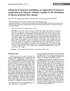
E o E •4 •3
•3 Q.
30
60 90 Time in Minutes
120
150
FIG. 1. The influence of magnesium (15 mg/kg) and calcium (15 mg/kg) infusion on CT secretion. Each point represents the mean value for CT, magnesium and calcium respectively in the plasma from 5 pigs at the times indicated ± SEM. The mean CT value of the samples taken during the calcium and magnesium infusion are compared statistically to one another and to the mean of the three control samples taken during the first 30 min of each experiment.
The Endocrine Society. Downloaded from press.endocrine.org by [${individualUser.displayName}] on 15 January 2017. at 08:10 For personal use only. No other uses without permission. . All rights reserved.
1178
NOTES AND COMMENTS
was implanted surgically in the jugular vein and placed in the area of the thyroid venous drainage. The catheter was held firmly with several layers of adhesive tape and remained in place for periods of one to two months. No evidence of slippage was noted by visual inspection, and lack of slippage was confirmed by the observation that basal levels of CT were similar from experiment to experiment in the individual animals. Infusion and plasma samplings were accomplished through this catheter. Contamination of blood samples was prevented by flushing the catheter with saline and discarding the first few ml of each sample. Blood samples were collected into heparinized tubes, immediately spun down, separated and frozen at —20 C. The animals were fasted from 18 to 24 hr before each experiment, and were under light sodium pentobarbital anesthesia during the experiments. At least 2 to 5 days were allowed to elapse between experiments on the same animal. Because the CT secretory response to a calcium infusion has been observed to vary widely in different animals (16), the experiments were designed to
compare the secretagogue influence of a strontium or magnesium infusion to a known stimulatory calcium infusion in the same experiment. These experiments were randomly performed on animals from a preselected group of 6 animals whose responsiveness to a calcium stimulation was predetermined and who were found to be highly sensitive (16). Because previous studies of calcium and magnesium influence on CT release indicated a stimulation of hormonal secretion (18,11), a directional hypothesis was tested using a /-test for correlated data in comparisons between two different time periods in the same experiment. The comparison of multiple means from different experiments was accomplished by means of the one-way analysis of variance. The individual group means were then compared to one another by Duncan's New Multiple Range Test
09). Results CT secretion was clearly sensitive most to the calcium ion (Fig. 1-2). A calcium infusion of
90 TIME
IN
Endo 1974 Vol 94 • No 4
150
MINUTES
FIG. 2. The influence of strontium (30 mg/kg) and calcium (IS mg/kg) infusion on CT secretion. Each point represents the mean value for CT, strontium and calcium respectively in the plasma from 5 pigs at the times indicated ± SEM.
The Endocrine Society. Downloaded from press.endocrine.org by [${individualUser.displayName}] on 15 January 2017. at 08:10 For personal use only. No other uses without permission. . All rights reserved.
NOTES AND COMMENTS 10 mg/kg caused a mean elevation in plasma CT levels of 3 ng/ml above base line, while the IS mg/kg infusion further stimulated CT secretion to a mean concentration of 8 ng/ml over base line (Fig. 1-2). Both rates of calcium infusion stimulated increases in CT which were significantly greater than CT levels during the preinfusion control period (first 30 min of each experiment) (p < 0.01). Further, the level of CT secretion during the calcium infusion was significantly greater than during the preceding Strontium or magnesium infusion periods (p < 0.025). The 7.5 mg/kg magnesium infusion produced only a slight rise in CT secretion (mean of 0.3 ng/ml over base line). The response to the 15 mg/kg infusion was variable. Several animals responded with a significant, 3-5 ng/ml rise in CT; while in other pigs the CT levels were only slightly elevated. The mean (3 ng/ml) response in all the animals was not highly significant (p < C.I5, Fig. 1). This variability in responsiveness among pigs has previously been observed in our laboratory (16) and by others in different species (20, 21). Strontium infusion at 15 mg/kg caused a small and statistically insignificant rise in plasma CT (a mean of 1 ng/ml over base line); however, the 30 mg/kg infusion caused a mean plasma CT elevation of 3 ng/ml above base line which was relatively consistent and statistically significant (p < 0.01) (Fig. 2). An analysis of variance of the 7.5 mg/kg magnesium infusion, 15 mg/kg calcium infusion and 30 mg/kg strontium infusion, which are approximately equivalent in molarity because of the differences in atomic weight, resulted in a highly significant F-test (p < 0.001). A multiple comparison of the individual means indicated that the 15 mg/kg calcium infusion produced a significantly greater CT stimulation than either the 7.5 mg/kg magnesium or 30 mg/kg strontium TABLE
1179
infusions (p < 0.01). Further, the 30 mg/kg strontium infusion was significantly more effective than the 7.5 mg/kg magnesium infusion (p < 0.05). Complete absence of a response to sodium chloride infusions of similar tonicity indicates that the responses observed in this study are specifically related to the alkaline earth cations infused and not to the hypertonicity of the infusate. Discussion The results of this study indicate clearly that calcium is not unique among the alkaline earth cations in its ability to stimulate CT secretion in the pig. Although the CT secretory mechanism appears to be most sensitive to fluctuations in serum calcium infusion of strontium and magnesium (which are approximately twice the atomic weight, and half the atomic weight of calcium respectively and which bracket this element in the periodic table) also enhance CT release. The relative sensitivity of the secretory mechanism is demonstrated by comparing the change in the plasma concentration of each cation necessary to elicit a significant release of the hormone. A 30 to 40% rise in plasma calcium levels produced a 3 to 9 ng/ml increase in plasma CT (Fig. 1-2). In contrast, even a 300% increase in plasma Mg only produced a slight, statistically insignificant, stimulation of CT release (mean rise of 1 ng/ml over base line, Fig. 1). Similar results have been reported by others using porcine thyroid slices in vitro (9,10). Finally, an increase in plasma strontium (whose normal plasma concentration is undetectable by atomic absorption spectrophotometry) to 1.3 mg per 100 ml was required to produce a significant increase in CT levels (mean rise of 3 ng/ml over baseline, Fig. 2). It would, therefore, appear that ol the ions
1. Increase in plasma calcitonin (ACT)* (ng/ml) as a function of cation infusion rate
Cation
0.190
0.250
Calcium Strontium Magnesium
— 1.0
3.0 —
0.3
—
Cation infusion rate (mM/kg/30 min) 0.310 — 3.0 —
0.375
0.625
8.0 —
— —
—
3.0
* ACT is the mean increase in plasma CT from baseline values (mean of three pre-infusion values).
The Endocrine Society. Downloaded from press.endocrine.org by [${individualUser.displayName}] on 15 January 2017. at 08:10 For personal use only. No other uses without permission. . All rights reserved.
NOTES AND COMMENTS
1180
tested only calcium is physiologically important for the regulation of CT secretion, since elevations of strontium or magnesium sufficient to influence CT release are unlikely to occur naturally. In the present study, magnesium infusions produced a minimal hypocalcemic response (Fig. 1), probably at least in part due to the enhanced CT secretion induced by this cation. Intravenous infusion of large doses of magnesium has previously been reported to depress plasma calcium in other species (3-6). The relative secretagogue influence of calcium, strontium and magnesium is summarized in Table 1. The highly significant statistical comparison between the 7.5 mg/kg magnesium, 15 mg/kg calcium and 30 mg/kg strontium infusion groups, suggests that the relative influence of these cations in their ability to enhance CT secretion in this study was calcium > strontium > magnesium. Acknowledgments The authors acknowledge the assistance of Mrs. Marilyn Harris, Mrs. Wanda DePaul, and Mr. Robert Schwartz in the preparation of this manuscript and the statistical advice of Dr. James Grogan. This study was supported by NIH training grant AM-05551. References 1. Copp, D. H., and E. C. Cameron, Science 134: 2038, 1961. 2. Hirsch, P. F., and P. L. Munson, Physiol Rev 49: 548, 1969.
Endo • 1974 Vol 94 • No 4
3. Kemeny, A., H. Boldizsar, and G. J. Pethes, N euro chemistry 7: 218, 1961. 4. Knippers, R., and U. Hehl, Z Gesamte Exp Med 139: 154, 1965. 5. Jones, K. H., and P. Fourman, Clin. Sci 30: 139, 1966. 6. Kelly, H. G., H. C. Cross, M. R. Turton, and J. D. Hatcher, Can Med Assoc J 82: 866, 1960. 7. Kielsen, S. P., Ada Endocrinol 64: 150, 1970. 8. Radde, I. C , E. R. Witterman, and S. Pensuwan, Endocrinology 83: 1285, 1968. 9. Bell, N. H., / Clin Invest 49: 1368, 1970. 10. Radde, I. C , D. K. Parkinson, E. R. Witterman, and B. Hoffken, In Taylor, S., and G. Foster (eds.), Calcitonin—Proceedings of the Second International Symposium, SpringerVerlag, New York, 1970, p. 376. 11. Littledike, E. T., and C. D. Arnaud, Proc Soc Exp Biol Med 136: 1000, 1971. 12. Care, A. D., T. Duncan, and D. Webster, / Endocrinol 37: 155, 1967. 13. Care, A. D., N. H. Bell, and R. F. L. Bates, J Endocrinol 5 1 : 381, 1971. 14. Rubin, R. P., Pharmacol Rev 22: 389, 1970. 15. Buckle, R. M., A. D. Care, C. W. Cooper, and H. J. Gitelman, / . Endocrinol 42: 529, 1968. 16. Pento, J. T., S. M. Glick, and A. Kagan, Metabolism 22: 735, 1973. 17. Perkin-Elmer Corp., Analytical Methods for Atomic Absorption Spectro photometry. PerkinElmer Corp., Norwalk, Conn. 1965. 18. Cooper, C. W., L. F. Deftos, and J. T. Potts, Endocrinology 88: 747, 1971. 19. Duncan, D. B., Biometrics 11: 1, 1955. 20. Tashjian, A. H., B. G. Howland, B. A. Kenneth, E. W. Melvin, and C. S. Hill, N Eng J Med 283: 890, 1970. 21. Lee, M. R., L. J. Deftos, and J. T. Pitts, Endocrinology 84: 36, 1969.
The Endocrine Society. Downloaded from press.endocrine.org by [${individualUser.displayName}] on 15 January 2017. at 08:10 For personal use only. No other uses without permission. . All rights reserved.