ORIGINAL CONTRIBUTIONS
ARTICLE 2
Clinical and radiographic success of mineral trioxide aggregate compared with formocresol as a pulpotomy treatment in primary molars A systematic review and meta-analysis ABSTRACT
Abdullah A. Marghalani, BDS, MSD; Samah Omar, DDS, MSD; Jung-Wei Chen, DDS, MS, PhD
T
he pulpotomy procedure is the most common pulp therapy for severely carious asymptomatic primary molars that have vital pulp. Multiple treatment protocols have been researched and implemented to determine which technique or material is superior. Two treatments of long standing involve the agents formocresol (FC) and mineral trioxide aggregate (MTA). FC, introduced in 1904,1 has been the preferred treatment for vital pulpotomies in primary molars since the 1930s.2 FC is available in two basic formulas, the full-strength Buckley FC (19 percent formaldehyde) and Sultan FC (48.5 percent formaldehyde). FC has demonstrated high success rates—up to 98 percent—within a 36- to 60-month follow-up period.3,4 Despite the success and popularity of FC, its possible cytotoxicity5 and evidence of systemic distribution,6-8 along with the suggested carcinogenicity of formaldehyde,9,10 have required dentistry to revisit its use and attempt to identify less toxic alternatives. Kahl and colleagues11 studied the presence of FC in the plasma of children who underwent oral rehabilitation under general anesthetic. They examined the children’s formaldehyde and cresol levels before, during and after the procedure; they found that formaldehyde and cresol plasma levels were undetectable above physiologic baseline levels. The researchers concluded that FC is unlikely to pose any risks to children if used in the typical dosage and manner as employed for the vital pulpotomy procedure. The International Agency for Research on Cancer12 and the National Toxicology
714
JADA 145(7)
http://jada.ada.org
Background. The authors conducted a systematic review and meta-analysis to compare the long-term clinical and radiographic success of using mineral trioxide aggregate (MTA) and formocresol (FC) as a pulp-dressing material in pulpotomy treatment in primary molars. Types of Studies Reviewed. The authors searched MEDLINE, Thomson Reuters Web of Science and the Cochrane Central Register of Controlled Trials for randomized controlled trials (RCTs) published from Jan. 1, 1990, to May 9, 2013. For an RCT to be included, the authors required that the primary molars treated with a pulpotomy procedure must have received stainless steel crowns as a final restoration and that rubber dam isolation was used during treatment; that the pulp must have been vital as determined clinically by means of hemorrhage control with a cotton pellet; and that the RCT must have included a follow-up period of at least two years. For each included RCT, two authors assessed the risk of bias independently. Results. The authors identified 20 trials and included five of them. A total of 377 primary molars were treated. The authors judged that none of the included RCTs had a low risk of bias. They noted no significant differences in clinical success (relative risk [RR] = 1.01; 95 percent confidence interval [CI], 0.98-1.05) and radiographic success (RR = 1.09; 95 percent CI, 0.97-1.21) for primary molars treated with MTA versus those treated with FC. Practical Implications. On the basis of the limited evidence, pulpotomy procedures performed in primary molars involving the use of MTA or FC showed comparable clinical success rates. Key Words. Formocresol; mineral trioxide aggregate; pulpotomy; primary molar; systematic review; meta-analysis; review literature; evidence-based dentistry; pediatric dentistry. JADA 2014;145(7):714-721. doi:10.14219/jada.2014.36
July 2014
Copyright © 2014 American Dental Association. All Rights Reserved.
ORIGINAL CONTRIBUTIONS
Program,13 an interagency of the U.S. Department of Health and Human Services, classified formaldehyde as a human carcinogen. Despite that fact, FC still is widely employed, its use still is taught in dental schools,14,15 and it still is recognized by the American Academy of Pediatric Dentistry16 (AAPD) as an acceptable pulp-dressing material for use in pulpotomy procedures performed in primary teeth. Mineral trioxide aggregate (MTA) was introduced in the dental literature in 1993,17 and its use as a pulpdressing material has been the topic of research since its development. MTA has demonstrated high success rates in primary and permanent teeth, perhaps as a result of its biocompatibility,18 alkanility19 and sealability,20,21 as well as its unique ability to form a dentin bridge.22 Researchers have compared MTA with other pulp-dressing materials, such as FC,23-25 ferric sulfate26 and calcium hydroxide.27,28 However, the number of high-quality randomized controlled trials (RCTs) in which researchers have evaluated these treatments for use in primary teeth is limited. Success of the pulpotomy procedure is not merely a matter of the dressing agent used. Rather, it is success of the entire treatment modality. For instance, a pulpotomy involving the use of FC typically requires placement of a zinc oxide-eugenol mixture on the treated radicular pulp. The pulp’s preoperative vitality status and extent of inflammation, as well as the final restoration, may be main indicators for the success of pulpotomy treatment. In 2003, Nadin and colleagues29 published a Cochrane review regarding pulp treatment for carious primary molars. They could not compare MTA with FC as a pulp-dressing agent owing to an inadequate number of published reports of RCTs. The most recent systematic review and meta-analysis in which investigators compared MTA with FC in pulpotomy treatment of primary molars was published in 2006; its authors concluded that clinical and radiographic outcomes were significantly better for the teeth treated with MTA.30 Some of the included trials lasted for less than 12 months. Since 2006, reports of several trials with follow-up periods longer than 12 months have been published.24,31-39 Our objective in this systematic review and metaanalysis was to evaluate the long-term clinical and radiographic success of two pulp-dressing materials, MTA and FC, in pulpotomy in carious primary molars in children. METHODS
We used the population, intervention, comparison, outcomes and study design (PICOS) method to develop a search strategy and to establish inclusion and exclusion criteria. We defined the target population as healthy pediatric patients who required pulpotomy treatment for vital and asymptomatic carious primary molars; the intervention group as primary molars treated with MTA as a pulpotomy dressing material; and the comparison group as primary molars treated with FC. We categorized
outcomes as clinical and radiographic success rates after an observation period of at least 24 months. We limited study design to RCTs in which investigators compared the two previously mentioned treatments. We did not include systematic reviews. Included trials for this review were limited to those in which investigators implemented standardized clinical procedures involving the use of a rubber dam for isolation, achievement of complete hemostasis after coronal pulp removal and before application of the dressing material to the vital pulp stumps, and use of stainless steel crowns as a final restoration for the treated primary molars. We used these limits to minimize chances of failure related to inadequate isolation and bacterial contamination, preexisting pulpal pathology or restoration leakage. Therefore, the number of confounders that may affect the success rate of these different treatment modalities was reduced. We defined the criteria for clinical success as the absence of pain, swelling, pathological mobility, tenderness to palpation or percussion, and abscess of discharge or development of a fistula. For radiographic success, we defined the criteria as the absence of pathological external root resorption, internal root resorption, furcation radiolucency and periapical bone destruction. We searched three databases: MEDLINE through PubMed, Thomson Reuters Web of Science and Cochrane Central Register of Controlled Trials. We structured the search strategy to involve the following key terms, as Medical Subject Headings terms or free text words, joined by “or”: “MTA,” “mineral trioxide aggregate,” “formocresol,” “pulpotomy” and “primary molars.” We limited the search to articles published from Jan. 1, 1990, through May 9, 2013. We placed no restriction on language of the searched trials; however, all eligible articles were written in English. We scanned the titles and abstracts of the identified trials for inclusion or exclusion in this systematic review. We also used the citation lists of published reviews for trial identification. When we could not make a decision on the basis of the abstract, we accessed a full report. We contacted one author of a published abstract40 for further information, but she preferred not to share the data and indicated interest in writing a complete report in the future. We agreed on the inclusion and exclusion criteria and databases. Two of the authors (A.A.M. and S.O.) conducted the search, study selection, data extraction and risk-of-bias assessment independently. Disagreements were resolved through discussion and through consideration given by the third author (J.-W.C.). We performed the risk-of-bias assessment for the included trials by usABBREVIATION KEY. AAPD: American Academy of Pediatric Dentistry. FC: Formocresol. MTA: Mineral trioxide aggregate. PCO: Pulp canal obliteration. RCT: Randomized controlled trial.
JADA 145(7) Copyright © 2014 American Dental Association. All Rights Reserved.
http://jada.ada.org
July 2014
715
ORIGINAL CONTRIBUTIONS
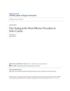
Records identified through database search (N = 8,077)
Records screened (N = 8,077)
Records discarded as not relevant on the basis of title or title and abstract (n = 8,057)
Articles excluded with reasons (n = 15)
Full-text articles assessed for eligibility (n = 20)
Articles included for qualitative and quantitative synthesis (n = 5) Figure 1. Flowchart of study search process.
ing the Cochrane Collaboration’s risk-of-bias assessment tool, which incorporated six domains: random sequence generation, allocation concealment, masking, completeness of outcome data, risk of selective outcome reporting and risk of other bias.41 Next, we determined a summary assessment of the risk of bias as follows: low risk of bias if all of the domains were judged to have low risk of bias, unclear risk of bias if at least one domain was judged to have unclear risk of bias, and high risk of bias if at least one domain was judged to have high risk of bias. We performed a meta-analysis by using a systematic review preparation software program (Review Manager [RevMan] Version 5.1, Nordic Cochrane Centre, Cochrane Collaboration, Copenhagen). We used randomeffects models to obtain pooled relative risks (RRs) with 95 percent confidence intervals (CIs) for clinical and radiographic success. We obtained the number needed to treat (NNT) for each outcome by reciprocating the pooled risk difference. We used the inverse variance method for all analyses. We also assessed heterogeneity by using the I2 statistic and the Cochrane test for heterogeneity, with P < .1 considered to be statistically significant. We assessed publication bias if the number of included RCTs exceeded 10. Figure 1 illustrates the trial selection process. RESULTS
Risk-of-bias assessment. We originally identified 20 trials as eligible for this review. After completing a fulltext reading of the eligible trials, we included only five RCTs on the basis of the stated predetermined inclusion and exclusion criteria (Figure 1; Table 132,35,38,39,42;
716
JADA 145(7)
http://jada.ada.org
Table 232,35,38,39,42). We excluded a trial reported by Hugar and Deshpande33 in 2010 owing to lack of randomization and lack of reporting about whether hemostasis was established after the removal of coronal pulp. We excluded other trials if the duration of follow-up was less than 24 months,23,25,37,43-47 if a stainless steel crown was not used as a final restoration for the pulpotomized primary molars24,31,34,36,48 or if the reporting was poor.49 In the included RCTs, methods of randomization to the treatment groups included a coin toss in two trials,38,42 a randomly numbered table in two trials32,35 and an envelope draw in one trial.25,39 One of the included RCTs involved a split-mouth design; children were enrolled if they had at least two primary molars that required pulpotomy procedures.38 None of the authors of the included trials indicated whether commercial funding was used to conduct the study. The follow-up duration of the included RCTs was 24 months. Using the predetermined six domains for risk-of-bias assessment, we determined three of the five RCTs35,39,42 to have a high risk of bias, whereas we judged two32,38 to have an unclear risk of bias and none to have a low risk of bias (Table 1). In total, 156 and 161 primary molars were treated with MTA and FC, respectively. Table 2 presents further description of the included RCTs. Clinical success. We observed a 100 percent success rate in primary molars treated with MTA (n = 156), with no reported clinical failures. For the molars treated with FC (n = 161), the success rate was 98.8 percent, with only two documented clinical failures. We noted no significant heterogeneity among the included RCTs (P = .99; I2 = 0 percent). Additionally, we noted no significant
July 2014
Copyright © 2014 American Dental Association. All Rights Reserved.
ORIGINAL CONTRIBUTIONS
TABLE 1
Description of the included trials. AUTHOR, YEAR
STUDY CHARACTERISTICS
Design
BASELINE NO. OF PARTICIPANTS (NO. OF TEETH), PERCENTAGE OF BOYS; AGE RANGE
SUCCESS RATE AT 24 RISK-OF-BIAS MONTHS IN PERCENTAGES, ASSESSMENT ACCORDING TO TREATMENT Clinical
Radiographic
MTA*
FC†
MTA
FC
100 children (120 teeth), percentage of boys not reported; age range: 3 to 8 years
100
97.2
100
86.1
High risk
Location; No. and Type of Center
Farsi and Colleagues, 42 2005
RCT‡ : two parallel Saudi Arabia; one center, arms university based
Noorollahian, 35 2008
RCT: two parallel arms
Iran; one center, university based
46 children (60 teeth), 63 percent boys; age range: 5 to 7 years
100
100
94.4
100
High risk
Subramaniam and Colleagues, 38 2009
RCT: two parallel arms, split mouth
India; one center, university based
19 children (40 teeth), 68 percent boys; age range: 6 to 8 years
100
100
95
85
Unclear risk
Sushynski and Colleagues, 39 2012
RCT: two parallel arms
United States; two centers, university and hospital based
168 children (252 teeth), 52 percent boys; age range: 5 to 8 years
100
98.5
95.4
75.8
High risk
Fernandez and Colleagues, 32 2013
RCT: four parallel arms
Spain; one center, university based
81 children (100 teeth), 62 percent boys; age range: 5 to 9 years
100
100
93.3
95.2
Unclear risk
* MTA: Mineral trioxide aggregate. † FC: Formocresol. ‡ RCT: Randomized controlled trial.
TABLE 2
Risk-of-bias assessment of the included trials. AUTHOR, YEAR
POSSIBLE SOURCE OF BIAS (TYPE OF BIAS) Random Sequence Generation (Selection)
Allocation Concealment (Selection)
Masking of Participants and Personnel (Performance)
Masking of Outcome Assessment (Detection) *
Incomplete Outcome Data (Attrition)
Selective Reporting (Reporting)
Other Sources
Farsi and Colleagues, 42 2005
Low risk
Unclear risk
Unclear risk
Unclear risk
High risk
Low risk
Unclear risk
Noorollahian, 35 2008
Low risk
Unclear risk
Unclear risk
Low risk
High risk
Low risk
Unclear risk
Subramaniam and Colleagues, 38 2009
Low risk
Unclear risk
Unclear risk
Unclear risk
Low risk
Low risk
Unclear risk
Sushynski and Colleagues, 39 2012
Low risk
Unclear risk
Unclear risk
Low risk
High risk
Low risk
Unclear risk
Fernandez and Colleagues, 32 2013
Low risk
Unclear risk
Unclear risk
Low risk
Unclear risk
Low risk
Unclear risk
* Masking was evaluated only for radiographic success (not clinical success).
difference in the likelihood of clinical success between primary molars treated with MTA and primary molars treated with FC (RR = 1.01; 95 percent confidence interval [CI], 0.98-1.05) during the observational period, as shown in Figure 2. The NNT was 100; this means that when 100 patients were treated with MTA, we observed one more clinical success among those patients during the study period than among patients treated with FC. Figure 2 includes a forest plot comparing clinical success for primary molars treated with MTA and with FC.
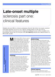
Radiographic success. We observed a radiographic success rate of 96.2 percent for primary molars treated with MTA (n = 156); only six radiographic failures were documented. For the FC group (n = 161), we calculated an 84.5 percent success rate and observed 25 radiographic failures. We noted significant heterogeneity among the included RCTs (P = .05; I2 = 58 percent). There was no significant difference in the likelihood of radiographic success’ occurring for primary molars treated with MTA as compared with those treated with FC (RR = 1.09;
JADA 145(7) Copyright © 2014 American Dental Association. All Rights Reserved.
http://jada.ada.org
July 2014
717
ORIGINAL CONTRIBUTIONS
STUDY, YEAR
MTA
FC
Events Total Farsi and Colleagues,42 2005 Noorollahian,35 2008 Subramaniam and Colleagues,38 2009 Sushynski and Colleagues,39 2012 Fernandez and Colleagues,32 2013
38 18 20 65 15
Total
38 18 20 65 15
Events
Total
Weight (%)
IV, Random, 95% CI
35 18 20 65 21
36 18 20 66 21
17.1 8.9 10.8 55.0 8.2
1.03 (0.95-1.11) 1.00 (0.90-1.11) 1.00 (0.91-1.10) 1.02 (0.97-1.06) 1.00 (0.90-1.11)
100
1.01 (0.98-1.05)
156
Total Events
RISK RATIO
161
156
Risk Ratio IV, Random, 95% CI
159
Heterogeneity: τ2 = 0.00; χ2 = 0.36, degrees of freedom = 4 (P = .99); I2 = 0%. Test for overall effect: Z = 0.83 (P = .41).
0.5
0.7 1 1.5 2 Favors [FC] Favors [MTA]
Figure 2. Forest plot comparing clinical success for primary molars treated with mineral trioxide aggregate (MTA) and for those treated with formocresol (FC). CI: Confidence interval. IV: Inverse variance.
STUDY, YEAR
MTA
FC
Events Total Farsi and Colleagues,42 2005 Noorollahian,35 2008 Subramaniam and Colleagues,38 2009 Sushynski and Colleagues,39 2012 Fernandez and Colleagues,32 2013
38 17 19 62 14
Total Total Events
38 18 20 65 15
Events
Total
Weight (%)
IV, Random, 95% CI
31 18 17 50 20
36 18 20 66 21
22.4 21.1 15.4 21.6 19.5
1.16 (1.01-1.33) 0.95 (0.81-1.10) 1.12 (0.91-1.38) 1.26 (1.09-1.46) 0.98 (0.83-1.16)
100
1.09 (0.97-1.21)
156 150
RISK RATIO
161
Risk Ratio IV, Random, 95% CI
136
0.5
Heterogeneity: τ2 = 0.01; χ2 = 9.50, degrees of freedom = 4 (P = .05); I2 = 58%. Test for overall effect: Z = 1.50 (P = .13).
0.7 1 1.5 2 Favors [FC] Favors [MTA]
Figure 3. Forest plot comparing radiographic success for primary molars treated with mineral trioxide aggregate (MTA) with that for those treated with formocresol (FC). CI: Confidence interval. IV: Inverse variance.
95 percent CI, 0.97-1.21) (Figure 3). The NNT was 13; thus, when 13 primary molars were treated with MTA, we observed one more radiographic success among those molars during the study period than among primary molars treated with FC. Figure 3 shows a forest plot comparing radiographic success for primary molars treated with MTA and with FC. DISCUSSION
This systematic review included five RCTs with a total of 317 primary molars (156 treated with MTA and 161 with FC). Primary molars treated with MTA had a clinical success rate approximately 1 percent higher and a radiographic success rate approximately 9 percent higher than the respective success rates for molars treated with FC. However, these differences were not statistically significant. The obtained evidence was consistent for clinical success but less consistent for radiographic success. We judged none of the included RCTs to have a low risk of bias. Owing to the limited number of included RCTs, we did not assess publication bias. In our systematic review, we noted demographic variations among the included trials. The fact that pro-
718
JADA 145(7)
http://jada.ada.org
cedures were performed by multiple operators might have increased the heterogeneity of the results. Investigators in three RCTs32,35,39 reported using Buckley’s FC with 1:5 dilution. Investigators in one RCT42 did not report the FC formula or dilution used, and those in the other RCT38 did not report the formula but did report that they used 1:5 dilution. For mixing MTA with a liquid, investigators in three RCTs32,39,42 used sterile saline and those in two other RCTs35,38 used distilled water. The findings of our systematic review indicated that the rate of radiographic failure was higher than that of clinical failure; therefore, it is plausible to conclude that radiographic changes perhaps are more sensitive to the aforementioned factors than are clinical changes. This might partially explain the lower consistency of the radiographic success. The results of four of the RCTs32,38,39,42 included in this analysis demonstrated over a two-year period a similar or a slightly higher rate of radiographic success in primary molars treated with MTA as compared with those treated with FC. On the contrary, Noorollahian35 documented a higher rate of radiographic success in molars treated with FC than in the group treated with
July 2014
Copyright © 2014 American Dental Association. All Rights Reserved.
ORIGINAL CONTRIBUTIONS
MTA (Figure 3). Noorollahian did not clearly explain the reverse findings she observed relative to the other studies and was the only researcher who mentioned using white MTA.35 Sushynski and colleagues39 reported using gray MTA, whereas investigators in the other three included RCTs did not specify whether they used white or gray MTA.32,38,42 Fernandez and colleagues32 and Subramaniam and colleagues38 reported using ProRoot MTA (Dentsply Tulsa Dental Specialties, Johnson City, Tenn.). Farsi and colleagues42 did not specify the type or the manufacturer of the MTA used. White MTA was developed to enhance esthetic appeal over that of gray MTA, particularly when used in anterior teeth.23 Compositional differences of white MTA versus gray MTA include lack of tetracalcium aluminoferrite and inclusion of finer particles.23,50 The effect of the type of MTA (gray versus white) on the clinical and radiographic success of pulpotomy procedures has been investigated.23,51 Cardoso-Silva and colleagues51 determined that the application of gray MTA resulted in radiographic success greater, but not significantly greater, than that of white MTA (100 percent versus 93 percent). Agamy and colleagues23 demonstrated a significantly higher radiographic success rate for gray MTA than for white MTA (100 percent versus 84 percent) over a one-year follow-up period. However, the histologic examination showed that both types of MTA successfully induced the formation of a thick dentin bridge at the amputation site. FC induced thin, poorly calcified dentin.23 Pulp canal obliteration (PCO) was evident in primary molars treated with FC and with MTA. PCO has been reported as a common radiographic finding in teeth that have undergone pulp treatments, including both MTA and FC.30 Typically, PCO results from odontoblastic activity; thus, the teeth usually are vital and treatment is not indicated. A common finding in nearly all RCTs included in this systematic review was that molars treated with MTA had a higher incidence of PCO than did those treated with FC; as such, it was considered a physiological response and was not reported as a failure. In the included RCTs, internal root resorption was one of the most commonly reported radiographic failures. Internal resorption usually is self-limiting and, according to the AAPD recommendations,16 no treatment is warranted unless the resorption extends to the supporting bone. This explains the difference between the radiographic success rate and the clinical success rate, as not all radiographic failures are accompanied by clinical symptoms and may not warrant treatment. The investigators in a 2006 meta-analysis of six trials that involved 381 teeth found statistically significantly superior clinical (RR = 0.32; 95 percent CI, 0.11-0.90) and radiographic (RR = 0.31; 95 percent CI, 0.13-0.74) outcomes for MTA when compared with those for FC.30 Compared with our review, this 2006 review had more
flexible inclusion and exclusion criteria; the duration of some included trials was less than 24 months. Additionally, the use of rubber dam isolation, achievement of hemostasis after coronal pulp removal and the restoration of the treated molars with stainless steel crowns were not listed among the inclusion criteria. A subsequent systematic review of six RCTs with no meta-analysis was published in 2010; the authors of that review favored the use of MTA over FC. This likely was because of the potential toxicity of FC.52 Investigators in two reviews discussed the new directions and materials used for vital pulp therapy in primary molars. Fuks’53 review contained a critical assessment of trials in which investigators compared the most commonly used pulp-dressing agents. The assessment concluded that MTA produced better results than did FC, even though the material did not produce statistically significantly different results in some cases. Chen and Jorden54 highlighted the absence of a direct association between FC use in pulpotomies and adverse health risks on the basis of the current literature. Moreover, Chen and Jorden considered FC to be a “viable” treatment option. The inclusion of RCTs with extended follow-up windows distinguishes our review from previous research and adds to the body of knowledge in the field. Correspondingly, we limited the included RCTs to those in which investigators implemented the use of rubber dam isolation, achieved hemostasis after coronal pulp removal and used stainless steel crowns as a final restoration after the completion of the pulpotomy procedures for both the intervention and comparison groups. Also, the use of the robust risk-of-bias assessment tool adopted by the Cochrane Collaboration41 is another strength of this systematic review. Homogeneity among trials is indicated mainly by consistency in study methodology, materials used and procedures used. Some degrees of interperformance and intraperformance variation—that is, heterogeneity—were expected, because the majority of the included trials were university based and involved multiple operators and intraexaminers or, more importantly, because interexaminer reliability measures were not performed in all trials. Investigators in the included trials did not adequately consider and subsequently adjust for potential confounders, especially patient-related factors of success or failure. For instance, age, sex, oral health status and caries risk sometimes were mentioned but not compared between children receiving active treatment and those receiving control treatment. Moreover, implementation of a split-mouth design may reduce this type of bias. The methods of RCTs are greatly improved when the Cochrane Collaboration’s risk-of-bias assessment tool is used and when the Consolidated Standards of Reporting Trials (CONSORT) statement for reporting RCTs is followed.41,55 Of the 20 eligible trials, we considered only
JADA 145(7) Copyright © 2014 American Dental Association. All Rights Reserved.
http://jada.ada.org
July 2014
719
ORIGINAL CONTRIBUTIONS
five in this review. Moreover, three of the five trials had a high risk of bias and none had a low risk of bias. Investigators in this field are encouraged to conduct RCTs with superior methodology, as it generates more valid and reliable conclusions. Besides considering the generated findings of this review, clinicians should take into account other factors when making clinical decisions. For instance, concerns with regard to the toxicity and mutagenicity of FC, as well as the cost of MTA compared with that of FC, continue to be among the major limiting factors.54 Additionally, multiple appointments should be required when MTA is used; this factor may limit its use in patients who require general anesthesia. Evidencebased dentistry is maximized when more of these factors are considered and implemented. Strict inclusion criteria may be a limitation of this study, as we excluded some well-designed trials with valuable information owing to the short observational time, the lack of a clear clinical protocol or the lack of randomization. Another limitation of this review was the absence of an identified RCT that had a low risk of bias. CONCLUSION
We judged none of the included RCTs to have a low risk of bias. On the basis of the limited evidence, we found that MTA and FC demonstrated comparable clinical success rates. To infer strong evidence, investigators are encouraged to conduct high-quality RCTs with longer follow-up periods than those used by the investigators in the trials included in this review, superior methodology and lower risk of bias. Q Dr. Marghalani is a doctoral student, Department of Epidemiology, Biostatistics and Population Medicine, School of Public Health, Loma Linda University, Calif. He also is a faculty member, Department of Preventive Dentistry, Umm Al-Qura University, Makkah, Saudi Arabia. Address correspondence to Dr. Marghalani at the Department of Epidemiology, Biostatistics and Population Medicine, School of Public Health, Loma Linda University, 24951 N. Circle Drive, Nicole Hall, Room 2005, Loma Linda, Calif. 92350, e-mail
[email protected]. Dr. Omar is an assistant professor and the associate program director, Advanced Specialty Education Program in Pediatric Dentistry, School of Dentistry, Loma Linda University, Loma Linda, Calif. Dr. Chen is a professor and the program director, Advanced Specialty Education Program in Pediatric Dentistry, School of Dentistry, Loma Linda University, Loma Linda, Calif. Disclosure. None of the authors reported any disclosures. The authors extend their appreciation to Drs. Lauren L. Gutenberg and Riste Simnjanovski for their helpful editorial comments. 1. Buckley J. The chemistry of pulp decomposition with a rational treatment for this condition and its sequelae. Amer Dent J 1904;3(11):764-771. 2. Sweet CA Jr. Procedure for treatment of exposed and pulpless deciduous teeth. JADA 1930;17(6):1150-1153. 3. Morawa AP, Straffon LH, Han SS, Corpron RE. Clinical evaluation of pulpotomies using dilute formocresol. ASDC J Dent Child 1975;42(5): 360-363. 4. Strange DM, Seale NS, Nunn ME, Strange M. Outcome of formocresol/ZOE sub-base pulpotomies utilizing alternative radiographic success criteria. Pediatr Dent 2001;23(4):331-336. 5. de Menezes JV, Takamori ER, Bijella MF, Granjeiro JM. In vitro tox-
720
JADA 145(7)
http://jada.ada.org
icity of MTA compared with other primary teeth pulpotomy agents. J Clin Pediatr Dent 2009;33(3):217-221. 6. Pashley EL, Myers DR, Pashley DH, Whitford GM. Systemic distribution of 14C-formaldehyde from formocresol-treated pulpotomy sites. J Dent Res 1980;59(3):602-608. 7. Ranly DM, Fulton R. Reaction of rat molar pulp tissue to formocresol, formaldehyde, and cresol. J Endod 1976;2(6):176-181. 8. Myers DR, Shoaf HK, Dirksen TR, Pashley DH, Whitford GM, Reynolds KE. Distribution of 14C-formaldehyde after pulpotomy with formocresol. JADA 1978;96(5):805-813. 9. Kerns WD, Pavkov KL, Donofrio DJ, Gralla EJ, Swenberg JA. Carcinogenicity of formaldehyde in rats and mice after long-term inhalation exposure. Cancer Res 1983;43(9):4382-4392. 10. Yodaiken RE. The uncertain consequences of formaldehyde toxicity. JAMA 1981;246(15):1677-1678. 11. Kahl J, Easton J, Johnson G, Zuk J, Wilson S, Galinkin J. Formocresol blood levels in children receiving dental treatment under general anesthesia. Pediatr Dent 2008;30(5):393-399. 12. International Agency for Research on Cancer (IARC), World Health Organization. IARC Monographs on the Evaluation of Carcinogenic Risks to Humans. monographs.iarc.fr/ENG/Classification/index.php. Accessed April 24, 2014. 13. National Toxicology Program, U.S. Department of Health and Human Services. 12th Report on Carcinogens. June 10, 2011. ntp.niehs.nih. gov/?objectid=03C9AF75-E1BF-FF40-DBA9EC0928DF8B15. Accessed April 24, 2014. 14. Walker LA, Sanders BJ, Jones JE, et al. Current trends in pulp therapy: a survey analyzing pulpotomy techniques taught in pediatric dental residency programs. J Dent Child (Chic) 2013;80(1):31-35. 15. Dunston B, Coll JA. A survey of primary tooth pulp therapy as taught in US dental schools and practiced by diplomates of the American Board of Pediatric Dentistry. Pediatr Dent 2008;30(1):42-48. 16. Pulp Therapy Subcommittee, Clinical Affairs Committee, American Academy of Pediatric Dentistry (AAPD). Guideline on pulp therapy for primary and immature permanent teeth. In: Reference Manual. Vol. 34, No. 6. Chicago: AAPD; 2012:222-229. www.aapd.org/media/Policies_ Guidelines/G_Pulp.pdf. Accessed April 24, 2014. 17. Lee SJ, Monsef M, Torabinejad M. Sealing ability of a mineral trioxide aggregate for repair of lateral root perforations. J Endod 1993;19(11): 541-544. 18. Torabinejad M, Parirokh M. Mineral trioxide aggregate: a comprehensive literature review, part II—leakage and biocompatibility investigations. J Endod 2010;36(2):190-202. 19. Parirokh M, Torabinejad M. Mineral trioxide aggregate: a comprehensive literature review, part III—clinical applications, drawbacks, and mechanism of action. J Endod 2010;36(3):400-413. 20. Torabinejad M, Rastegar AF, Kettering JD, Pitt Ford TR. Bacterial leakage of mineral trioxide aggregate as a root-end filling material. J Endod 1995;21(3):109-112. 21. Torabinejad M, Higa RK, McKendry DJ, Pitt Ford TR. Dye leakage of four root end filling materials: effects of blood contamination. J Endod 1994;20(4):159-163. 22. Eskandarizadeh A, Shahpasandzadeh MH, Shahpasandzadeh M, Torabi M, Parirokh M. A comparative study on dental pulp response to calcium hydroxide, white and grey mineral trioxide aggregate as pulp capping agents. J Conserv Dent 2011;14(4):351-355. 23. Agamy HA, Bakry NS, Mounir MM, Avery DR. Comparison of mineral trioxide aggregate and formocresol as pulp-capping agents in pulpotomized primary teeth. Pediatr Dent 2004;26(4):302-309. 24. Ansari G, Ranjpour M. Mineral trioxide aggregate and formocresol pulpotomy of primary teeth: a 2-year follow-up. Int Endod J 2010;43(5): 413-418. 25. Zealand CM, Briskie DM, Botero TM, Boynton JR, Hu JC. Comparing gray mineral trioxide aggregate and diluted formocresol in pulpotomized human primary molars. Pediatr Dent 2010;32(5):393-399. 26. Odabas ME, Alacam A, Sillelioglu H, Deveci C. Clinical and radiographic success rates of mineral trioxide aggregate and ferric sulphate pulpotomies performed by dental students. Eur J Paediatr Dent 2012;13(2):118-122. 27. Liu H, Zhou Q, Qin M. Mineral trioxide aggregate versus calcium hydroxide for pulpotomy in primary molars. Chin J Dent Res 2011;14(2): 121-125.
July 2014
Copyright © 2014 American Dental Association. All Rights Reserved.
ORIGINAL CONTRIBUTIONS
28. Leye Benoist F, Gaye Ndiaye F, Kane AW, Benoist HM, Farge P. Evaluation of mineral trioxide aggregate (MTA) versus calcium hydroxide cement (Dycal) in the formation of a dentine bridge: a randomised controlled trial. Int Dent J 2012;62(1):33-39. 29. Nadin G, Goel BR, Yeung CA, Glenny AM. Pulp treatment for extensive decay in primary teeth. Cochrane Database Syst Rev 2003;(1): CD003220. 30. Peng L, Ye L, Tan H, Zhou X. Evaluation of the formocresol versus mineral trioxide aggregate primary molar pulpotomy: a meta-analysis. Oral Surg Oral Med Oral Pathol Oral Radiol Endod 2006;102(6):e40-e44. 31. Erdem AP, Guven Y, Balli B, et al. Success rates of mineral trioxide aggregate, ferric sulfate, and formocresol pulpotomies: a 24-month study. Pediatr Dent 2011;33(2):165-170. 32. Fernandez CC, Martinez SS, Jimeno FG, Lorente Rodriguez AI, Mercade M. Clinical and radiographic outcomes of the use of four dressing materials in pulpotomized primary molars: a randomized clinical trial with 2-year follow-up. Int J Paediatr Dent 2013;32(6):400-407. 33. Hugar SM, Deshpande SD. Comparative investigation of clinical/ radiographical signs of mineral trioxide aggregate and formocresol on pulpotomized primary molars. Contemp Clin Dent 2010;1(3):146-151. 34. Moretti AB, Sakai VT, Oliveira TM, et al. The effectiveness of mineral trioxide aggregate, calcium hydroxide and formocresol for pulpotomies in primary teeth. Int Endod J 2008;41(7):547-555. 35. Noorollahian H. Comparison of mineral trioxide aggregate and formocresol as pulp medicaments for pulpotomies in primary molars. Br Dent J 2008;204(11):E20. 36. Sonmez D, Sari S, Cetinbas T. A comparison of four pulpotomy techniques in primary molars: a long-term follow-up. J Endod 2008; 34(8):950-955. 37. Srinivasan D, Jayanthi M. Comparative evaluation of formocresol and mineral trioxide aggregate as pulpotomy agents in deciduous teeth. Indian J Dent Res 2011;22(3):385-390. 38. Subramaniam P, Konde S, Mathew S, Sugnani S. Mineral trioxide aggregate as pulp capping agent for primary teeth pulpotomy: 2 year follow up study. J Clin Pediatr Dent 2009;33(4):311-314. 39. Sushynski JM, Zealand CM, Botero TM, et al. Comparison of gray mineral trioxide aggregate and diluted formocresol in pulpotomized primary molars: a 6- to 24-month observation. Pediatr Dent 2012;34(5): 120-128. 40. Ritwik P, Cuisia ZV, Dabir P, Musselman RJ. MTA pulpotomies in the primary molars of children: results after 3 years. Poster presented at: 19th Congress of the International Association of Paediatric Dentistry; Oct. 15-19, 2003; New Orleans. 41. Higgins J, Green S, eds. Cochrane Handbook for Systematic Reviews of Interventions Version 5.1.0. (updated March 2011). Oxford, England :
The Cochrane Collaboration; 2011. www.cochrane-handbook.org. Accessed April 24, 2014. 42. Farsi N, Alamoudi N, Balto K, Mushayt A. Success of mineral trioxide aggregate in pulpotomized primary molars. J Clin Pediatr Dent 2005;29(4):307-311. 43. Aeinehchi M, Dadvand S, Fayazi S, Bayat-Movahed S. Randomized controlled trial of mineral trioxide aggregate and formocresol for pulpotomy in primary molar teeth. Int Endod J 2007;40(4):261-267. 44. Naik S, Hegde AH. Mineral trioxide aggregate as a pulpotomy agent in primary molars: an in vivo study. J Indian Soc Pedod Prev Dent 2005;23(1):13-16. 45. Jabbarifar SE, Khademi AA, Ghasemi D. Success rate of formocresol pulpotomy versus mineral trioxide aggregate in human primary molar tooth. J Res Med Sci 2004;9(6):304-307. 46. Eidelman E, Holan G, Fuks AB. Mineral trioxide aggregate vs. formocresol in pulpotomized primary molars: a preliminary report. Pediatr Dent 2001;23(1):15-18. 47. Godhi B, Sood PB, Sharma A. Effects of mineral trioxide aggregate and formocresol on vital pulp after pulpotomy of primary molars: an in vivo study. Contemp Clin Dent 2011;2(4):296-301. 48. Holan G, Eidelman E, Fuks AB. Long-term evaluation of pulpotomy in primary molars using mineral trioxide aggregate or formocresol. Pediatr Dent 2005;27(2):129-136. 49. Airen P, Shigli A, Airen B. Comparative evaluation of formocresol and mineral trioxide aggregate in pulpotomized primary molars: 2 year follow up. J Clin Pediatr Dent 2012;37(2):143-147. 50. Camilleri J, Montesin FE, Di Silvio L, Pitt Ford TR. The chemical constitution and biocompatibility of accelerated Portland cement for endodontic use. Int Endod J 2005;38(11):834-842. 51. Cardoso-Silva C, Barberia E, Maroto M, Garcia-Godoy F. Clinical study of mineral trioxide aggregate in primary molars: comparison between grey and white MTA—a long term follow-up (84 months). J Dent 2011;39(2):187-193. 52. Simancas-Pallares MA, Diaz-Caballero AJ, Luna-Ricardo LM. Mineral trioxide aggregate in primary teeth pulpotomy: a systematic literature review. Med Oral Patol Oral Cir Bucal 2010;15(6):e942-e946. 53. Fuks AB. Vital pulp therapy with new materials for primary teeth: new directions and treatment perspectives. Pediatr Dent 2008;30(3): 211-219. 54. Chen JW, Jorden M. Materials for primary tooth pulp treatment: the present and the future. Endod Topics 2010;23(1):41-49. 55. Schulz KF, Altman DG, Moher D; CONSORT Group. CONSORT 2010 statement: updated guidelines for reporting parallel group randomised trials. BMC Med 2010;8(1):18.
JADA 145(7) Copyright © 2014 American Dental Association. All Rights Reserved.
http://jada.ada.org
July 2014
721