Clin Orthop Relat Res DOI 10.1007/s11999-010-1431-4
SYMPOSIUM: PAPERS PRESENTED AT THE ANNUAL MEETINGS OF THE KNEE SOCIETY
The Peel in Total Knee Revision Exposure in the Difficult Knee Carlos Lavernia MD, Juan Salvador Contreras MD, Jose Carlos Alcerro MD
Ó The Association of Bone and Joint Surgeons1 2010
Abstract Background The femoral peel to expose a difficult knee was first described by Windsor and Insall in the mid-1980s. This surgical exposure consists of a complete soft tissue subperiosteal peel of the femur. It includes the detachment of the origin of the medial and lateral collateral ligaments. Questions/purposes We investigated the utility of a surgical exposure, the modified femoral peel, for total knee revision. Methods We retrospectively reviewed all 101 patients who had revision TKA (132 revisions) with the femoral peel technique from January 2000 to September 2007. Of the 101 patients, three patients died, eight patients were excluded, and three patients were lost to followup. Eightyseven patients (116 procedures) met the inclusion criteria. Outcome measures assessed included Knee Society knee score, Knee Society function score, Hospital for Special Surgery knee score, complications, and number of
reoperations. The minimum followup was 2 years (mean, 3.5 years; range, 2–9 years). Results Mean Knee Society knee scores improved from 47 to 85. Hospital for Special Surgery knee scores improved from 56 to 80. Quality of Well-Being and WOMAC all dimensions improved. Overall orthopaedic complication rate was 17%. Flexion contracture was improved. Conclusions This surgical approach, which results in a complete soft tissue degloving of the distal femur and proximal tibia, allowed satisfactory exposure in all cases and the complication rate related to this exposure method was comparable with other series using diverse methods of knee exposure. Level of Evidence Level IV, therapeutic study. See Guidelines for Authors for a complete description of levels of evidence.
Introduction Each author certifies that he or she has no commercial associations (eg, consultancies, stock ownership, equity interest, patent/licensing arrangements, etc) that might pose a conflict of interest in connection with the submitted article. Each author certifies that his or her institution approved the human protocol for this investigation, that all investigations were conducted in conformity with ethical principles of research, and that informed consent for participation in the study was obtained. This work was performed at Orthopaedic Institute at Mercy Hospital. C. Lavernia (&) Orthopaedic Institute at Mercy Hospital, 3659 S Miami Avenue, Suite 4008, Miami, FL 33133, USA e-mail:
[email protected];
[email protected] C. Lavernia, J. S. Contreras, J. C. Alcerro Arthritis Surgery Research Foundation, Inc, Miami, FL, USA
Adequate exposure in difficult primary or revision TKA is a key step for a successful outcome. Revision TKA surgery requires adequate visualization to remove the old prosthetic components and insert the new device, while preserving function of the extensor mechanism and surrounding soft tissues [3]. Without adequate exposure, technical errors can lead to potential complications, such as malposition of components and sizing errors. In the process of obtaining adequate exposure, skin, patellar tendon, or collateral ligaments are at risk. Intraoperative fractures of the tibial plateau, tibial tuberosity, or femoral condyles can occur [5, 10, 17]. Most surgeons on the initial portion of the approach use the traditional medial parapatellar incision utilized in primary TKA. In the presence of a severely
123
Clinical Orthopaedics and Related Research1
Lavernia et al.
ankylosed or stiff knee (\ 60° or 70° of flexion), an alternative secondary technique is often required [18]. Multiple approaches have been described for the secondary portion in the exposure of a difficult knee, including the quadriceps snip [9], V-Y patellar turndown or quadricepsplasty [10], tibial tubercle osteotomy [28, 30], ‘‘the wandering resident’’ surgical exposure [11], femoral peel [31], banana peel [15], and the Engh medial epicondylectomy [7]. The femoral peel was first described by Windsor and Insall [31] in 1988 for revision TKA. In 2008, Huff and Russel [12] redescribed the technique. This surgical exposure utilizes a partial or complete subperiosteal dissection of the distal femur and provides satisfactory exposure of the knee. The fundamental concept is ‘‘skeletonization’’ of the distal femur [12, 31]. We evaluated a modified femoral peel technique during revision TKA by (1) examining the utility of the exposure; (2) reporting the degree of flexion contracture and extension lag obtained as a result of the procedure; (3) documenting patient’s perceived outcomes; and (4) reviewing the complications associated with this technique.
Patients and Methods We performed 235 consecutive revision TKAs in 164 patients. Of these, 101 patients had 132 revision TKAs performed using a modified femoral peel. This technique was utilized on all cases in which a standard medial parapatellar approach with extensive medial and lateral gutter de´bridement did not provide the visualization necessary for the safe and accurate revision procedure. The femoral peel was chosen intraoperatively when the standard median parapatellar approach combined with extensive gutter releases did not provide adequate exposure. We know of no contraindication to use this approach. The mean age of the patients at the time of index revision surgery was 67 years (standard error, ± 1.16 years; range, 34–94 years); 61% were female. At the time of
index revision procedure, patellectomy was performed in three of the 116 cases (due to severe patellar osteolysis, chronic infection, and patellar dislocation); three cases underwent additional quadriceps tendon repair (secondary to quadriceps tendon defect and extension lag of 50° and 77°, respectively), two underwent quadricepsplasty (due to fracture of the insertion of the patellar tendon), two underwent patellar tendon allograft (one patient had a patellar tendon rupture; the other patient had a combined quadriceps tendon defect and extension lag of 77°), and one underwent quadriceps muscle reconstruction (due to patellar dislocation). Index revision TKA was the first in 57 cases, second in 28 cases, third in 15 cases, fourth in nine cases, fifth in four cases, sixth in two cases, and eighth in one case. Of the 101 patients, three patients (three knees) died for reasons unrelated to surgery. Eight patients (10 knees) underwent resection arthroplasty and were excluded because no reimplantation was performed at the time of last followup. Three patients were lost to followup. This left 87 patients (116 procedures). There were more operative procedures than patients in this series as 18 patients were treated for chronic infection and required two or more procedures, and six patients underwent bilateral revision TKAs. The patient’s hospital and clinical charts were reviewed to identify any complications that occurred. Complications were classified as orthopaedic (directly related to the procedure) or medical (systemic complications that occurred perioperatively or postoperatively). Operative notes were reviewed to confirm the surgical approach utilized. The minimum followup was 2 years (mean, 3.5 years; range, 2–9 years). The study was approved by the Institutional Review Board and all patients provided informed consent. All surgeries were performed by the senior author (CL). The most frequent procedure performed was revision of the femoral and tibial components (74 cases), followed by second-stage reimplantation of previously infected TKA (24 cases), irrigation and de´bridement for infection (11 cases), and resection arthroplasty for infection (seven cases) (Table 1). The modified femoral peel technique used
Table 1. Primary indications for revision TKA and type of implant utilized Indication
Number of cases
CCK
Hinged
Distal femur
CR/PS
Spacer
Chronic infection
47
24
11
2
3
7
Mechanical loosening of prosthetic joint
33
19
7
2
5
Unstable total knee
12
9
3
Periprosthetic fracture
3
1
12
1
7
Failed tibiofemoral component
4
3
1
Stiffness
4
1
3
Patellofemoral pain Extensor mechanism rupture
3 1
3 1
CCK = constrained condylar knee; CR/PS = cruciate-retaining, posterior-stabilized.
123
The Peel in Total Knee Revision
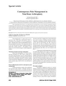
is as follows. As previously described by Windsor and Insall [31], extensive skin incision was performed in all cases. Every attempt was made to incorporate any previous incisional scar. The extension of any existing skin incision was performed, avoiding large lateral based flaps (Fig. 1). Standard medial parapatellar arthrotomy was performed in all cases. Fibrous adhesions in the medial and lateral gutters were lysed using a combination of sharp, blunt, and cautery dissection until the suprapatellar pouch along with the gutters were fully developed and the patella could be safely displaced laterally. The polyethylene insert was generally removed at this point to facilitate mobilization of the bones and to help relax the soft tissues circumferentially. The leg was gently flexed and a sharp subperiosteal dissection was begun at the distal origin of the medial
Fig. 1 Extensive exposure is shown in an intraoperative photograph.
collateral ligament. We modified the previously described technique to include dissection with an electric cautery using a rake retractor or manual tension to pull on the ligament capsular flap. The medial collateral ligament was elevated until the distal femur and the proximal tibia could be mobilized and the proper exposure achieved (Fig. 2A–B). This skeletonization was performed as extensively as necessary. A bone hook was placed in the prosthetic intercondylar notch to pull on the distal femur superiorly to expose and liberate the posterior capsule and fibrous tissues from the back of the femur as well. Manual tension was utilized in the posterior femur to avoid damaging the neurovascular structures; a Cobb elevator was not utilized in the posterior femoral area. The tibia could also be skeletonized in a similar manner, beginning on the medial side. External rotation of the tibial shaft and subperiosteal elevation in a single sleeve containing the medial soft tissues up to the insertion of the pes anserinus distally and beyond the insertion of the deep medial collateral ligament and the semimembranosus tendon posteriorly was performed if required. After completing the reconstruction, the oscillating saw was gently used in a ‘‘brushlike fashion’’ to remove the surface layer of fibrous tissue and cortical bone on the origin and insertion of ligaments (Fig. 3). A TC-3 or a constrained condylar knee (CCK)-type device was used in most reconstructions. A lateral release was sometimes required to improve patellar tracking at this point. The implants utilized were CCK in 61 cases: NexGen1 CCK (Zimmer, Inc, Warsaw, IN) (57); AMK1 CCK (DePuy, Warsaw, IN) (two cases); and Profix1 CCK (Smith and Nephew, London, UK) (two cases). Hinged knees were used in 32 cases: NexGen1 Rotating Hinge
Fig. 2A–B A diagram illustrates (A) the beginning of the medial collateral ligament release and (B) completion of the release.
123
Clinical Orthopaedics and Related Research1
Lavernia et al.
Fig. 3 A diagram illustrates use of the saw to ‘‘freshen up’’ the bony origins of the ligaments.
(Zimmer) (27); Coordinate Rotating Hinged (DePuy) (two); Modular Rotating Hinge (Howmedica, Kalamazoo, MI) (two); and MOST Options System (Centerpulse Orthopedics Inc, Austin, TX) (one). Distal femoral prostheses were used in seven cases: Kinematic Rotating Hinge Distal Femur Prosthesis (Howmedica) (five); Segmental System (Zimmer) (one); and MOST Options System (Centerpulse Orthopedics Inc, Austin, TX) (one). Cruciateretaining posterior-stabilized (CR/PS) knees were used in nine cases: NexGen1 (Zimmer) (six); MJSTM (Advantim Encore, Portland, OR) (one); Advanced1 (Wright Technologies, Inc, Arlington, TN) (one); and Scorpio1 PS (Howmedica) (one). In all revision TKAs, cultures were obtained routinely and prophylactic antibiotics were discontinued once results of cultures were known. If cultures were positive, 6 weeks of antibiotics were administered. Deep venous thrombosis and pulmonary embolism prophylaxis was used on all cases using warfarin sodium with a target international normalized ratio of 1.5 to 2.5, with the exception of those who underwent bilateral revision TKAs 1 week apart who received low-molecular-weight heparin (enoxaparin) between procedures. All patients also had thromboembolism-deterrent hoses and pneumatic pump devices. All patients received multimodal preemptive pain control. Weightbearing status was determined based on findings during surgery and the stability and strength of the final construct. Most patients were allowed full weightbearing using a knee immobilizer during Postoperative Day 1. Immediately after surgery, the leg was placed fully extended in a knee immobilizer. A knee immobilizer was used until straight leg raise was performed without
123
assistance. Physical therapy protocols began on Postoperative Day 1 with assisted continuous passive motion from 0° to 30° in 30-minute sessions twice a day; flexion range gradually increased until reaching 90°, usually on Postoperative Day 3. The ROM was limited to 90° through the first 6 weeks initially, but after three complications, it was modified to 8 weeks. Routine followup evaluation was scheduled at postoperative intervals of 3 months, 6 months, 1 year, and yearly thereafter. Patients were evaluated utilizing a long-arm goniometer for knee ROM (extension/flexion), flexion contracture, and extension lag. Standing AP and lateral radiographs were also taken during visits. Patient-perceived outcomes and orthopaedic scores were taken on all patients before and after surgery. Knee Society (KS) [13], Hospital for Special Surgery (HSS) knee score [20], WOMAC Index [4], Quality of Well-Being (QWB) [14], and SF-36 were used to assess clinical outcomes [1, 16]. For the SF-36, special attention was focused on the scales of physical function, bodily pain (extent to which health limited physical activities such as walking and climbing stairs), role-physical (extent to which physical health interfered with work or other daily activities), and physical component (extent to which responsiveness to treatments changed physical morbidity). Radiographic analysis of component position and alignment was performed by one independent observer (JSC) on all patients preoperatively and postoperatively based on the Knee Society Radiological Evaluation System [8] using a digital radiography PACS system (eFilm WorkstationTM, Merge HealthCare Version 12.0; Merge HealthCare, Inc, Milwaukee, WI). Low interobserver variability has been reported when knee component position is assessed using PACS systems [26]. To assess for differences between preoperative and postoperative scores for all dependant measures, we used paired t tests. To control for attribute variables, gender (male/female), ethnicity (Hispanic, non-Hispanic), and type of implant (CCK, hinged, CR/PS, distal femur), we used three separate ANOVAs (blocked design) to assess for differences. Statistical analysis was performed using SPSS1 Version 15.0 (SPSS Inc, Chicago, IL) and Excel1 (Microsoft1 Office1 2007; Microsoft Corp, Redmond, WA).
Results Adequate exposure was obtained in all 116 cases. One case began as quadriceps snip and was converted to femoral peel. No osteotomies, rectus snips, or V-Y quadricepsplasty were performed for lack of visualization. In seven cases, a partial peel was performed. Average tourniquet
The Peel in Total Knee Revision
time was 112.96 ± 7.91(SE) minutes. Patient’s height and weight had no effect on the technique. All patient-perceived outcome measures improved postoperatively (Table 2). Flexion contracture was improved in all patients (20 patients had mean preoperative flexion contracture of 6.3°[range, 5°–55°]). Seven cases developed flexion contracture after the intervention (mean postoperative flexion contracture of 0.92° [range, 4°–25°]). Five of the seven cases with postoperative flexion contracture had preoperative flexion contracture. Although there was no difference preoperatively and postoperatively for extension lag, all 16 patients (22.2%) with preoperative extension lag (mean, 3.5°; range, 3°–45°) did not present with extension lag postoperatively. Seven patients (9.7%) developed a postoperative extension lag (mean, 3°; range, 4°–65°). One patient who had previous patellectomy had a 20° extension lag. All knees had good radiographic results. Tibiofemoral
alignment and the alignment of the tibial component in the coronal plane improved (Table 2). Controlling for gender, women presented lower preoperative scores compared to men for WOMAC pain (11 versus 8; p = 0.004), function (44 versus 33; p = 0.002), and total (59 versus 44; p = 0.001). Postoperatively, there were no differences between women and men for any outcome measures (p = 0.08–0.93). Pertaining to ethnicity, there was no difference between Hispanics and nonHispanics on all outcome measures preoperatively or postoperatively (p = 0.06–0.96). Controlling for implant class, there were no differences between groups preoperatively or postoperatively for any outcome measures (p = 0.07–0.98). Radiographic parameters measured did not vary between gender, ethnicity, or implant class. Overall complication rate was 17% (Table 3). A total of 14 orthopaedic complications occurred in 14 of the 116 knees (12%). All three dislocations (two CCK, one CR/PS)
Table 2. Preoperative and postoperative patient outcome measures Outcome measure
Preoperative
Postoperative
p Value
QWB SF-36
0.498 (± 0.006)
0.606 (± 0.013)
B 0.0001
Physical function
14.45 (± 2.07)
46.56 (± 2.94)
B 0.0001
Role physical
15.15 (± 3.77)
51.89 (± 5.87)
B 0.0001
Bodily pain
42.64 (± 2.89)
65.33 (± 2.88)
B 0.0001
Physical component
27.31 (± 0.914)
38.53 (± 1.27)
B 0.0001
Function
40.24 (± 2.71)
10.3 (± 1.61)
B 0.0001
Pain
WOMAC 10.54 (± 0.593)
2.13 (± 0.434)
B 0.0001
Stiffness
3.16 (± 0.281)
0.63 (± 0.174)
B 0.0001
Total
53.9 (± 2.31)
13.1 (± 2.11)
B 0.0001
9.84 (± 1.88)
3.5 (± 1.46)
0.002
1.24 (± 0.569)
B 0.0001
ROM (°) Knee active extension Knee passive extension
6.4 (± 1.54)
Knee active flexion
89.56 (± 2.71)
99.56 (± 2.34)
0.001
Knee passive flexion Flexion contracture
95.37 (± 2.87) 6.3 (± 1.52)
108.79 (± 2.06) 0.92 (± 0.458)
B 0.0001 B 0.0001
3.49 (± 1.06)
2.59 (± 1.37)
0.5
Extensor lag KS ROM (°) Function score Knee score HSS score
86.45 (± 3.84)
106.9 (± 2.07)
B 0.0001
27.5 (± 2.47)
44.29 (± 2.71)
B 0.0001
47.85 (± 3.14)
85 (± 1.91)
B 0.0001
56.4 (± 1.61)
80 (± 1.22)
B 0.0001
6.26 (± 0.359)*
0.021
Tibiofemoral angle (°)
3.87 (± 0.957)*
Femoral component coronal (°)
6.29 (± 0.725)*
6.66 (± 0.275)*
0.655
Femoral component sagittal (°)
1.6 (± 1.19)
0.74 (± 0.356)
0.502
Tibial component coronal (°)
1.97 (± 0.537)à
0.61 (± 0.263)à
0.037
Tibial component sagittal (°)
0.84 (± 1.24)§
2.92 (± 0.76)§
0.065
à
§
Values are expressed as means ± standard errors in parentheses; * valgus; flexion; varus; posterior; QWB = Quality of Well-Being; KS = Knee Society; HSS = Hospital for Special Surgery.
123
Clinical Orthopaedics and Related Research1
Lavernia et al. Table 3. Complications Complication
Number of cases
Intraoperative Vascular injury
1
Intrahospital (medical) Diarrhea negative Clostridium difficile
1
Arrhythmia
1
Urinary tract infection
1
Syndrome of inappropriate antidiuretic hormone secretion
1
Bronchitis
1
At followup (orthopaedic) Tibiofemoral dislocation
3
Deep infection [ 6 weeks
3
Periprosthetic fracture
3
Patellar tendon rupture
2
Superficial infection
1
Wound ulceration
1
Stiffness requiring manipulation under anesthesia
1
occurred early in this series and were treated initially with closed reduction. Before the dislocations, we treated all revision TKAs performed with the ‘‘peel’’ as primaries, allowing unlimited ROM in the first 6 weeks. After the third dislocation, we modified the postoperative protocol so that ROM was restricted to 90° for the first 8 weeks. There were no more dislocations since we adopted the modified physical therapy protocol. Three periprosthetic fractures occurred postoperatively. Two of them were managed with implantation of a hinged prosthesis. One was managed with a distal femur arthroplasty. Both patellar tendon ruptures occurred in the first 2 months postoperatively. The first occurred in a patient after a forceful flexion of the knee. The patient has opted against further surgical intervention. He is currently with 80° of active flexion and no flexion contracture or extension lag. The second patellar tendon rupture occurred after an avulsion fracture of the insertion of the patellar tendon in the tibia, managed with a patellar tendon allograft and quadricepsplasty. The patient is able to ambulate with an assistive device and perform many activities of daily living with minimal discomfort.
Discussion The femoral peel to expose a difficult knee was first described by Windsor and Insall [31] in the mid-1980s. Our objective was to assess the femoral peel technique for the complex revision TKA and the complications associated with this surgical technique.
123
There are limitations to this investigation. First, although not a prospective randomized study, we believe, because of our relatively large number of cases, our study provides feasible information. Second, it is uncontrolled and all cases were performed by a single surgeon. Under such circumstances, the surgical approach was inevitably subject of selection bias. However, the efforts to account for baseline differences in the cohort with the use of multivariate analysis provide some reassurance that these findings are valid. Third, we had a relatively high number of patients with infections, which may not reflect the average surgeon’s practice. We believe these limitations do not reduce the value of this series since large centers frequently have similar distribution of diagnoses. In addition, revision knee arthroplasty surgeries are mostly performed in large specialized centers by a few surgeons. The orthopaedic scores (KS, HSS) in our study were similar to those in the literature (Table 4). In our series, seven cases presented with flexion contracture postoperatively (five had prior contractures); only two developed new flexion contracture of 5°. Similar to our findings, the mean total ROM of previous series was 77° to 106° (we reported 107°), accentuating the importance in preserving the integrity of the extensor mechanism. The quadriceps turndown violates the extensor mechanism and blood supply of the patella. This becomes apparent from the relatively high incidence of extension lag, quadriceps weakness [2, 19, 23, 27], and insult to the patellar blood supply [17, 25] reported. Experience with V-Y quadricepsplasty and quadriceps snip averages 4° and 6° of extension lag, respectively [19, 23]. In contrast to other reports, most of our patients with preoperative extension lag presented with no postoperative extension lag. The ‘‘self-tightening’’ of the soft tissue sleeve that occurs postoperatively could result in these corrections. Our complication rate was 17%. This was less than those reported by Elia and Lotke [6], Lahav and Hofmann [15], and Wolff et al. [32]. The peel avoids the high complication rates of tibial tubercle osteotomy and does not require tendon or muscle disruption. In addition, there is no need for internal fixation. Postoperatively, the soft tissue sleeve adheres to bone, restoring some of the varus-valgus stability while preserving motion in the flexion-extension plane. We believe the ‘‘refreshing’’ of the epicondylar surfaces of the femur improves bony attachment. The complication rate in revision TKA has been reported between 10% and 30% while the prevalence of further surgery has been between 7% and 10% [6, 9, 24]. Infection was the most common cause of failure in our study, especially when a previously infected TKA was the cause for revision. The overall complication and failure rates in our series are comparable to those from other
17% 6.26° valgus
* Values are expressed as means; HSS = Hospital for Special Surgery; pre = preoperative; post = postoperative.
107° 85 (post)
47.85 (pre) 27.5 (pre) 116
Lavernia et al. [current study]
40 2003
2003
Rand et al. [21]
2003 Saleh et al. [22]
Elia and Lotke [6]
1995 Haas et al. [9]
44.29 (post)
32.8 (pre) 74.9 (post) 30.4 (pre) 57.4 (post) 76
1989
26
2007 Lahav and Hofmann [15]
Wolff et al. [32]
103
1985 Scott and Siliski [23]
7
80 (post)
56.4 (pre)
0°
5.5° valgus
4.5° valgus
30%
13% 49 (pre) 95°
84 (post)
77°
5.5° valgus
4.4° valgus 0°
24°
106°
6°
8°
75.2 (post) 2005 Rajgopal et al. [19]
81
1993 Trousdale et al. [27]
14
84.7 (post)
Knee Society functional score* Number of cases Year Study
Table 4. Literature review summary
73 (post)
4° 43 (pre) 85°
Knee Society knee score*
Knee Society ROM*
HSS score*
Extension lag*
Tibiofemoral angle*
23%
7.36%
Complication rate
The Peel in Total Knee Revision
reports (Table 4). Although we did not have patellar fractures, we had two patellar tendon ruptures. The radiographic analysis revealed average AP tibiofemoral, femoral, and tibial component alignment angles of 6.26° valgus, 6.66° valgus, and 0.61° valgus, respectively. Component position in our series is comparable to the results previously reported [6, 15, 21] (Table 4). The complete detachment of the medial and lateral collateral ligaments is considered by many authors to cause severe knee instability requiring the use of hinge-type implants [2, 29]. We observed in all reoperations complete collateral ligament adhesion to bone. We believe the complete tissue sleeve (including the ligaments) heals and partially or wholly adheres to bone, as long as the ligaments are not transected. One of the advantages of the technique is that a reintervention after a peel is not a difficult procedure. Our results demonstrate this modified femoral peel provides adequate exposure with an acceptable rate of complications. We reported perceived measures of function, pain, and quality of life improved with surgery. ROM, limb alignment, and stability improved postoperatively. At an approximate mean followup of 3.5 years, gender, ethnicity, or implant class did not influence patient-perceived outcomes. Acknowledgments We thank Artit Laoruengthana, MD, Diego Cardona, MD, David Gallego, MD, Jesus Villa, MD, and Luis Boquin, MD, for their technical assistance and support.
References 1. Arocho R, McMillan CA, Sutton-Wallace P. Construct validation of the USA-Spanish version of the SF-36 health survey in a Cuban-American population with benign prostatic hyperplasia. Qual Life Res. 1998;7:121–126. 2. Barrack RL. Evolution of the rotating hinge for complex total knee arthroplasty. Clin Orthop Relat Res. 2001;392:292–299. 3. Barrack RL, Smith P, Munn B, Engh G, Rorabeck C. The Ranawat Award. Comparison of surgical approaches in total knee arthroplasty. Clin Orthop Relat Res. 1998;356:16–21. 4. Bellamy N, Buchanan WW, Goldsmith CH, Campbell J, Stitt LW. Validation study of WOMAC: a health status instrument for measuring clinically important patient relevant outcomes to antirheumatic drug therapy in patients with osteoarthritis of the hip or knee. J Rheumatol. 1988;15:1833–1840. 5. Berend ME, Ritter MA, Meding JB, Faris PM, Keating EM, Redelman R, Faris GW, Davis KE. Tibial component failure mechanisms in total knee arthroplasty. Clin Orthop Relat Res. 2004;428:26–34. 6. Elia EA, Lotke PA. Results of revision total knee arthroplasty associated with significant bone loss. Clin Orthop Relat Res. 1991;271:114–121. 7. Engh GA, Ammeen D. Results of total knee arthroplasty with medial epicondylar osteotomy to correct varus deformity. Clin Orthop Relat Res. 1999;367:141–148. 8. Ewald FC. The Knee Society total knee arthroplasty roentgenographic evaluation and scoring system. Clin Orthop Relat Res. 1989;248:9–12.
123
Lavernia et al. 9. Haas SB, Insall JN, Montgomery W 3rd, Windsor RE. Revision total knee arthroplasty with use of modular components with stems inserted without cement. J Bone Joint Surg Am. 1995; 77:1700–1707. 10. Haleem AA, Berry DJ, Hanssen AD. Mid-term to long-term followup of two-stage reimplantation for infected total knee arthroplasty. Clin Orthop Relat Res. 2004;428:35–39. 11. Hendel D, Weisbort M, Garti A. ‘‘Wandering resident’’ surgical exposure for 1- or 2-stage revision arthroplasty in stiff aseptic and septic knee arthroplasty. J Arthroplasty. 2004;19:757–759. 12. Huff TW, Russel E. Difficult exposure in total knee arthroplasty: the femoral peel. Curr Orthop Pract. 2008;19:272–275. 13. Insall JN, Dorr LD, Scott RD, Scott WN. Rationale of the Knee Society clinical rating system. Clin Orthop Relat Res. 1989; 248:13–14. 14. Kaplan RM, Alcaraz JE, Anderson JP, Weisman M. Qualityadjusted life years lost to arthritis: effects of gender, race, and social class. Arthritis Care Res. 1996;9:473–482. 15. Lahav A, Hofmann AA. The ‘‘banana peel’’ exposure method in revision total knee arthroplasty. Am J Orthop. 2007;36:526–529; discussion 529. 16. Lieberman JR, Dorey F, Shekelle P, Schumacher L, Kilgus DJ, Thomas BJ, Finerman GA. Outcome after total hip arthroplasty: comparison of a traditional disease-specific and a quality-of-life measurement of outcome. J Arthroplasty. 1997;12:639–645. 17. McMahon MS, Scuderi GR, Glashow JL, Scharf SC, Meltzer LP, Scott WN. Scintigraphic determination of patellar viability after excision of infrapatellar fat pad and/or lateral retinacular release in total knee arthroplasty. Clin Orthop Relat Res. 1990;260:10–16. 18. Nicholls DW, Dorr LD. Revision surgery for stiff total knee arthroplasty. J Arthroplasty. 1990;5(suppl):S73–S77. 19. Rajgopal A, Ahuja N, Dolai B. Total knee arthroplasty in stiff and ankylosed knees. J Arthroplasty. 2005;20:585–590. 20. Ranawat CS, Insall J, Shine J. Duo-condylar knee arthroplasty: hospital for special surgery design. Clin Orthop Relat Res. 1976;120:76–82.
123
Clinical Orthopaedics and Related Research1 21. Rand JA, Morrey BF, Bryan RS. Patellar tendon rupture after total knee arthroplasty. Clin Orthop Relat Res. 1989;244:233– 238. 22. Saleh KJ, Hoeffel DP, Kassim RA, Burstein G. Complications after revision total knee arthroplasty. J Bone Joint Surg Am. 2003;85(Suppl 1):S71–S74. 23. Scott RD, Siliski JM. The use of a modified V-Y quadricepsplasty during total knee replacement to gain exposure and improve flexion in the ankylosed knee. Orthopedics. 1985;8:45–48. 24. Sharkey PF, Homesley HD, Shastri S, Jacoby SM, Hozack WJ, Rothman RH. Results of revision total knee arthroplasty after exposure of the knee with extensor mechanism tenolysis. J Arthroplasty. 2004;19:751–756. 25. Smith PN, Parker DA, Gelinas J, Rorabeck CH, Bourne RB. Radiographic changes in the patella following quadriceps turndown for revision total knee arthroplasty. J Arthroplasty. 2004; 19:714–719. 26. Trickett RW, Hodgson P, Forster MC, Robertson A. The reliability and accuracy of digital templating in total knee replacement. J Bone Joint Surg Br. 2009;91:903–906. 27. Trousdale RT, Hanssen AD, Rand JA, Cahalan TD. V-Y quadricepsplasty in total knee arthroplasty. Clin Orthop Relat Res. 1993;286:48–55. 28. Whiteside LA. Exposure in difficult total knee arthroplasty using tibial tubercle osteotomy. Clin Orthop Relat Res. 1995;321: 32–35. 29. Whiteside LA. Ligament balancing in revision total knee arthroplasty. Clin Orthop Relat Res. 2004;423:178–185. 30. Whiteside LA, Ohl MD. Tibial tubercle osteotomy for exposure of the difficult total knee arthroplasty. Clin Orthop Relat Res. 1990;260:6–9. 31. Windsor RE, Insall JN. Exposure in revision total knee arthroplasty: the femoral peel. Tech Orthop. 1988;3:1–4. 32. Wolff AM, Hungerford DS, Krackow KA, Jacobs MA. Osteotomy of the tibial tubercle during total knee replacement: a report of twenty-six cases. J Bone Joint Surg Am. 1989;71:848–852.