The omega-3 index as a risk factor for coronary heart disease1–3 William S Harris
THE OMEGA-3 INDEX
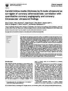
The omega-3 index is the sum of 2 prominent long-chain nҀ3 fatty acids [ie, eicosapentaenoic acid (EPA) and docosahexaenoic acids (DHA)] in erythrocyte membranes and is expressed as a percentage of total erythrocyte fatty acids (FAs). The choice of membrane nҀ3 FA concentrations (rather than plasma nҀ3 FA composition or concentration) as a biomarker of intake is based on the fact that the effects of these FAs on basic cellular function appear to arise primarily from their effects on and in membranes. They act in at least 4 separate ways, but the relative importance of each, their coordinated interaction, and their sufficiency to explain the clinical observations have yet to be precisely determined. First, because of their highly unsaturated nature, nҀ3 FAs may alter membrane biophysical properties (1). This alteration can have the secondary effect of changing the microenvironment of transmembrane proteins (eg, receptors), altering the manner in which they interact with their ligands (2) (Figure 1). Changing membrane FA composition can also affect the affinity of
membrane-associated proteins for the membrane and consequently their interaction with other multiprotein complexes involved with cell signaling systems (3). In addition, a variety of cell stressors (eg, inflammatory mediators) interact with transmembrane receptors and subsequently initiate intracellular Gprotein–linked responses, one of which is the activation of phospholipase A2. This enzyme hydrolyzes long-chain nҀ6 and nҀ3 FAs esterified to inner leaflet phospholipids, liberating them and making them available for conversion to a wide variety of eicosanoids via cyclooxygenase, lipoxygenase, and cytochrome P-450 monooxygenases (4). These molecules powerfully influence cellular metabolism. Phospholipase A2–liberated nҀ3 FAs may directly modify the activity of the ion channel themselves, resulting in altered resting membrane potentials (5). Finally, intracellular nҀ3 FAs liberated from membranes are also able to serve as ligands for a variety of nuclear receptors [eg, peroxisome proliferation-activated receptors, sterol receptor element– binding protein-1c, retinoid X receptor, farnesol X receptor, and hepatocyte nuclear factor-␣ (6, 7)], which have an impact on inflammatory responses and lipid metabolism. Presumably via an orchestrated set of membrane effects, nҀ3 FAs can diminish the activity of inflammatory cells and reduce concentrations of certain inflammatory mediators (4), which may ultimately result in reduced arterial plaque fragility (8). Alterations in the susceptibility of excitable membranes to inappropriate electrical patterns appear to be due to the effects of nҀ3 FAs released from membranes (5). The mild effects of these FAs on blood pressure (9) are likely to be the result of an improvement in arterial compliance (10) and endothelial function (11), possibly arising from enhanced nitric oxide availability (12). The decrease in serum triacylglycerol concentrations that is produced by intakes of 3– 4 g/d EPA ѿ DHA (13) appears to be due to increased hepatic -oxidation and decreased lipogenesis (14), which themselves are the result of modulation of the nuclear receptor actions noted above. Although the precise mechanisms by which nҀ3 FAs reduce the risk for cardiac events are not known in detail, their presence in membranes and the coordinated downstream effects outlined here undoubtedly play a role. 1
From the Metabolism and Nutrition Research Center, Sanford Research/ USD, and the Sanford School of Medicine of the University of South Dakota, Sioux Falls, SD. 2 Presented at the symposium “Beyond Cholesterol: Prevention and Treatment of Coronary Heart Disease with nҀ3 Fatty Acids,” held in New York, NY, June 9, 2007. 3 Address reprint requests and correspondence to WS Harris, 1100 East 21st, Suite 700, Sioux Falls, SD 57105. E-mail:
[email protected]
Am J Clin Nutr 2008;87(suppl):1997S–2002S. Printed in USA. © 2008 American Society for Nutrition
1997S
Downloaded from www.ajcn.org by William Harris on June 28, 2008
ABSTRACT Because blood concentrations of nҀ3 (or omega-3) fatty acids (FAs) (eicosapentaenoic and docosahexaenoic acids) are a strong reflection of dietary intake, it is proposed that a nҀ3 FA biomarker, the omega-3 index (erythrocyte eicosapentaenoic acid plus docosahexaenoic acid), be considered as a potential risk factor for coronary heart disease mortality, especially sudden cardiac death. The omega-3 index fulfills many of the requirements for a risk factor including consistent epidemiologic evidence, a plausible mechanism of action, a reproducible assay, independence from classic risk factors, modifiability, and, most important, the demonstration that raising levels will reduce risk for cardiac events. Measuring membrane concentrations of nҀ3 FAs is a rational approach to biostatus assessment as these FAs appear to exert their beneficial metabolic effects because of their actions in membranes. They alter membrane physical characteristics and the activity of membrane-bound proteins, and, once released by intracellular phospholipases from membrane stores, they can interact with ion channels, be converted into a wide variety of bioactive eicosanoids, and serve as ligands for several nuclear transcription factors, thereby altering gene expression. The omega-3 index compares very favorably with other risk factors for sudden cardiac death. Proposed omega-3 index risk zones are (in percentages of erythrocyte FAs): high risk, 쏝4%; intermediate risk, 4 – 8%; and low risk, 쏜8%. Before assessment of nҀ3 FA biostatus can be used in routine clinical evaluation of patients, standardized laboratory methods and quality control materials must become available. Am J Clin Nutr 2008;87(suppl):1997S–2002S.
1998S
HARRIS
n–3 EPIDEMIOLOGY
Primary compared with secondary prevention of coronary heart disease (CHD) with nⴚ3 fatty acids Although there are secondary prevention trials demonstrating CHD and total mortality benefits of nҀ3 FAs (15, 16), there are no primary prevention, prospective randomized controlled trials with nҀ3 FAs including clinical CHD endpoints, with the possible exception of the primary prevention subset of the Japan EPA Lipid Intervention Study (JELIS) (17). This was a prospective, open-label, randomized trial with masked endpoint evaluation. The study cohort consisted 18 645 hypercholesterolemic men and women who either did (20%) or did not (80%) have a history of CHD. All patients were given statins (10 mg pravastatin or 5 mg simvastatin) and then randomly assigned to a control or EPA group, with the latter receiving 1.8 g/d EPA ethyl esters (Epadel, Mochida, Japan). Patients were followed for a mean of 4.6 y for clinical endpoints. In the entire cohort, the major adverse coronary event rate was 19% lower in patients who received EPA (2.8% compared with 3.5%; P ҃ 0.011). In the primary prevention subset, the event rate reduction was 18%, but it was not statistically significant (P ҃ 0.13). The event rate reduction in the secondary prevention subset was 19% (P ҃ 0.048). Because the effect sizes were virtually the same in both subsets, these findings suggest that EPA is as effective in reducing relative risk for CHD events in both primary and secondary prevention settings. The study was complicated, however, by
OMEGA-3 INDEX COMPARED WITH OTHER CHD RISK FACTORS
The relative predictive value for sudden death of blood nҀ3 FA concentrations compared with other more established CHD risk factors can be estimated by comparing 2 reports from the Physicians’ Health Study. Albert et al (26) published the relative risk for sudden cardiac death across quartiles of C-reactive protein (CRP), total cholesterol, LDL cholesterol,
Downloaded from www.ajcn.org by William Harris on June 28, 2008
FIGURE 1. Modifications of membrane composition with increased eicosapentaenoic acid (EPA) and docosahexaenoic acid (DHA) intake. When EPA and DHA become incorporated into membranes, they affect the properties of both lipid rafts (left) and caveolae (right), reducing the sphingomyelin content of the former and the cholesterol and caveolin content of the latter. These changes can influence the function and binding of membraneassociated proteins, ultimately altering cell function. PUFA, polyunsaturated fatty acids. From Ma et al (2).
the very high background intake of both EPA and DHA, which is estimated to be between 800 and 1000 mg/d in the adult Japanese population (18). Thus, the application of these findings to Western populations consuming lower amounts of nҀ3 FAs is not clear. In addition, the authors did not measure the omega-3 index in this study (only serum concentrations of EPA), so the relations between the index and risk cannot be directly ascertained. In the absence of prospective randomized intervention trials in primary prevention populations, we may turn to prospective observational cohort studies in which either dietary estimates of nҀ3 FA intake or, apropos to the omega-3 index, blood or blood cell membrane nҀ3 FA concentrations were measured. The former studies have been summarized by He et al (19), who performed a meta-analysis of 13 cohorts including 쏜222 000 individuals followed for CHD death for an average of 앒12 y. They found that the consumption of only 1 fish meal/wk compared with 쏝1/mo was associated with a statistically significant 15% reduction in risk. When subjects were classified into categories of increasing fish consumption (쏝1/mo, 1–3/mo, 1/wk, 2– 4/wk, and 욷5/wk), those in the highest intake group showed a 40% reduction in risk. An inverse relation between fish intake and risk for CHD has also been recently reported in Greek (20) and in Japanese cohorts (18). Relations between nҀ3 FA biomarkers and risk for CHD events was recently analyzed by Harris et al (21). We showed in a meta-analysis that across a wide variety of FA biomarkers (plasma, erythrocyte, adipose tissue, and others) DHA and DHA ѿ EPA were inversely related to risk. A recent casecontrol study in which the omega-3 index was measured in patients admitted to the hospital with acute coronary syndromes showed that this biomarker was a significant predictor of case status, independent of multiple clinical covariates including plasma lipids and lipoproteins (22). Furthermore, across the 3 proposed omega-3 index risk categories, there was an approximate 70% reduction in odds for acute coronary syndrome status (Figure 2). Siscovick et al (23) were the first to show in a case-control setting that erythrocyte EPA ѿ DHA was a powerful discriminator of cases experiencing primary cardiac arrest compared with controls (Figure 3). Long-chain nҀ3 FAs measured in whole blood samples collected at baseline in the Physicians’ Health Study were compared between participants who ultimately experienced sudden cardiac death and matched control subjects who did not (24) (Figure 3). Here again, this biomarker of nҀ3 FA status [which correlates highly with the omega-3 index (25)] was shown to be an independent predictor of risk. Both of these studies were conducted in subjects who were free of known CHD at the time of blood sampling, and thus they constitute evidence that a higher omega-3 index is predictive of CHD risk in the primary prevention setting.
OMEGA-3 INDEX AS A RISK FACTOR FOR CHD
1999S
FIGURE 2. Odds ratios for acute coronary syndrome case status is inversely associated with proposed omega-3 index categories. Low (쏝4%), intermediate, and high (쏜8%) omega-3 index values are expressed as a percentage of total blood cell membrane fatty acids (FAs). Odds ratios were calculated in a multivariable logistic regression model including the following additional factors: age, race, sex, diabetes, hypertension, family history of coronary artery disease, history of myocardial infarction, and serum lipids (LDL, HDL, and log-triacylglycerols). Odds P values above columns reference the 쏝4% category. Odds ratios were higher in the intermediate versus the high omega-3 index categories (P ҃ 0.012). EPA, eicosapentaenoic acid; DHA, docosahexaenoic acid; RBC, erythrocyte. From Block et al (22).
CRP, which was more of a dichotomous predictor. In addition, the risk reduction at the highest levels of the omega-3 index (90%) was greater than that associated with the lowest concentrations of CRP (65%). Therefore, in the case of sudden cardiac death [which is responsible for about half of all CHD deaths (27)], the omega-3 index may be more informative than other currently accepted risk factors. PROPOSED CUTOFFS FOR THE OMEGA-3 INDEX
In proposing that the omega-3 index may serve as a new risk marker for CHD mortality, von Schacky and I (25) estimated what a reasonable target “healthy” omega-3 index might be. We surveyed the literature to determine what estimated omega-3 index values were associated with the lowest risk for CHD mortality by either extrapolating from other nҀ3 FA biomarkers (eg, whole blood or plasma phospholipids) or, in studies that reported nҀ3 FA intakes only, by estimating how such intakes would affect the omega-3 index. Using this approach, we arrived at a proposed cardioprotective target concentration of 8% of erythrocyte FAs as EPA ѿ DHA. It also appeared that an omega-3 index of 울4% was associated with the greatest risk for CHD death. WHY ERYTHROCYTES?
FIGURE 3. Relations between the omega-3 index and risk for primary cardiac arrest (left) and the estimated (below) omega-3 index and sudden cardiac death (right). The former data were derived from Siscovick et al (23) from a population-based case-control study, and the latter from Albert, et al (24) from a case-control study nested in the prospective Physicians’ Health Study. The omega-3 index was estimated from the latter data set from the whole-blood long-chain nҀ3 fatty acid content with use of equations described by Harris and von Schacky (25). *P 쏝 0.05 compared with quartile 1. OR, odds ratio; RR, relative risk.
Recommendations to use erythrocyte FA composition as a marker of dietary FA intake date back to 1963 (28). The phospholipids comprising the cell membranes become enriched with nҀ3 FAs not only during reticulocyte maturation in bone marrow but also via direct exchange in plasma (29). This enrichment appears to occur via transfer of DHA (and presumably also EPA)– containing lysophosphatidylcholine, which circulates associated with serum albumin (30), and these molecules are derived from the hydrolysis of nҀ3 FA– containing phospholipids in lipoproteins. Plasma exchange explains why erythrocytes can become enriched with nҀ3 FAs faster than would be predicted if they only appeared with new erythrocytes (31). We recommend measuring the EPA ѿ DHA content of erythrocyte membranes for several reasons (Table 1), not the least of
Downloaded from www.ajcn.org by William Harris on June 28, 2008
HDL cholesterol, triacylglycerols, homocysteine, and the total cholesterol-to-HDL cholesterol ratio. In a second paper (24) they reported the relations between blood nҀ3 FA content and the same endpoint in the same study (Figure 4). Only 2 risk factors demonstrated statistically significant relations with risk for sudden cardiac death after controlling for age and smoking status: CRP and nҀ3 FAs. Unfortunately, neither of these was adjusted for the other nor was either adjusted for homocysteine or other lipid/lipoproteins classes. Thus, whether either of these add predictive value beyond the other is unclear. Nevertheless, blood nҀ3 FA content was related to risk in a dose-dependent manner, which was not the case for
2000S
HARRIS
which is the demonstration that the omega-3 index is correlated with human cardiac membrane EPA ѿ DHA levels (r ҃ 0.81, P 쏝 0.0001), and in cardiac transplant patients supplemented with nҀ3 FAs, both cardiac and erythrocyte EPA ѿ DHA concentrations increase to the same extent (59). Depending on the dose, it takes 3– 6 mo for a new steady-state omega-3 index to be established (60, 61). In addition, the EPA ѿ DHA content of eryth-
rocyte membranes is highly correlated with other plasma-based measures of EPA ѿ DHA content (Figure 5). FUTURE RESEARCH NEEDS
There is a pressing need for standardization in FA analysis. At present, although gas chromatography is used for virtually all FA
TABLE 1 Omega-3 index (and other n-3 biomarkers) as potential risk markers Reference numbers Consistency of epidemiological data for fish intake/omega-3 biomarkers and CHD risk Between populations Within populations Prospective cohorts Case-control studies Raising biomarker levels with fish or n-3 FAs reduces risk Strong association between biomarker and disease Biomarker independent of other known risk factors Adds discriminative value to currently-available risk markers Biologically plausible relation between biomarker and risk Biomarker is modifiable (safely, quickly, and cheaply) Omega-3 index is highly correlated with intake of oily fish and fish oil supplements Other advantages of RBC for n-3 FA analysis Standardized methodology - (In development in our laboratory) Low biological variability High analytical reproducibility, CV ҃ 4%–7% Pre-analytical stability; 7-d at room temperature, unpublished observation; can be mailed to lab unrefrigerated Unaffected by fasting/fed state RBC FA composition stable for at least 4 years frozen at Ҁ80 °C RBC FA composition is less influenced by day-to-day variations and by dyslipidemias than are plasma FAs [(coefficient of variation for plasma phospholipid EPAѿDHA ҃ 32% vs 22% for RBC EPAѿDHA (N҃207; unpublished observation) Better reflection of long-term dietary n-3 FA intake than plasma The RBC is a readily-available byproduct of usual phlebotomy that, if shown to be useful, can be obtained without subjecting the patient to additional procedures The half-life of RBC EPAѿDHA is 4–6 times longer than that of serum EPAѿDHA RBC EPAѿDHA is highly correlated with other n-3 biomarkers such as whole serum, serum phospholipids, and whole blood (which can be measured in a dried blood spot; Figure 5
36–39 23; 40 24; 41–51 22; 53; 54 15; 16 24 23; 24; 49 22 5 33; 57 58; 25
31 59 Unpublished observation 59; 60 61
33; 62
31 35
Downloaded from www.ajcn.org by William Harris on June 28, 2008
FIGURE 4. The multivariable-adjusted relative risk for sudden cardiac death (RR SCD) by quartile (Q) of blood nҀ3 fatty acids (FAs) compared with other, more traditional circulating risk factors. The quartiles at presumed highest risk (black bars) are set at a relative risk of 1.0. Each subsequent lighter bar represents the risk at each decreasing (or, for HDL and nҀ3 FAs, increasing) quartile. CRP, C-reactive protein; Hcy, homocysteine; TC, total cholesterol; Tg, triacylglycerols. Adapted from Albert et al (24, 26).
OMEGA-3 INDEX AS A RISK FACTOR FOR CHD
2001S
analyses, there are a myriad of pre- and postanalytic variables that can all affect the outcome. Some of these factors include the choice of methylation reagent and time, extraction solvents, direct compared with postlipid extraction methylation, gas chromatography columns and program conditions, carrier gases, and postrun control for response factors. There is also a need for standardized qualitycontrol materials that multiple laboratories can use to harmonize results. The use of dried blood spots on specially prepared filter papers (62) may facilitate the development of such interlaboratory assessment systems. Obviously, if the results of studies from various laboratories are to be compared (eg, to identify cardioprotective target nҀ3 FA biomarker concentrations), it will be essential to have standardized methods. Furthermore, if the analysis of blood nҀ3 FAs is ever to enter clinical practice, uniformity and standardization must be established. The day will probably come when a test such as the omega-3 index will be routinely included in lipid panels so that the clinician can address not only cholesterol, triacylglycerol, and lipoprotein abnormalities but also deficiencies in nҀ3 FAs. Opportunities to make a significant impact on not only CHD risk but also risk for cognitive decline, neuropsychiatric disorders, macular degeneration, inflammatory diseases, and other disorders await the development of harmonized methods of assessing nҀ3 FA status. The author is a consultant to GlaxoSmithKline and Monsanto.
REFERENCES 1. Stillwell W, Wassall SR. Docosahexaenoic acid: membrane properties of a unique fatty acid. Chem Phys Lipids 2003;126:1–27. 2. Ma DW, Seo J, Switzer KC, et al. n–3 PUFA and membrane microdomains: a new frontier in bioactive lipid research. J Nutr Biochem 2004;15:700 – 6. 3. Li Q, Wang M, Tan L, et al. Docosahexaenoic acid changes lipid composition and interleukin-2 receptor signaling in membrane rafts. J Lipid Res 2005;46:1904 –13.
4. Calder PC. n–3 polyunsaturated fatty acids, inflammation, and inflammatory diseases. Am J Clin Nutr 2006;83:1505S–19S. 5. Leaf A, Kang JX, Xiao YF, Billman GE. Clinical prevention of sudden cardiac death by n–3 polyunsaturated fatty acids and mechanism of prevention of arrhythmias by n–3 fish oils. Circulation 2003;107:2646 –52. 6. Davidson MH. Mechanisms for the hypotriglyceridemic effect of marine omega-3 fatty acids. Am J Cardiol 2006;98:27i–33i. 7. Deckelbaum RJ, Worgall TS, Seo T. n–3 fatty acids and gene expression. Am J Clin Nutr 2006;83:1520S–5S. 8. Thies F, Garry JM, Yaqoob P, et al. Association of n–3 polyunsaturated fatty acids with stability of atherosclerotic plaques: a randomised controlled trial. Lancet 2003;361:477– 85. 9. Geleijnse JM, Giltay EJ, Grobbee DE, Donders AR, Kok FJ. Blood pressure response to fish oil supplementation: metaregression analysis of randomized trials. J Hypertens 2002;20:1493–9. 10. Nestel P, Shige H, Pomeroy S, Cehun M, Abbey M, Raederstorff D. The n–3 fatty acids eicosapentaenoic acid and docosahexaenoic acid increase systemic arterial compliance in humans. Am J Clin Nutr 2002;76:326 –30. 11. Goodfellow J, Bellamy MF, Ramsey MW, Jones CJH, Lewis MJ. Dietary supplementation with marine omega-3 fatty acids improve systemic large artery endothelial function in subjects with hypercholesterolemia. J Am Coll Cardiol 2000;35:265–70. 12. Lopez d, Orta X, Casos K, et al. Upregulation of endothelial nitric oxide synthase in rat aorta after ingestion of fish oil-rich diet. Am J Physiol Heart Circ Physiol 2004;287:H567–72. 13. Harris WS. n–3 Fatty acids and serum lipoproteins: human studies. Am J Clin Nutr 1997;65(suppl):1645S–54S. 14. Harris WS, Bulchandani D. Why do omega-3 fatty acids lower serum triglycerides? Curr Opin Lipidol 2006;17:387–93. 15. Burr ML, Fehily AM, Gilbert JF, et al. Effects of changes in fat, fish, and fibre intakes on death and myocardial reinfarction: Diet and Reinfarction Trial (DART). Lancet 1989;2:757– 61. 16. Marchioli R, Barzi F, Bomba E, et al. Early protection against sudden death by n–3 polyunsaturated fatty acids after myocardial infarction: time-course analysis of the results of the Gruppo Italiano per lo Studio della Sopravvivenza nell’Infarto Miocardico (GISSI)-Prevenzione. Circulation 2002;105:1897–903. 17. Yokoyama M, Origasa H, Matsuzaki M, et al. Effects of eicosapentaenoic acid on major coronary events in hypercholesterolaemic patients
Downloaded from www.ajcn.org by William Harris on June 28, 2008
FIGURE 5. Correlations between the omega-3 index and (A) blood cell membranes isolated by ultracentrifugation from frozen whole blood [as used in (22)], (B) dried whole blood on filter paper (35), (C) plasma phospholipids, and (D) liquid whole blood. Sample size and correlation coefficients are given in each graph (WS Harris, unpublished observations). EPA, eicosapentaenoic acid; DHA, docosahexaenoic acid; RBC, erythrocyte; PL, phospholipids; FA, fatty acid.
2002S
18. 19. 20. 21. 22. 23. 24. 25.
27. 28. 29. 30. 31.
32. 33. 34. 35.
36. 37. 38. 39. 40.
(JELIS): a randomised open-label, blinded endpoint analysis. Lancet 2007;369:1090 – 8. Iso H, Kobayashi M, Ishihara J, et al. Intake of fish and n–3 fatty acids and risk of coronary heart disease among Japanese: the Japan Public Health Center-Based (JPHC) Study Cohort I. Circulation 2006;113:195–202. He K, Song Y, Daviglus ML, et al. Accumulated evidence on fish consumption and coronary heart disease mortality: a meta-analysis of cohort studies. Circulation 2004;109:2705–11. Panagiotakos DB, Pitsavos C, Zampelas A, et al. Fish consumption and the risk of developing acute coronary syndromes: the CARDIO2000 study. Int J Cardiol 2005;102:403–9. Harris WS, Poston WC, Haddock CK. Tissue n–3 and n– 6 fatty acids and risk for coronary heart disease events. Atherosclerosis 2007;193:1–10. Block RC, Harris WS, Reid KJ, Sands SA, Spertus JA. EPA and DHA in blood cell membranes from acute coronary syndrome patients and controls. Atherosclerosis 2008;197:821– 8. Siscovick DS, Raghunathan TE, King I, et al. Dietary intake and cell membrane levels of long-chain n–3 polyunsaturated fatty acids and the risk of primary cardiac arrest. JAMA 1995;274:1363–7. Albert CM, Campos H, Stampfer MJ, et al. Blood levels of long-chain n–3 fatty acids and the risk of sudden death. N Engl J Med 2002;346:1113– 8. Harris WS, von Schacky C. The Omega-3 Index: a new risk factor for death from coronary heart disease? Prev Med 2004;39:212–20. Albert CM, Ma J, Rifai N, Stampfer MJ, Ridker PM. Prospective study of C-reactive protein, homocysteine, and plasma lipid levels as predictors of sudden cardiac death. Circulation 2002;105:2595–9. Zheng ZJ, Croft JB, Giles WH, Mensah GA. Sudden cardiac death in the United States, 1989 to 1998. Circulation 2001;104:2158 – 63. Farquhar JW, Ahrens EH. Effect of dietary fats on human erythrocyte fatty acid patterns. J Clin Invest 1963;42:675– 85. Renooij W, Van Golde LM, Zwaal RF, Roelofsen B, Van Deenen LL. Preferential incorporation of fatty acids at the inside of human erythrocyte membranes. Biochim Biophys Acta 1974;363:287–92. Brossard N, Croset M, Normand S, et al. Human plasma albumin transports [13C]docosahexaenoic acid in two lipid forms to blood cells. J Lipid Res 1997;38:1571– 82. Katan MB, Deslypere JP, van Birgelen AP, Penders M, Zegwaard M. Kinetics of the incorporation of dietary fatty acids into serum cholesteryl esters, erythrocyte membranes, and adipose tissue: an 18-month controlled study. J Lipid Res 1997;38:2012–22. Harris WS, Sands SA, Windsor SL, et al. Omega-3 fatty acids in cardiac biopsies from heart transplant patients: correlation with erythrocytes and response to supplementation. Circulation 2004;110:1645–9. Cao J, Schwichtenberg KA, Hanson NQ, Tsai MY. Incorporation and clearance of omega-3 fatty acids in erythrocyte membranes and plasma phospholipids. Clin Chem 2006;52:2265–72. Leaf A, Jorgensen MB, Jacobs AK, et al. Do fish oils prevent restenosis after coronary angioplasty? Circulation 1994;90:2248 –57. Marangoni F, Colombo C, Galli C. A method for the direct evaluation of the fatty acid status in a drop of blood from a fingertip in humans: applicability to nutritional and epidemiological studies. Anal Biochem 2004;326:267–72. Dewailly E, Blanchet C, Lemieux S, et al. n–3 fatty acids and cardiovascular disease risk factors among the Inuit of Nunavik. Am J Clin Nutr 2001;74:464 –73. Davidson M, Bulkow LR, Gellin BG. Cardiac mortality in Alaska’s indigenous and non-native residents. Int J Epidemiol 1993;22:62–71. Kromann N, Green A. Epidemiological studies in the Upernavik District, Greenland. Acta Med Scand 1980;208:401– 6. Zhang J, Sasaki S, Amano K, Kesteloot H. Fish consumption and mortality from all causes, ischemic heart disease, and stroke: an ecological study. Prev Med 1999;28:520 –9. Burchfiel CM, Reed DM, Strong JP, Sharp DS, Chyou P-H, Rodriguez BL. Predictors of myocardial lesions in men with minimal coronary atherosclerosis at autopsy. The Honolulu Heart Program. Ann Epidemiol 1996;6:137– 46.
41. Kromhout D, Bosschieter EB, de Lezenne Coulander C. The inverse relation between fish consumption and 20-year mortality from coronary heart disease. N Engl J Med 1985;312:1205–9. 42. Rodriguez BL, Sharp DS, Abbott RD, et al. Fish intake may limit the increase in risk of coronary heart disease morbidity and mortality among heavy smokers. The Honolulu Heart Program. Circulation 1996;94:952– 6. 43. Daviglus ML, Stamler J, Orencia AJ, et al. Fish consumption and the 30-year risk of fatal myocardial infarction. N Engl J Med 1997;336:1046–53. 44. Yuan JM, Ross RK, Gao YT, Yu MC. Fish and shellfish consumption in relation to death from myocardial infarction among men in Shanghai, China. Am J Epidemiol 2001;154:809 –16. 45. Oomen CM, Feskens EJ, Rasanen L, et al. Fish consumption and coronary heart disease mortality in Finland, Italy, and The Netherlands. Am J Epidemiol 2000;151:999 –1006. 46. Dolecek TA. Epidemiological evidence of relationships between dietary polyunsaturated fatty acids and mortality in the Multiple Risk Factor Intervention Trial. Proc Soc Exp Bio Med 1992;200:177– 82. 47. Albert CM, Hennekens CH, O’Donnell CJ, et al. Fish consumption and risk of sudden cardiac death. JAMA 1998;279:23– 8. 48. Hu FB, Bronner L, Willett WC, et al. Fish and omega-3 fatty acid intake and risk of coronary heart disease in women. JAMA 2002;287:1815–21. 49. Mozaffarian D, Lemaitre RN, Kuller LH, Burke GL, Tracy RP, Siscovick DS. Cardiac benefits of fish consumption may depend on the type of fish meal consumed: the Cardiovascular Health Study. Circulation 2003;107:1372–7. 50. Hu FB, Cho E, Rexrode KM, Albert CM, Manson JE. Fish and longchain omega-3 fatty acid intake and risk of coronary heart disease and total mortality in diabetic women. Circulation 2003;107:1852–7. 51. Lemaitre RN, King IB, Mozaffarian D, Kuller LH, Tracy RP, Siscovick DS. n–3 polyunsaturated fatty acids, fatal ischemic heart disease and non-fatal myocardial infarction in older adults. The Cardiovascular Health Study. Am J Clin Nutr 2003;77:319 –25. 52. Guallar E, Hennekens CH, Sacks FM, Willett WC, Stampfer MJ. A prospective study of plasma fish oil levels and incidence of myocardial infarction in U.S. male physicians. J Am Coll Cardiol 1995;25:387–94. 53. Lemaitre RN, King IB, Raghunathan TE, et al. Cell membrane transfatty acids and the risk of primary cardiac arrest. Circulation 2002;105: 697–701. 54. Erkkila AT, Lehto S, Pyorala K, Uusitupa MI. n–3 fatty acids and 5-y risks of death and cardiovascular disease events in patients with coronary artery disease. Am J Clin Nutr 2003;78:65–71. 55. Simon JA, Hodgkins ML, Browner WS, Neuhaus JM, Bernert JT Jr, Hulley SB. Serum fatty acids and the risk of coronary heart disease. Am J Epidemiol 1995;142:469 –76. 56. Burr ML, Ashfield-Watt PA, Dunstan FD, et al. Lack of benefit of dietary advice to men with angina: results of a controlled trial. Eur J Clin Nutr 2003;57:193–200. 57. von Schacky C, Angerer P, Kothny W, Theisen K, Mudra H. The effect of dietary -3 fatty acids on coronary atherosclerosis. A randomized, doubleblind, placebo-controlled trial. Ann Intern Med 1999;130:554 – 62. 58. Mensink RP, Katan MB. Effect of dietary trans fatty acids on highdensity and low-density lipoprotein cholesterol levels in healthy subjects. N Engl J Med 1990;323:439 – 45. 59. Giltay EJ, Geleijnse JM, Schouten EG, Katan MB, Kromhout D. High stability of markers of cardiovascular risk in blood samples. Clin Chem 2003;49:652–5. 60. Hodson L, Skeaff CM, Wallace AJ, Arribas GL. Stability of plasma and erythrocyte fatty acid composition during cold storage. Clin Chim Acta 2002;321:63–7. 61. Tynan MB, Nicholls DP, Maguire SM, et al. Erythrocyte membrane fatty acid composition as a marker of dietary compliance in hyperlipidaemic subjects. Atherosclerosis 1995;117:245–52. 62. Sun Q, Ma J, Campos H, Hankinson SE, Hu FB. Comparison between plasma and erythrocyte fatty acid content as biomarkers of fatty acid intake in US women. Am J Clin Nutr 2007;86:74 – 81.
Downloaded from www.ajcn.org by William Harris on June 28, 2008
26.
HARRIS