The Ne w E n g l a nd Jo u r n a l o f Me d ic i ne
Review Article
Primary Care
O CCULT G ASTROINTESTINAL B LEEDING DON C. ROCKEY, M.D.
O
CCULT gastrointestinal bleeding typically refers to bleeding that is not apparent to the patient. The potential for occult bleeding is emphasized by the finding that for melena to be produced consistently, 150 to 200 ml of blood must be present in the stomach.1 Moreover, patients with gastroduodenal blood loss of 100 ml per day may have stools that appear normal.2 Thus, occult bleeding is usually identified only by tests that detect fecal blood or, if bleeding is sufficient, when it becomes manifest as iron deficiency. Occult gastrointestinal bleeding can also refer to bleeding that is clinically evident but from an obscure source. Obscure gastrointestinal bleeding is the least common form of occult gastrointestinal bleeding but represents a tremendous diagnostic and therapeutic challenge. This article reviews important concepts in the evaluation and care of patients with each type of occult gastrointestinal bleeding. FECAL OCCULT BLOOD
The amount of blood lost from the gastrointestinal tract is normally approximately 0.5 to 1.5 ml per day,3-5 an amount that is typically not detected by occult-blood tests. Nonetheless, occult blood is commonly detected in the stool by fecal occult-blood tests when there has been no clinical evidence of bleeding or iron deficiency. In screening studies, 2 to 16 percent of the patients tested had positive tests, although many tests may have been falsely positive.6,7 A variety of fecal occult-blood tests have been designed, primarily to screen for colon cancer.8,9 However, they also detect blood from other lesions in the gastrointestinal tract (Fig. 1). The likelihood that fecal occult-blood tests will detect gastrointestinal blood
From the Division of Gastroenterology, Duke University Medical Center, Durham, N.C. Address reprint requests to Dr. Rockey at the Liver Center, Duke University Medical Center, Sands Bldg., Rm. 336, Research Dr., Box 3083, Durham, NC 27710, or at
[email protected]. ©1999, Massachusetts Medical Society.
38 ·
is affected by the anatomical level of bleeding, factors relating to the patient — such as stool transit time, stool mixing, and intraluminal hemoglobin degradation — and the intrinsic features of the bleeding of gastrointestinal tract lesions (e.g., irregular bleeding).10 Fecal Occult-Blood Tests
Guaiac-based fecal occult-blood tests make use of the pseudoperoxidase activity of hemoglobin. Guaiac turns blue after oxidation by oxidants or peroxidases in the presence of an oxygen donor such as hydrogen peroxide. Several different guaiac-based tests are available, the characteristics of which vary considerably. For example, of the two most commonly used tests, Hemoccult II and Hemoccult II Sensa (SmithKline Diagnostics, Palo Alto, Calif.), the latter is much more sensitive than the former for detecting fecal heme.11,12 The likelihood that a guaiac-based test will be positive is generally proportional to the quantity of fecal heme, which in turn is related to the size and location of the bleeding lesion (Fig. 1).13,14 Guaiacbased tests are generally best at detecting large, more distal lesions. However, many factors contribute to the variation in the effectiveness of guaiac-based tests for the detection of fecal blood.15 The inconsistency of fecal occult-blood tests in detecting fecal blood is emphasized by the finding that fecal hemoglobin levels must exceed 10 mg per gram of stool (10 ml of daily blood loss) for Hemoccult II tests to be positive 50 percent of the time,16 but stools containing hemoglobin levels of less than 1 mg per gram can result in positive tests.2 Such data have raised questions about the accuracy of guaiac-based tests for detecting colonic lesions.15,17 Many variables influence guaiac-based tests, including dietary factors (Table 1). For this reason, it is important to consider (and modify) diet when performing guaiac-based tests. In addition, fecal rehydration markedly raises the sensitivity of guaiac-based tests but reduces specificity.7 Many believe that oral iron causes positive results on guaiac-based tests. Although the dark-green or black appearance of iron in stool can be confused with the blue typical of a positive guaiac-based test, iron administered orally does not cause positive guaiac reactions.18 Antacids and antidiarrheal drugs containing bismuth also render the stool dark and may confound the reading of guaiacbased tests. Immunochemical tests, which use antibodies directed against human globin epitopes, detect colonic blood with great sensitivity (at a level of as little as 0.3 ml of blood added to stool)19 and do not detect
Jul y 1 , 19 9 9 Downloaded from www.nejm.org at BOTSFORD GENERAL HOSPITAL LIB on February 1, 2006 . Copyright © 1999 Massachusetts Medical Society. All rights reserved.
PR IMA RY C A R E
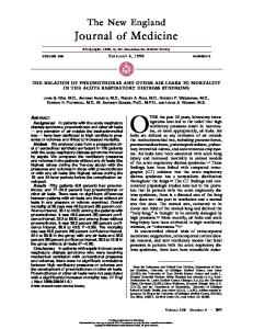
Sites of Gastrointestinal Bleeding
Relative Likelihood of a Positive Fecal Occult-Blood Test
Upper gastrointestinal tract Porphyrins, partially degraded heme, degraded globin
Middle gastrointestinal tract Porphyrins, partially degraded heme, partially degraded globin
Lower gastrointestinal tract Intact heme and intact globin
Guaiacbased
Heme– porphyrin
Immunochemical
Figure 1. Sites of Gastrointestinal Bleeding, Intraluminal Metabolism of Hemoglobin, and Accuracy of Fecal Occult-Blood Tests. In the upper gastrointestinal tract, hemoglobin is cleaved to form heme and globin by gastric pepsin proteases, pancreatic proteases, or both types of proteases in the proximal small intestine. Some intraluminal heme (generally less than 15 percent) is reabsorbed in the small intestine. A portion of heme that is not absorbed and is converted to porphyrins and iron through poorly understood mechanisms has been termed the “intestinal converted fraction” of heme. This fraction is not detected by guaiac-based tests but is detected by the heme–porphyrin assay (e.g., HemoQuant), which measures both heme and porphyrins and is therefore a highly accurate indicator of bleeding, regardless of the level. Globin in the upper gastrointestinal tract is digested by pepsin and pancreatic and intestinal proteases and is thus not detected by immunochemical fecal occult-blood tests. The biology of intraluminal hemoglobin degradation suggests that a guaiac-based test combined with an immunochemical test could help differentiate occult bleeding in the upper gastrointestinal tract from that in the lower gastrointestinal tract. The red cells indicate the sites of blood loss.
small quantities of blood from the upper gastrointestinal tract (Fig. 1).12 Thus, they have a theoretical advantage over guaiac-based tests in terms of localizing bleeding to the colon. Unfortunately, the tests are limited by loss of globin antigenicity at room temperature and require processing in a laboratory. Newer slide tests may help circumvent these problems. The heme–porphyrin test, HemoQuant (Mayo Medical Laboratories, Rochester, Minn.), measures hemoglobin-derived porphyrin spectrofluorometrically and allows exact measurement of total hemoglobin in stools. Moreover, substances that may interfere with or cause false positive guaiac-based tests (for example, vegetable peroxidases) do not affect the test. Unfortunately, the need for laboratory processing and the high false positive rate of this test have limited its clinical application. Differential Diagnosis and Approach to Evaluation
Although many variables influence the results of fecal occult-blood tests, including variables that cause false positive results and those that cause false negatives, once it is determined that a patient has a positive test, the clinical focus is generally first on colonic
TABLE 1. CHARACTERISTICS OF DIFFERENT CLASSES OF FECAL OCCULT-BLOOD TESTS.* CHARACTERISTIC
TEST†
Bedside availability Time to develop Cost‡ Reasons for false positive results Nonhuman hemoglobin Dietary peroxidases Rehydration Iron Reasons for false negative results Hemoglobin degradation Storage Vitamin C
GUAIAC-
HEME–
IMMUNO-
BASED
PORPHYRIN
CHEMICAL
++++ 1 min $18
0 1 hr $33
0 to ++ 5 min to 24 hr $18–$35
++++ +++ +++ 0
++++ 0 0 0
0 0 0 0
+++ ++ ++
0 0 0
+++ ++ 0
*Relative comparisons are shown on a scale of 0 to ++++, with ++++ indicating highly likely and 0 highly unlikely. †Commonly used tests are Hemoccult II and Hemoccult II Sensa for guaiac-based tests, HemoQuant for heme–porphyrin tests, and HemeSelect and FlexSure OBT (SmithKline Diagnostics, Palo Alto, Calif.) for immunochemical tests. ‡The dollar amounts shown, which represent 1998 values, are for reimbursements. The amount shown for the guaiac-based test is the price for three cards.
Vol ume 341
Numb e r 1
Downloaded from www.nejm.org at BOTSFORD GENERAL HOSPITAL LIB on February 1, 2006 . Copyright © 1999 Massachusetts Medical Society. All rights reserved.
·
39
The Ne w E n g l a nd Jo u r n a l o f Me d ic i ne
imaging. The choice of imaging mode — colonoscopy or air-contrast barium enema — is controversial.20-22 Flexible sigmoidoscopy is mandatory for patients undergoing air-contrast barium enema to evaluate the rectosigmoid colon fully.23 Some studies have found that air-contrast barium enema accurately detects colon cancer and large adenomas,24 but most studies have found air-contrast barium enema to be less accurate than colonoscopy.20,25 Both tests can miss important neoplastic lesions.20,26,27 In addition, other factors are important when choosing an evaluation strategy; not only is the accuracy of the test an issue, but cost, acceptance by the patient, and complication rates differ between the two methods. On the basis of the available data, colonoscopy is recommended for the evaluation of the colon in patients with fecal occult blood; however, the use of air-contrast barium enema with flexible sigmoidoscopy may be an acceptable alternative. Computed tomographic studies of the colon (virtual colonoscopy) could eventually play a part in colonic evaluation,28 but clinical experience is limited. Further studies are required to assess colonic-imaging techniques in patients with fecal occult blood. Many gastrointestinal lesions can bleed and cause a positive fecal occult-blood test (Table 2); indeed, patients with fecal occult blood detected by guaiacbased tests may have serious disease of the upper gastrointestinal tract. Upper endoscopic examinations detected abnormalities in 25 to 41 percent of patients with fecal occult blood, many of whom were asymptomatic.29-32 Although this finding is surprising given the intraluminal metabolism of hemoglobin (Fig. 1), currently available guaiac-based tests detect small amounts of blood in the upper gastrointestinal tract,12,33 and many upper gastrointestinal tract lesions bleed enough to produce positive results on guaiac-based tests.3,12 The highly sensitive Hemoccult II Sensa test is substantially more likely to detect blood in the upper gastrointestinal tract than is the Hemoccult II test.12 Therefore, the upper gastrointestinal tract must be considered as a potential source of bleeding in patients with normal results on colonoscopy, especially when highly sensitive guaiac-based tests are positive. In the patient with a positive guaiac-based fecal occult-blood test and a negative colonic examination, symptoms of upper gastrointestinal disorders (severe reflux, dyspepsia, and abdominal pain), weight loss, and iron deficiency should be assessed. If such signs and symptoms are present, the upper gastrointestinal tract should be studied. Whether asymptomatic patients should undergo evaluation of the upper gastrointestinal tract has not yet been determined. Appropriate evaluation of patients with fecal occult blood detected by digital rectal examination is controversial, because obtaining stool by this means may lead to false positive tests. False positive tests 40 ·
OF
TABLE 2. DIFFERENTIAL DIAGNOSIS OCCULT GASTROINTESTINAL BLEEDING.* Mass lesions Carcinoma (any site)† Large (>1.5 cm) adenoma (any site) Inflammation Erosive esophagitis† Ulcer (any site)† Cameron lesions‡ Erosive gastritis Celiac disease Ulcerative colitis Crohn’s disease Colitis (nonspecific) Idiopathic cecal ulcer Vascular disorders Vascular ectasia (any site)† Portal hypertensive gastropathy or colopathy Watermelon stomach Varices (any site) Hemangioma Dieulafoy’s vascular malformation§ Infectious diseases Hookworm Whipworm Strongyloidiasis Ascariasis Tuberculous enterocolitis Amebiasis Surreptitious bleeding Hemoptysis Oropharyngeal bleeding (including epistaxis) Other causes Hemosuccus pancreaticus Hemobilia Long-distance running Factitious cause
*Potential lesions leading to all forms of occult gastrointestinal bleeding are listed here. Some lesions that may lead to recurrent obscure bleeding are not listed. †These abnormalities are the most common. ‡These are linear erosions within a hiatus hernia. §This is a large superficial artery underlying a small mucosal defect.
may, in theory, result from anorectal trauma induced by the gloved finger during digital rectal examination. In addition, patients generally have not had dietary modification implemented at the time of digital rectal examination. However, in both symptomatic and asymptomatic patients with fecal occult blood detected by digital rectal examination, the number of new lesions identified by gastrointestinal evaluation is substantial.32,34 Thus, evaluation of these patients is indicated and, if symptoms are present, the investigation should be focused on the site or sites of the symptoms. Occult gastrointestinal bleeding is often attributed to therapy with anticoagulants or aspirin. However, fecal blood levels in patients treated with anticoagulants or low-dose aspirin are normal or only minimally elevated, respectively.35,36 Neither warfarin nor low-dose aspirin alone appears to cause positive
Jul y 1, 19 9 9 Downloaded from www.nejm.org at BOTSFORD GENERAL HOSPITAL LIB on February 1, 2006 . Copyright © 1999 Massachusetts Medical Society. All rights reserved.
PRIMA RY C A R E
Normal Balance of Iron
Iron Pool
Iron Deficiency
Dietary iron
Dietary iron
(5–15 mg elemental, 1–5 mg heme)
Tissues 300 mg
300 mg
(5–15 mg elemental, 1–5 mg heme) absorption increases 2–3 times
Storage 100–400 mg in women 1000 mg in men
None
Absorption of 1 mg of iron
Absorption increases
Red cells
Normal 2500 mg Loss of 1 mg of iron
Deficient 2000 mg
3–5 mg of iron (i.e., gastrointestinal, menses)
Loss of 1 mg of iron
Obligate loss: ~1 mg of iron=~1 ml of blood (~0.5 mg of iron)+~0.5 mg of nonblood iron
Figure 2. Gastrointestinal Blood Loss and Iron Balance. Normal obligate daily iron loss is from blood loss (presumably from gastrointestinal mucosal microerosions or microulcerations) and iron in sloughed epithelial cells of the gut. Total daily iron loss is approximately 1 mg. The usual Western diet contains mostly elemental iron, of which about 10 percent is absorbed. Heme iron, derived primarily from myoglobin in meats, is preferentially absorbed and accounts for 60 to 80 percent of the iron absorbed per day. Under normal circumstances, iron homeostasis is tightly regulated, and daily iron loss is precisely balanced by iron absorption. Iron deficiency results only when the dynamic, but limited, absorptive capacity of the small intestine is exceeded by iron loss. The time required for the development of iron deficiency depends on the size of initial iron stores, the rate of bleeding, and intestinal iron absorption. Iron deficiency generally occurs only with loss of more than 5 ml of blood per day. Anemia is a late manifestation of the iron-depleted state. The red cells indicate bleeding and potential sites of blood loss.
guaiac-based fecal occult-blood tests.36 In a prospective study evaluating the gastrointestinal tract in patients taking anticoagulants who had positive guaiacbased fecal occult-blood tests, 15 of 16 patients had new lesions, 20 percent of which were malignant.37 In addition, there was no difference in the frequency of lesions between patients receiving warfarin and those receiving standard heparin.37 Thus, positive fecal occult-blood tests should not be attributed solely to anticoagulant therapy — whether with therapeutic or prophylactic warfarin, heparin (including lowdose heparin), or aspirin — but, rather, should lead to formal evaluation. In patients with fecal occult blood (or any occult gastrointestinal bleeding), too high a degree of anticoagulation diminishes the yield of a gastrointestinal tract evaluation. In those with intrinsic coagulopathy (hemophilia or von Willebrand’s disease), particularly if bleeding is chronic, abnormalities of the gastrointestinal tract are also less likely to be identified; nevertheless, in both clinical scenarios, gastrointestinal evaluation must be given serious consideration.
Treatment and Outcome
The care of patients with fecal occult blood is based on the abnormalities identified. Likewise, outcomes are directly related to specific findings. Since nonsteroidal antiinflammatory drugs may lead to gastrointestinal injury, these should be discontinued if possible. Particularly problematic are vascular ectasias, which are often multiple and bleed chronically. (The medical management of vascular ectasias is discussed below.) The prognosis of patients with positive fecal occult-blood tests but no identifiable gastrointestinal disorder is generally favorable. ANEMIA DUE TO IRON DEFICIENCY
In the United States, 5 to 11 percent of women and 1 to 4 percent of men have iron deficiency, and approximately 5 percent and 2 percent of women and men, respectively, have iron-deficiency anemia.38 In women, iron-deficiency anemia — a result of chronic iron loss (Fig. 2) — is most common during the reproductive years, because of menstrual and pregnancy-associated iron losses.39 Among other patients, Vol ume 341
Numb e r 1
Downloaded from www.nejm.org at BOTSFORD GENERAL HOSPITAL LIB on February 1, 2006 . Copyright © 1999 Massachusetts Medical Society. All rights reserved.
·
41
The Ne w E n g l a nd Jo u r n a l o f Me d ic i ne
however, iron-deficiency anemia has been associated with chronic occult gastrointestinal bleeding.40 Differential Diagnosis and Approach to Evaluation
Because a diagnosis of iron-deficiency anemia necessitates extensive and often costly evaluation, the diagnosis must be established carefully. Although the gold standard is bone marrow biopsy, iron deficiency is most often indicated by a serum ferritin level of less than 45 µg per liter; iron-deficiency anemia is most often diagnosed on the basis of a similarly low ferritin level plus a hemoglobin level of less than 12 g per deciliter for women and less than 13 g per deciliter for men.41 Iron deficiency without anemia also requires further investigation, because it may be associated with serious abnormalities of the gastrointestinal tract.42,43 Cancers of the right side of the colon have traditionally been considered a leading cause of occult bleeding leading to iron-deficiency anemia, although practically any lesion in the gastrointestinal tract can bleed in an occult fashion (Table 2). Several crosssectional studies have documented prominent abnormalities of the upper gastrointestinal tract. In studies of 381 patients with iron-deficiency anemia, lesions of the gastrointestinal tract consistent with chronic blood loss were identified in the following locations: the esophagus, stomach, or duodenum (in the form of severe esophagitis, presumably mediated by reflux, and ulcers) (41 percent), the small intestine (3 percent), and the colon (in the form of colon cancer and large adenoma) (22 percent).44-47 In this group of patients, 34 percent had no identifiable lesion consistent with blood loss, and only 5 percent had disease identified in both upper and lower gastrointestinal tracts. Focused evaluation of the gastrointestinal tract should be considered in patients with iron-deficiency anemia. Gastrointestinal symptoms help direct the workup,47,48 although symptoms may not localize the disease.45,46 If epigastric pain, a change in stool caliber, and reflux symptoms are present, the initial investigation should be directed toward these symptoms, especially if the onset was recent. Because multiple lesions are rare, the identification of an abnormality clearly consistent with bleeding, such as a mass lesion, large ulceration, or severe inflammation, generally makes further evaluation unnecessary. In the absence of gastrointestinal symptoms, particularly in elderly patients, evaluation should begin with the colon and, if the colonic evaluation is negative, proceed to the upper gastrointestinal tract. Large mass lesions, ulcerative gastrointestinal lesions, and even erosive gastritis associated with Helicobacter pylori infection commonly lead to serious occult blood loss (up to 60 ml per day).3,5,49 However, trivial lesions such as mild inflammation and small adenomas do not bleed substantially.3,10 Fur42 ·
thermore, not all patients with iron-deficiency anemia and gastrointestinal lesions have elevated levels of hemoglobin in gastrointestinal-lavage fluid.50 Thus, it is unlikely that every identified gastrointestinal tract lesion is associated with occult bleeding; caution is therefore urged in attributing iron-deficiency anemia to trivial lesions. The role of gastrointestinal evaluation in premenopausal women with iron-deficiency anemia is not yet settled. A recent retrospective study found that 23 of 186 premenopausal women with iron-deficiency anemia (12 percent) had serious gastrointestinal tract abnormalities, including lesions of the upper gastrointestinal tract (12 patients) and the lower gastrointestinal tract (11 patients).48 Surprisingly, gastric cancer was the most common lesion of the upper gastrointestinal tract (five patients) and colon cancer the most prevalent colonic lesion (six patients). Of 15 variables examined, the only clinical features predictive of a detectable lesion were severe anemia (hemoglobin,