Clinical Neurology and Neurosurgery 108 (2006) 227–233
The multiple sclerosis trait and the development of multiple sclerosis: Genetic vulnerability and environmental effect Charles M. Poser ∗ Department of Neurology, Harvard Medical School and Beth Israel Deaconess Medical Center, 330 Brookline Avenue, Boston, MA 02118, USA
Abstract The remarkably low rate of concordance of multiple sclerosis (MS) in monozygotic twins has never been fully explained but it implies the possibility of a systemic condition called the multiple sclerosis trait (MST), which is quite different from asymptomatic MS. It results from the action of an antigenic challenge on the immune system of a genetically vulnerable person that does not cause damage to the nervous parenchyma; it may never evolve into the disease MS. A subsequent environmental viral-antigenic event in some MST-carriers can change the trait into the disease. This event could be an infection, which need not be symptomatic, or a vaccination. The MS may become symptomatic, remain asymptomatic, or manifested only by lesions visible by MRI. It is likely that the development of the MST, called activation, occurs early in life, while the transition from MST to MS, called acquisition, takes place at puberty in most patients. Differences in prevalence between pre-puberal migrants, and the locally born children of migrants, and their population of origin may also be explained by the MST. © 2005 Elsevier B.V. All rights reserved. Keywords: Multiple sclerosis; Multiple sclerosis trait; Twins; Genetic susceptibility; Environment; Migration; Molecular mimicry; Puberty
1. Introduction It is well recognized that both genetic endowment and environmental influences play important roles in the pathogenesis of MS [1,2], the former being the stronger, and the nature of the latter still unidentified. The astonishingly low degree of concordance of the disease in monozygotic (MZ) twins is puzzling in persons sharing an identical genetic endowment. In a recent study of MS in twins, clinical concordance was noted in only 62 of 308 pairs of MZ twins, 20.1% (range 0–40%), and in 23 of 452 pairs of dizygotic (DZ) twins, 5.1% (range 0–28.6%). Brain MRI lesions suggestive of MS (according to the criteria of Fazekas et al. [3]) were noted in only 13% of discordant twins [4]. Even when abnormal MRI and visual evoked response studies are included, the concordance rate in MZ twins is only about 25% [5]. Several reasons have been given for this remarkably low rate of concordance including somatic muta∗
Tel.: +1 617 667 2063; fax: +1 617 536 9517. E-mail address:
[email protected].
0303-8467/$ – see front matter © 2005 Elsevier B.V. All rights reserved. doi:10.1016/j.clineuro.2005.11.019
tions, uneven X chromosome inactivation [6], and changes of the epigenome [7]. Another possibility is that the unaffected twin has asymptomatic disease or carries the MS trait [8].
2. The multiple sclerosis trait The MST is a systemic non-pathological condition not involving the nervous system parenchyma that may affect some persons who are genetically susceptible to MS [8]. In concept, it is analogous to the sickle cell trait, acute intermittent porphyria and glucose-6-phosphate dehydrogenase deficiency, in that it requires a trigger to develop into the disease MS. It is the premorbid stage of MS, “a disease waiting to happen.” It is not simply asymptomatic MS, since myelin and axons are intact. The MST will not develop in all MS-susceptible persons; thus it will not be present in all siblings and DZ twins of patients with MS. None the less, it is highly probable that a clinically unaffected, genetically identical MZ twin would
228
C.M. Poser / Clinical Neurology and Neurosurgery 108 (2006) 227–233
carry it. The MST may never develop into clinical MS, it may do so but remain asymptomatic (with or without MRI lesions), or it may emerge into the symptomatic form. The MST is characterized by the following: an exaggerated response to a number of viral antigens, the presence of oligoclonal bands in the CSF, and an increased vulnerability of the blood–brain barrier (BBB). 2.1. Antibody response Increased serum antibody titers against several viral antigens are noted in patients with MS as well as in their clinically unaffected siblings [9]) and discordant MZ- and DZ-twins [10–12]. Panelius et al. [13] showed that in blast transformation tests, lymphocytes from MS patients and their siblings reacted in a similar way. Konttinen et al. [14] described a phytoagglutinin-induced lymphocyte activation sequence in MZ- and DZ-discordant twin pairs in a quiescent phase of disease. The level and kinetics of interleukin-2 receptor expression, DNA synthesis, ␥-interferon secretion and major HLA II complex expression were similar in both. Immunological hyperreactivity is one of the manifestations of the MST. 2.2. CSF oligoclonal bands The presence of oligoclonal bands (OCB) in the CSF of unaffected twins [15–17] is another characteristic of the MST. 2.3. Increased vulnerability of the blood–brain barrier (BBB) In 1986, Poser [8] suggested that the alteration of the BBB was the obligatory first step in the pathogenesis of MS, which was confirmed by Kermode et al. [18] by means of gadolinium-enhanced MRI. Adams et al. [19] first described an inflammatory lymphocytic infiltrate in the walls of venules and capillaries in the normal-appearing white matter (NAWM) of MS brains. Utilizing much more sophisticated immunohistochemical techniques, Gay and Esiri [20] were able to demonstrate the disruption of the vessel wall and the lack of pathological changes in the surrounding parenchyma. Magnetization transfer imaging (MTI) of NAWM in MS patients have also revealed these changes [21] in MS patient. Siger-Zajdel et al. [22] demonstrated changes in the NAWM by MTI in 30 asymptomatic first-degree relatives of both familial and sporadic MS patients; in addition, their MRIs were completely normal. The failure of gadolinium-enhanced MRI to reveal NAWM lesions suggests that the changes in the blood-vessel wall are not severe enough to lead to plaque formation, although they might facilitate the penetration of B-cells. The abnormalities of the blood vessels in the NAWM of MS patients represent the remaining evidence of the MST. These minor alterations of the BBB increase its vulnerability to subsequent insults.
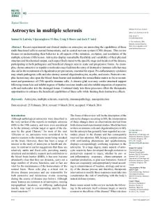
3. The origin of the MST The increased immune response that underlies the pathogenesis of MS is the response to various antigenic challenges, either from infections, most likely viral, but possibly also from some viral vaccinations. This challenge is probably non-specific, i.e. that a number of agents may be responsible, although it is likely that for each individual specific antigens are necessary. The specificity of the immunogenic challenge may be determined by the person’s genetic susceptibility, i.e. the type of receptors available; there must be compatibility between the antigen and one of the immune receptors (Fig. 1a). The possible role of vaccines in triggering MS, if any, is unresolved. Vaccines, by definition, are powerful antigenic challenges. No thorough study has ever been carried out to discover if the introduction of compulsory routine vaccinations against the exanthemata and other common infectious diseases of early childhood has had any effect on the incidence and prevalence of MS in countries with a high degree of compliance, a condition which is lacking in most areas of Africa and Latin America, where the low prevalence of MS has been ascribed primarily to the nearabsence of genetic vulnerability to the disease. Susceptibility to vaccination-induced autoimmunity is also determined by genetic factors [23]. The MST is clinically and radiologically silent; thus its time of activation is impossible to recognize. It is most likely that in genetically vulnerable children it occurs shortly after the first serious immunological challenge as a consequence of one or more of the exanthemata, or of vaccination against them.
4. The transition from the MS trait to MS: disease acquisition The pathogenic mechanism of MS is initiated by an inflammatory phenomenon that results in a much more severe alteration of the BBB. The transformation of MST into MS – the acquisition of the disease – requires a second viral antigenic challenge, its identity also determined by the type of receptors in the host. When the MST-carrier enters a different environment, “new” antigens are encountered, either viruses or vaccine components, among which are some that exhibit molecular mimicry [24,25] with the original MST immune activator, and are also compatible with one of the person’s immune receptors (Fig. 1b). 4.1. Age of acquisition and the role of gender There is ample evidence from migration studies to suggest that the critical age for acquisition of MS is around 15 years, a finding that has generally been interpreted as meaning puberty [26–30]. MS is almost twice more common in women, who have a greater immune reactivity and stronger reactions against infections and immunizations,
C.M. Poser / Clinical Neurology and Neurosurgery 108 (2006) 227–233
229
Fig. 1. (a) The genetically MS-vulnerable (GV) individuals A and B are exposed to a variety of antigens (a–k). Appropriate receptors are available for antigens c in A, and for h in B; as a result, an immune response results and the MST develops in both. (b) Both MST-carriers change environment at about puberty and are now exposed to a “new” set of antigens (m–s). An appropriate receptor is available only for antigen m which exhibits the required molecular mimicry with antigen c which had activated the MST in A; A acquires MS from the MST. None of the new antigens elicit a response from B, who remains at the MST stage. In the migration scenario, A or his parents had migrated from an area of “low risk” (e.g. the West Indies) to one of “high risk” (e.g. London). B or his parents had moved from a “high risk” (e.g. England) to a “low risk” area (e.g. South Africa). Note: molecular mimicry is depicted by the similarity of patterns between parts of the antigens.
than in men because of the marked effect of sex hormones [31–35]. Gender emerges as one of the most important epidemiological risk factors for the development of autoimmune dis-
eases. Women have higher levels of circulating immunoglobulins and a more frequent production of a variety of autoreactive antibodies. Sex hormones have been shown to modulate a large variety of mechanisms involved in the immune
230
C.M. Poser / Clinical Neurology and Neurosurgery 108 (2006) 227–233
response, including cell trafficking, cytokine production, lymphocyte proliferation, expression of adhesion molecules and HLA-class II receptors. Estrogens have a stimulating effects on B-cell functions, which seem to dependent on the inhibition of suppressor T-cells. Numerous studies demonstrate that sex hormones can directly influence the immune system through specific receptors identified in the thymus and peripheral immunocompetent cells, but these may be modulated by other sex steroid-induced changes in other biological systems capable of influencing the immune system [35]. Estradiol is the major form of estrogen present in women. At puberty the estradiol levels increase in girls and remain elevated during periods of each menstrual cycle. In boys there is an increase of estradiol levels until their growth spurt at midpuberty, which then decreases [36]. In addition, the pituitary hormones prolactin and growth hormone also stimulate autoimmunity [37,38]. Serum levels of prolactin and GH are higher in women, differences which develop at puberty. How these gender biases in immune system function affect the clinical expression of autoimmune disease is not understood. Most antigens require internalization and processing by B cells and subsequent recognition by CD4+ cells. The recognition of processed antigen by T helper (CD4+) cells and the subsequent activation of these cells constitute the critical events in the humoral (antibody-mediated) immune response. MS is associated with predominantly Th1-type (pro-inflammatory) cytokines. Women are more likely to develop a Th1 response after challenge with an infectious agent. Sex steroid hormones may alter the Th1-Th2 balance during autoimmune disease states, thus influencing disease susceptibility and severity [38]. It is also noteworthy that changes in environment, and thus potential exposure to new antigenic challenges, not infrequently occur at about the time of puberty: a move from grammar-elementary school to middle or junior high school, from one city, or even country, new sports and recreational activities. The developed world has become enormously more mobile. Long distance air travel and commercial, industrial and military relocations are increasingly more common. The probability of exposure to new antigenic challenges has markedly increased.
who acquired MS after antirabies vaccination were genetically vulnerable persons who had become MST-carriers. DEM developed when this vaccine was administered to an immuno-active host who was not genetically MS-vulnerable, while normal antibody production occurred in the immunona¨ıve person (Fig. 2). A few other similar cases of MS have been reported following other viral infections and immunizations [41–45]. Several of these patients were adults, in fact one was 58 years old [45], implying that disease acquisition may in unusual circumstances happen long after puberty.
6. The MST and migrations The unexpected changes in incidence and prevalence of MS resulting from migrations before puberty can be explained by the fact that leaving a “low risk” area for a “high risk” one [46] would “unmask” MST-carriers by exposing them to – for them – new or unusual viral antigenic challenges, while those going to a “low risk” [29] area were escaping from exposure to ones that would have triggered the acquisition of MS from the MST (Fig. 1a and b). The same epidemiological changes have been found to apply to the locally born children of immigrants to developed, industrialized countries; those children would also most likely move from a rural to an urban environment and receive early childhood vaccinations.
7. The Hawaii effect The puzzling differences in prevalence between different ethnic groups in Hawaii provides yet another illustration of the possible role of the MST. Caucasians born and raised in the islands have a prevalence of MS only one-third that of those raised in California; on the other hand, Hawaii-born Japanese have triple the prevalence of Japan-born and -raised MS patients [47]. This suggests that the MST-carriers among the latter encountered a “new” viral antigen in the Hawaiian Island that triggered the acquisition of MS, whereas for some of the Hawaii-raised Caucasian MST-carriers, no compatible second antigenic challenge was encountered to cause them to acquire the disease (Fig. 3).
5. Postinfectious and postvaccinal MS: the role of the MST
8. Monozygotic twins – from MST to MS
In extremely rare instances exanthemata or vaccinations produce typical MS instead of causing disseminated encephalomyelitis (DEM). The best-known examples of this phenomenon occurred in several Japanese after receiving antirabies vaccination, whereas many more other vaccinees developed classical DEM [39]. True MS is rare in the Japanese, probably due to low genetic vulnerability; on the other hand, neuromyelitis optica, a variant of DEM [40] is more common. The most reasonable explanation is that those
It has been generally assumed that twins are exposed to identical environmental factors during childhood and adolescence, and that the identical genetic endowment of MZ twins translates into the same susceptibility to MS. Thus, they would both develop the MST, probably at the same time. DZ twins and siblings are less likely to acquire the MST, thus accounting for the lesser incidence of MS among them. These assumptions now seem to be incorrect. At some time in their lives twins no longer seem to share the same environment.
C.M. Poser / Clinical Neurology and Neurosurgery 108 (2006) 227–233
231
Fig. 2. The differential effect of vaccination: (A) the non-specific viral activator connects with the appropriate receptor of the genetically MS-vulnerable (GV) individual inducing the MST. A protein in the vaccine exhibits molecular mimicry with the original activator and triggers a response from the MST-carrier causing the acquisition of MS. (B) The vaccine connects with a receptor of the immuno-active host and, due to molecular mimicry, produces DEM. (C) The vaccine connects with a receptor of the immuno-na¨ıve host, resulting in the production of antibodies.
In a study of events preceding the clinical onset of MS in a series of MZ twins there was a remarkable difference in the frequencies of definable events occurring preferentially in the later affected twin [48]. There was virtually no difference in early childhood or development, or in other environmental factors in preschool years. But from age 6 years on, increasingly more environmental events were noted among the affected twins than the unaffected ones. The events were recorded for all twins even if concurrently present in both members of the set. They then grouped all other definable phenomena that had occurred at one time to one twin and not the other regardless of diagnosis. These events included head
and other trauma, tonsillectomy and appendectomy, allergies, and ether anesthesia. Others were infections and animal contacts. The French Research Group on Multiple Sclerosis [5] also indicated that the MS twin concordance rate is related to environmental factor loading. They noted that the mean time interval between twin separations and onset of MS in the affected twins was 13.4 years, a length of time which is remarkably similar to the latency of MS, which is approximately 15 years [49]. It is certainly possible for only one of the MZ twins to have been exposed to a viral antigen that transformed the MST into MS. A recently published study of age-dependent changes of the genome of MZ twins suggests
232
C.M. Poser / Clinical Neurology and Neurosurgery 108 (2006) 227–233
Fig. 3. (A) is a Hawaii-born Japanese who is genetically MS-vulnerable (GV) and develops the MST as a reaction to antigen h. At about the time of puberty, he is exposed to antigen s and acquires MS. (B) is a Hawaii-born GV Caucasian who develops the MST by reacting with antigen q; none of the new antigens that he encounters at the time of puberty trigger a response and he remains at the MST stage.
that the epigenetic profiles may represent a link between environmental factors and phenotypic differences [7]: the epigenetic markers were more distinct in MZ twins who were older, had different lifestyles, and had spent less of their lives together.
9. Discussion No attempt has yet been made to identify MST-carriers in the blood relatives of MS patients. It is not possible to determine when the MST changes into MS in an individual, unless symptoms develop or MRI lesions become apparent, both of which may not occur until a decade or more afterward. Furthermore, many persons never develop symptoms of MS despite having its lesions in the CNS [50–53]. Identifying persons with the MST would obviously be of enormous importance for the purpose of initiating prophylactic treatment. This would have to be done very early, preferably before puberty, and protecting the BBB would be the goal of therapy. One important objective of research in MS would be to develop means of identifying MST-carriers in early childhood. Twins are the ideal subjects but there are relatively few of them; unaffected blood relatives of MS patients are considerably more numerous. The increasing sophistication of molecular genetics and immunological investigations gives hope that studies
of family members of MS families may throw light on the problem.
References [1] Sadovnick A, Ebers G, Dyment D, et al. Evidence for the genetic basis of multiple sclerosis. Lancet 1996;347:1728–30. [2] Poser C. The epidemiology of multiple sclerosis: a general overview. Ann Neurol 1994;36(Suppl. 2):S189–93. [3] Fazekas F, Offenbacher H, Fuchs S. Criteria for an increased specificity of MRI interpretation in elderly subjects with suspected multiple sclerosis. Neurology 1988;38:1822–5. [4] Willer C, Dyment D, Risch N, et al. Twin concordance and sibling recurrence rate in multiple sclerosis. Proc Natl Acad Sci 2003;100:12877–82. [5] French Research Group on Multiple Sclerosis. Multiple sclerosis in 54 twinships: concordance rate is independent of zygosity. Ann Neurol 1992;32:724–7. [6] McFarland H. Twin studies and multiple sclerosis. Ann Neurol 1992;32:722–3. [7] Fraga M, Ballestar E, Paz M, et al. Epigenetic differences arise during the lifetime of monozygotic twins. Proc Natl Acad Sci Early Ed July 5, 2005. [8] Poser C. The pathogenesis of multiple sclerosis: a critical reappraisal. Acta Neuropathol 1986;71:1–10. [9] Brody J, Sever J, Henson T. Virus antibody titers in MS patients, siblings and controls. JAMA 1971;216:1441–6. [10] Panelius M, Salmi A, Halonen P, et al. Virus antibodies in serum specimens from patients with MS, from siblings, and matched controls. Acta Neurol Scand 1973;49:85–107.
C.M. Poser / Clinical Neurology and Neurosurgery 108 (2006) 227–233 [11] Woyciechowska J, Dambrozia J, Leinikki P, et al. Viral antibodies in twins. Neurology 1985;35:1176–80. [12] Kinnunen E, Valle M, Piiranen L, et al. Viral antibodies in MS: a nationwide co-twin study. Arch Neurol 1990;47:743–6. [13] Panelius M, Nikoskelainen E, Ilonen J. Genetic and immunological case studies in familial MS. Acta Neurol Scand 1984;98(Suppl.):348. [14] Konttinen Y, Kinnunen E, Kemppinen P, et al. Lymphocyte activation in discordant twin pairs. J Neuroimmunol 1990;27:1–8. [15] Xu X, McFarlin D. Oligoclonal bands in CSF: twins with MS. Neurology 1984;34:769–74. [16] Woyciechowska J, Dambrozia J, Chu A, et al. Correlation of oligoclonal IgG bands and viral antibodies in twins with multiple sclerosis. In: Cazullo C, Caputo D, Ghezze A, et al., editors. Virology and immunology in multiple sclerosis: rationale for therapy. Berlin: Springer; 1987. p. 45–9. [17] Duquette P. Familial subclinical MS. Neurology 1991;41:159. [18] Kermode A, Thompson A, Tofts P, et al. Breakdown of the blood–brain barrier precedes symptoms and other MRI signs of new lesions in multiple sclerosis: pathogenetic and clinical implications. Brain 1990;113:1477–89. [19] Adams C, Poston R, Buk S, et al. Inflammatory vasculitis in acute MS plaques. J Neurol Sci 1985;69:269–83. [20] Gay D, Esiri M. Blood–brain damage in acute MS plaques. Brain 1991;114:557–72. [21] Grossman R. Magnetization transfer in multiple sclerosis. Ann Neurol 1994;36:S97–9. [22] Siger-Zajdel M, Filippi M, Selmaj K. MTR discloses subtle changes in the normal-appearing tissue from relatives of patients with MS. Neurology 2002;58:317–20. [23] Shoenfeld Y, Aron-Maor A. Vaccination and autoimmunity – ‘Vaccinosis’: a dangerous liaison? J Autoimmun 2000;14:1–10. [24] Jahnke U, Fischer E, Alvord E. Sequence homology between certain viral proteins and proteins related to encephalomyelitis and neuritis. Science 1985;229:282–4. [25] Atkins G, Daly E, Sheahan B, et al. MS and molecular mimicry. Neuropathol Appl Neurobiol 1990;16:179–80. [26] Schapira K, Poskanzer D, Miller H. Familial and conjugal multiple sclerosis. Brain 1963;86:315–22. [27] Kurtzke J. On the time of onset of multiple sclerosis. Acta Neurol Scand 1965;41:127–39. [28] Alter M, Leibowitz U, Speer J. Risk of multiple sclerosis related to age of immigration to Israel. Arch Neurol 1966;15:234–7. [29] Dean G, Kurtzke J. On the risk of multiple sclerosis according to age at immigration to South Africa. Br Med J 1971;3:725–9. [30] Alter M, Okihiro M. When is multiple sclerosis acquired? Neurology 1971;21:1030–6. [31] Ansar Ahmed S, Penhale W, Talal N. Sex hormones, immuner responses, and autoimmune diseases. Mechanisms of sex hormone action. Am J Pathol 1985;121:531–51. [32] Giltay E, Fonk J, von Blomberg B, et al. In vivo effects of sex steroids on lymphocyte responsiveness and immunoglobulin levels in humans. J Clin Endocrinol Metab 2000;85:1648–57.
233
[33] Cutolo M, Sulli A, Capellino S, et al. Sex hormones influence on the immune system: basic and clinical aspects of autoimmuniuty. Lupus 2004;13:635–8. [34] Sandborg C. Expression of autoimmunity in the transition from childhood to adulthood: role of cytokines and gender. J Adolesc Hlth 2002;30(Suppl. 1):76–80. [35] Da Silva J. Sex hormones and glucocorticoids: interactions with the immune system. Ann NY Acad Sci 1999;876:102–18. [36] Fechner P. Gender differences in puberty. J Adolesc Hlth 2002;30(Suppl. 1):44–8. [37] Berczi I. The role of the growth and lactogenic hormone family in immune function. Neuroimmunomodulation 1994;1:201–16. [38] Shames R. Gender differences in the development and function of the immune system. J Adolesc Hlth 2002;30(Suppl. 1):59– 70. [39] Uchimura I, Shiraki H. A contribution to the classification and the pathogenesis of demyelinating encephalomyelitis. J Neuropathol Exp Neurol 1957;16:139–203. [40] Poser C, Brinar V. The nature of multiple sclerosis. Clin Neurol Neurosurg 2004;106:159–71. [41] van Bogaert L. Histopathologische Studie u¨ ber die Encephalitis nach Windpocken (Encephalitis post-varicellosa). Z Neurol Psychiatr 1932;140:201–17. [42] Iizuka R, Jacob H, Solcher H. Multiple-sclerosis plaques nach rubeolenerkrankung. J Neurol Sci 1958;17:327–38. [43] Miller H, Cendrowski W, Schapira K. MS and vaccination. Br Med J 1967;2:210–3. [44] Carpenter S, Lampert P. Post-infectious perivenous encephalitis and acute hemorrhagic leukoencephalitis. In: Minckler J, editor. Pathology of the nervous system. New York: McGraw-Hill; 1972. p. 2260–9. [45] Toro G, Vergara I, Roman G. Neuroparalytic accidents of antirabies vaccination with suckling mouse brain vaccine. Arch Neurol 1977;34:694–700. [46] Elian M, Nightingale S, Dean G. Multiple sclerosis among the United Kingdom-born children of immigrants from the Indian subcontinent, Africa and the West Indies. J Neurol Neurosurg Psychiatr 1990;53:906–11. [47] Alter M, Okihiro M, Rowley W. MS among orientals and Caucasians in Hawaii. Neurology 1971;21:122–30. [48] Bobowick A, Kurtzke J, Brody J, et al. Twin study of multiple sclerosis: an epidemiologic study. Neurology 1978;28:978–87. [49] Fischman H. Multiple sclerosis: a new perspective on epidemiologic patterns. Neurology 1982;32:864–70. [50] Vost A, Wolochow D, Howell D. Incidence of infarcts of the brain in heart disease. J Pathol Bacteriol 1964;88:463–70. [51] Phadke J, Best P. Atypical and silent MS: a report of 12 cases discovered unexpectedly at necropsy. J Neurol Neurosurg Psychiatr 1983;46:414–20. [52] Gilbert J, Sadler M. Unsuspected MS. Arch Neurol 1983;40:533–6. [53] Engell T. A clinico-patho-anatomical study of clinically silent MS. Acta Neurol Scand 1989;79:428–30.