EXPERIMENTAL Comparative Healing of Surgical Incisions Created by the PEAK PlasmaBlade, Conventional Electrosurgery, and a Scalpel Shang A. Loh, M.D. Grace A. Carlson, M.D., M.B.A. Edward I. Chang, M.D. Eric Huang, M.D., Ph.D. Daniel Palanker, Ph.D. Geoffrey C. Gurtner, M.D. Stanford, Palo Alto, and San Francisco, Calif.
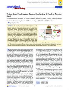
Background: The PEAK PlasmaBlade is a new electrosurgical device that uses pulsed radiofrequency to generate a plasma-mediated discharge along the exposed rim of an insulated blade, creating an effective cutting edge while the blade stays near body temperature. Methods: Full-thickness incisions were made on the dorsums of pigs with the PlasmaBlade, a conventional electrosurgical device, and a scalpel, and blood loss was quantified. Wounds were harvested at designated time points, tested for wound tensile strength, and examined histologically for scar formation and tissue damage. Results: Bleeding was reduced significantly (59 percent) in PlasmaBlade incisions compared with scalpel incisions, and acute thermal damage from the PlasmaBlade (66 ⫾ 5 m) was significantly less than both cut and coagulation mode electrosurgical incisions (456 ⫾ 35 m and 615 ⫾ 22 m, respectively). Histologic scoring for injury and wound strength was equivalent between the PlasmaBlade and scalpel incisions. By 6 weeks, the healed PlasmaBlade and scalpel incisions were approximately three times stronger, and scar cosmetic appearance was significantly better compared with electrosurgical incisions. Conclusions: The PlasmaBlade is a promising new surgical instrument that provides atraumatic, scalpel-like cutting precision and electrosurgical-like hemostasis, resulting in minimal bleeding, tissue injury, and scar formation. (Plast. Reconstr. Surg. 124: 1849, 2009.)
T
he most widely used cutting instrument in surgery is the scalpel; however, scalpel incisions are prone to bleeding that obscures the operative field. Electrosurgical cutting tools date to the 1920s and are now used in more than 17.5 million procedures per year in the United States alone.1 Conventional electrosurgery uses continuous-waveform radiofrequency energy delivered by means of an electrode to “incise” tissue by thermal ablation, which also produces simultaneous hemostasis.2,3 Although this hemostatic capability represents a major advance, there are a number of disadvantages associated with electrosurgery, including lack of surgical precision, thermal injury
to adjacent tissues such as nerves and blood vessels, and delayed wound healing.4 –12 Consequently, a number of new electrosurgical technologies have been introduced, such as insulated cutting electrodes, ultrasonic blades, and feedback-controlled radiofrequency generators.5–9,12 Some of these developments have demonstrated incremental improvements in reducing the thermal damage while preserving hemostatic ability. However, substantial room for improvement remains in electrosurgical technology that can approach the surgical precision and favorable wound-healing
From the Department of Surgery, Division of Plastic Surgery, and the Department of Ophthalmology and Hansen Experimental Physics Laboratory, Stanford University, PEAK Surgical, Inc., and the Department of Pathology and Laboratory Medicine, University of California, San Francisco. Received for publication February 17, 2009; accepted June 9, 2009. Copyright ©2009 by the American Society of Plastic Surgeons DOI: 10.1097/PRS.0b013e3181bcee87
Disclosure: Geoffrey C. Gurtner received a research grant to support study costs provided by PEAK Surgical, Inc.; Shang A. Loh and Edward I. Chang received education grants provided by PEAK Surgical, Inc.; Grace A. Carlson is Vice President of Clinical and Regulatory Affairs for PEAK Surgical, Inc.; and Daniel Palanker is a shareholder and consultant for PEAK Surgical, Inc.
www.PRSJournal.com
1849
Plastic and Reconstructive Surgery • December 2009 characteristics of the scalpel while also providing hemostasis. Whereas conventional electrosurgical devices cut with continuous waveforms, the pulsed electron avalanche knife13 operates with cyclic bursts lasting from 10 to 100 msec. To allow for efficient cooling, the burst repetition rate does not exceed 1 kHz and thus a typical duty cycle (fraction of time the energy is delivered) does not exceed 5 percent. The radiofrequency pulses induce a plasma-mediated discharge along the exposed edge of a thin (10 to 50 m), flat, 99.5 percent insulated electrode.14,15 This plasma rim provides a cutting edge for precise tissue dissection with simultaneous hemostasis. Because of the reduced duty cycle and the small exposed area of the electrode, pulsed electron avalanche knife technology yields a much lower average power output and uses less total energy than conventional electrosurgery to achieve an equivalent rate of tissue cutting, thus reducing collateral thermal damage.14,15 Although pulsed electron avalanche knife technology has been evaluated previously on ophthalmic tissues,13,16 –21 its effects on cutaneous wound healing remain unknown. In the present study, we hypothesized that the unique features of the PlasmaBlade (PEAK Surgical, Inc., Palo Alto, Calif.) would have a superior wound-healing profile compared with standard electrosurgery. Thus, we investigated the healing profile of incisions created by a traditional scalpel, conventional electrosurgery, and the PlasmaBlade using an established porcine skin model.12 We examined objective parameters including blood loss, wound tensile strength, histologic coagulation necrosis, and inflammation. In addition, we evaluated instrument operating temperature, wound cosmesis, and scarring.
MATERIALS AND METHODS Infrared Analysis of the Instrument Operating Temperatures Infrared temperature analysis was performed by Elastic Design, LLC (Redwood City, Calif.). Images were captured using a Thermavision SC600 camera (FLIR, Wilsonville, Ore.) in the mid-infrared (3 to 5 m) spectral range. The rate of motion during imaging was approximately 0.5 to 1 cm/second, similar to the cutting speed during skin surgery. Images were processed using MATLAB R2008 software (The MathWorks, Natick, Mass.). Surgical Procedure The study was conducted on six healthy Yucatan swine weighing 30 to 40 kg at study onset. The
1850
study protocol was approved by an institutional animal care and use committee and conducted in accordance with the Animal Welfare Act and the National Institutes of Health Guide for the Care and Use of Laboratory Animals. The animals were acclimated for 72 hours and fed a standard diet once daily. Anesthesia was induced using ketamine (15 mg/kg) (MWI Veterinary Supply, Meridian, Idaho) and atropine (0.04 mg/kg) (MWI Veterinary Supply), and Telazol (6 mg/kg) (MWI Veterinary Supply) for the final wounding before the animals were killed. After endotracheal intubation, anesthesia was maintained with inhaled isoflurane, and animals were prepared and draped in standard sterile fashion for each of the five surgical procedures on days 0, 21, 28, 35, and 42. The surgical procedure at each time point consisted of one incision made with a scalpel, the PlasmaBlade, and electrosurgical cut and coagulation modes. Each set of incisions was considered to be an independent study point. The timing of the surgical procedures allowed for wound-healing data points at 1, 2, 3, and 6 weeks as the animals were killed immediately after the surgical procedure on day 42 (i.e., incisions made on day 0 harvested at day 42 represent the 6-week time point). Incisional Wound Model Full-thickness skin incisions 3 cm in length were made on the dorsum of each animal. The medialmost incision was placed 3 cm from the dorsal spinal processes, with each subsequent incision placed 2 cm lateral to the prior incision, all in a parallel orientation. Differences in skin thickness were controlled for by rotating the instrument cutting order. Incisions were made with the PlasmaBlade using the PULSAR Generator (PEAK Surgical) on cut setting 3 (6 W), a no. 10 scalpel blade (Bard-Parker, Franklin Lakes, N.J.), and the Valleylab Electrosurgical Pencil using a Force 2 Generator (Valleylab, Boulder, Col.) on cut (40 W, Blend 2) and coagulation (40 W, Spray) modes. Extensive pilot studies were performed to determine the optimal power setting for the electrosurgery unit. Multiple power settings were used, including 20, 50, and 75 W. Interestingly, all three power settings had similar widths of thermal damage (data not shown). On the coagulation setting, the lower power setting led to increased thermal injury because of prolonged contact of the hot tip with the tissue (data not shown). The preliminary studies indicated that, given similar widths of thermal damage, a setting of 40 W was sufficient to
Volume 124, Number 6 • PEAK PlasmaBlade incise the thick porcine skin while creating a cutting environment and feel most similar to a real clinical setting. The PlasmaBlade settings were selected to provide an equivalent cutting rate of the skin. All incisions were made in a single stroke, with repetitive strokes made only if necessary to ensure a full-thickness wound. All wounds were closed with 3-0 nylon suture and covered with antibiotic ointment (MWI Veterinary Supply). Preoperative and postoperative antibiotics (Baytril, 5 mg/kg administered intramuscularly; MWI Veterinary Supply) were administered for 7 days. All wounds were inspected daily and sutures were removed on day 14. Blood Loss Evaluation Blood loss evaluation was performed on three animals (n ⫽ 15 wounds per device). A 110-mmdiameter filter paper (Schleicher & Schuell, West Chester, Pa.) was placed immediately over each incision for 60 seconds and then scanned (HP Scanjet; Hewlett Packard, Mountain View, Calf.) into JPEG digital image format. The digitized area of blood staining was quantified (in pixels) using ImageJ software (from vsbweb.nih.gov/ij/). Scar Formation Digital photographs of the wounds were taken immediately postoperatively and every 7 days thereafter. Scar width was measured using the ImageJ software program. Wound Strength After the procedure on day 42, animals were killed and all wound sites were harvested immediately to maintain all wounds in an equal postmortem state for objective comparison. A 2 ⫻ 1-cm, full-thickness strip of tissue with the incision at the midpoint was excised using a steel template. Wound burst strength was measured in poundfeet per inch as described previously7 using a Chatillon TCD200 digital force tester (Ametek, Largo, Fla.). Briefly, the incision line was aligned within the jaws of the clamp, and progressive force at an extension rate of 2 inches per minute was applied until wound disruption. Histologic Examination A 1 ⫻ 0.5-cm specimen containing the incision was excised and fixed in 10% buffered formalin (VWR, West Chester, Pa.) for 24 hours and embedded in paraffin. Representative 4-m sections were stained with hematoxylin and eosin and Masson trichrome stain. All specimens were coded
and evaluated by light microscopy (Olympus BX 40 microscope with a DP70 digital camera; Olympus, Center Valley, Pa.) by a single pathologist in a blinded manner. Acute thermal injury was determined as a maximum width of the zone of coagulation necrosis using hematoxylin and eosin sections. Immunohistochemistry All sections were examined for T lymphocytes (CD3 M7254; Dako, Carpinteria, Calif.), macrophages (CD68 NCL-L-CD68, Novacostra, Newcastle, United Kingdom), and myofibroblasts [␣-smooth muscle actin (SMA M0851; Dako)]. Sections were examined under high-power magnification (40⫻) and the number of cells counted by a blinded observer. Statistical Analysis All data are reported as mean ⫾ SEM. Data were compared using the t test. A value of p ⬍ 0.05 was considered significant. No correction was made for multiple testing.
RESULTS Instrument Operating Temperatures The operating temperature of each instrument was measured by an infrared camera, with power settings consistent with those used during the surgical procedures performed in this study. With the tissue at room temperature (25°C), the PlasmaBlade exhibited an average operating temperature of 45°C. In contrast, the average operating temperature of the electrosurgical instrument was 241°C in cut mode and 180°C in coagulation mode (Fig. 1). Blood Loss Evaluation The filter paper– based bleeding analysis demonstrated an average relative area units (relative area unit ⫽ 105 pixels) measurement of 1.03 ⫾ 0.27 for the PlasmaBlade versus 2.50 ⫾ 0.32 for the scalpel (p ⫽ 0.002) (Fig. 2), representing a 59 percent reduction in bleeding. Electrosurgical cut and coagulation also demonstrated reduced bleeding compared with scalpel incisions (0.52 ⫾ 0.33 and 0.29 ⫾ 0.29 relative area unit, respectively; p ⫽ 0.002) (Fig. 2); however, there was no statistically significant difference compared with the PlasmaBlade (p ⫽ 0.23 and p ⫽ 0.07, respectively). This demonstrates that the PlasmaBlade significantly reduces bleeding compared with the scalpel, and is comparable to traditional electrosurgical devices.
1851
Plastic and Reconstructive Surgery • December 2009
Fig. 1. Temperature maps of the PlasmaBlade and electrosurgical device in the cut mode, captured by the infrared camera. The PlasmaBlade average temperature was 45°C, whereas that of electrosurgery was 241ºC.
Fig. 2. Bleeding from incisions produced by the scalpel, PlasmaBlade (PB), and electrosurgical (ES) device in cut and coagulation modes, over 1 minute, measured as an area of blood staining the filter paper.
Zone of Coagulation Necrosis The zone of thermal coagulation necrosis in PlasmaBlade incisions (66 ⫾ 5 m) was significantly narrower than in electrosurgical cut or coagulation incisions (456 ⫾ 35 m and 615 ⫾ 22 m, respectively; p ⬍ 0.0001) (Figs. 3 and 4, a). This observation suggests that the PlasmaBlade may be used in closer proximity to adjacent fragile structures, such as nerves and blood vessels, with less risk of collateral injury. Inflammatory Markers At 1 week after surgery, T-cell counts based on CD3⫹ staining were increased in all samples (Fig. 5). By week 3, the PlasmaBlade incisions contained 46 percent fewer T cells than scalpel incisions (9.7 ⫾ 3.7 cells/high-power field versus 18.0
1852
⫾ 3.7 cells/high-power field; p ⬍ 0.04), 59 percent fewer T cells than in the electrosurgical cut incisions, and 56 percent fewer T cells than in the electrosurgical coagulation incisions (23.7 cells/ high-power field and 22.3 cells/high-power field, respectively; p ⬍ 0.05). At 6 weeks, the incisions created by the PlasmaBlade contained 52 percent fewer T cells than scalpel incisions (4.7 ⫾ 1.7 cells/high-power field versus 9.7 ⫾ 1.2 cells/highpower field; p ⬍ 0.04) (Fig. 5) and 66 percent and 72 percent fewer T cells than the electrosurgical cut and coagulation incisions (13.7 ⫾ 4.4 cells/ high-power field and 17.1 ⫾ 3.2 cells/high-power field, respectively; p ⬍ 0.03). This suggests the PlasmaBlade causes less inflammation compared with all other modalities. Macrophages identified as CD68⫹ cells were highest at 1 week (Fig. 6), with the lowest number seen in PlasmaBlade incisions, which contained 38 percent fewer macrophages than scalpel incisions, 41 percent fewer macrophages than electrosurgical cut incisions, and 32 percent fewer macrophages than electrosurgical coagulation incisions (19.3 ⫾ 5.5 cells/high-power field versus 31.3 ⫾ 5.7, 32.7 ⫾ 7.3, and 28.5 ⫾ 7.3 cells/high-power field, respectively; p ⬍ 0.05). By week 2, the electrosurgical cut incisions had approximately 50 percent more macrophages compared with all other modalities (32.7 ⫾ 7.3 cells/high-power field versus 16.5 ⫾ 6.9, 13.7 ⫾ 6.6, and 16.3 ⫾ 6.6 cells/highpower field for PlasmaBlade, scalpel, and electrosurgical coagulation, respectively; p ⬍ 0.05). However, at week 6, the electrosurgical coagulation incisions had significantly more macrophages than the other modalities (12.5 ⫾ 5.8 cells/ high-power field versus 1.2 ⫾ 0.4, 2.8 ⫾ 2.3, and 1.0 ⫾ 1.0 cells/high-power field for Plasma-
Volume 124, Number 6 • PEAK PlasmaBlade
Fig. 3. Width of the acute thermal injury zone created by the PlasmaBlade (66 ⫾ 5 m), electrosurgical cut mode (456 ⫾ 35 m), and electrosurgical coagulation mode (615 ⫾ 22 m).
Blade, scalpel, and electrosurgical cut, respectively; p ⬍ 0.05). These data further suggest that during the first and second weeks of wound healing, the PlasmaBlade induces significantly less inflammation than the electrosurgical device and possibly even the scalpel. Although the inflammatory response appeared to have stabilized among all modalities by week 3, a higher prevalence of macrophages remained up to 6 weeks in electrosurgical coagulation wounds, indicating a persistent, ongoing inflammatory response likely attributable to the presence of necrotic debris in the wound site. Scalpel incisions showed the lowest levels of smooth muscle actin–positive myofibroblasts throughout the 6-week time period (Fig. 7). The PlasmaBlade and scalpel incisions showed a similar prevalence of myofibroblasts at the 1-, 3-, and 6-week time points, with no statistically significant difference at any time point (19.3 ⫾ 10.6 versus 18.5 ⫾ 10.6 cells/high-power field at 1 week, 9.7 ⫾ 1.5 versus 5.3 ⫾ 2.0 cells/high-power field at 3 weeks, and 1.7 ⫾ 0.3 versus 1.3 ⫾ 0.7 cells/high-power field at 6 weeks for PlasmaBlade and scalpel, respectively; p ⬎ 0.15). However, both electrosurgical modes induced significantly more myofibroblasts throughout the entire time period, with the greatest numbers seen at 3 weeks after surgery, when PlasmaBlade incisions had only a fraction of myofibroblasts compared with electrosurgical cut (43 percent; 9.7 ⫾ 1.5 versus 22.7 ⫾ 12.0 cells/high-power field; p ⬍ 0.001) and electrosurgical coagulation (29 percent; 9.7 ⫾ 1.5 versus 38.3 ⫾ 1.7 cells/high-power field; p ⬍ 0.001) incisions, respectively. By week 6, the myofibroblast cell numbers normalized except for electrosurgical coagulation, although the difference was not statis-
Fig. 4. Acute histology of the skin incisions produced by (above) scalpel, (center) PlasmaBlade, and (below) electrosurgical cut modes. Arrows indicate the zone of coagulation necrosis. Scale bar ⫽ 2.0 mm.
tically significant. The increased presence of myofibroblasts in the electrosurgical incisions offers a potential explanation for the poor cosmesis seen in healed electrosurgical wounds. Scar Formation Measurement of the scar width at each time point showed no significant difference between
1853
Plastic and Reconstructive Surgery • December 2009
Fig. 5. Number of CD3⫹ lymphocytes in the wounds produced by the four instruments at 1, 2, 3, and 6 weeks after surgery. PlasmaBlade incisions exhibited the least amount of inflammation. PB, PlasmaBlade; ES, electrosurgical; Coag, coagulation.
Fig. 6. Number of macrophages (CD68⫹ cells) in the wounds at 1, 2, 3, and 6 weeks after surgery. The significant presence of macrophages in the electrosurgical coagulation wounds at 6 weeks suggests continued inflammation. PB, PlasmaBlade; ES, electrosurgical; Coag, coagulation.
PlasmaBlade and scalpel incisions (p ⫽ 0.25 to 0.84) (Figs. 8 –10, a), with both much narrower than the electrosurgical scars. At 1 week, the PlasmaBlade scar width was 24 percent and 22 percent the width of the electrosurgical scars (0.85 ⫾ 0.17 mm versus 3.50 ⫾ 0.76 mm for electrosurgical cut and 3.77 ⫾ 0.39 mm for electrosurgical coagulation; p ⫽ 0.0021 and p ⬍ 0.0001, respectively). Similarly, at 2 weeks, the PlasmaBlade scar width
1854
was 44 percent and 32 percent the width of the electrosurgical scars (1.02 ⫾ 0.30 mm versus 2.33 ⫾ 0.33 mm for cut and 3.17 ⫾ 0.41 mm for coagulation; p ⫽ 0.03 and p ⫽ 0.0017, respectively). This trend continued at 3 weeks, when the PlasmaBlade scar width was 27 percent and 18 percent the width of the electrosurgical scars (0.59 ⫾ 0.10 mm versus 2.17 ⫾ 0.4 mm for cut and 3.27 ⫾ 0.21 mm for coagulation; p ⫽ 0.0018 and p ⬍ 0.0001,
Volume 124, Number 6 • PEAK PlasmaBlade
Fig. 7. Number of myofibroblasts (smooth muscle actin–positive cells) in the wounds at 1, 2, 3, and 6 weeks after surgery. PB, PlasmaBlade; ES, electrosurgical; Coag, coagulation.
respectively). Finally, at 6 weeks, the PlasmaBlade scar width was 25 percent and 16 percent the width of the electrosurgical scars (0.38 ⫾ 0.06 mm versus 1.50 ⫾ 0.29 mm for cut and 2.33 ⫾ 0.20 mm for coagulation; p ⫽ 0.001 and p ⬍ 0.0001, respectively) (Figs. 8 –10, b). Visual evaluation of electrosurgical incisions demonstrated heavy scarring and poor cosmesis, whereas PlasmaBlade and scal-
pel incisions healed with minimal scarring and an improved aesthetic outcome (Figs. 8 –10, c). Wound Burst Strength Evaluation Scalpel and PlasmaBlade incisions exhibited greater tensile strength compared with electrosurgical incisions at all time points (Fig. 11),
Fig. 8. Histologic measurement of wound scar width. At any time point, there was no statistically significant difference between the PlasmaBlade and scalpel wounds, whereas both were much smaller than electrosurgical cut and electrosurgical coagulation wounds. PB, PlasmaBlade; ES, electrosurgical; Coag, coagulation.
1855
Plastic and Reconstructive Surgery • December 2009
Fig. 9. Histology of the wounds at 1, 2, and 6 weeks after incision. Scalpel and PlasmaBlade wounds appear healed with normal tissue morphology. In contrast, the electrosurgical cut wounds are wider and contain significantly more scar tissue.
although it was not until week 3 that statistical significance was achieved compared with the electrosurgical coagulation incisions (76.9 ⫾ 14.9 lb-ft/in for PlasmaBlade versus 20.1 ⫾ 2.9 lb-ft/in for electrosurgical coagulation; p ⫽ 0.006) and approached compared with electrosurgical cut incisions (32.6 ⫾ 1.6 lb-ft/in; p ⫽ 0.07). PlasmaBlade incisions demonstrated similar burst strength compared with scalpel incisions at 6 weeks (154.4 ⫾ 12.3 lb-ft/in versus 165.5 ⫾ 13.5 lb-ft/in; p ⫽ 0.56). In contrast, both electrosurgical modes were markedly weaker, withstanding only 59.6 ⫾ 20.9 lb-ft/in (cut) and 55.5 ⫾ 8.8 lb-ft/in (coagulation) at 6 weeks (p ⫽ 0.005 and p ⫽ 0.0002, respectively). This further supports our conclusion that the PlasmaBlade offers superior wound-healing and strength characteristics compared with conventional electrosurgical incisions.
1856
DISCUSSION Traditionally, the scalpel has been considered the surgical cutting tool of choice because of its precision, control, preservation of tissue integrity, and superior associated wound healing. However, its primary disadvantage is bleeding; consequently, numerous electrosurgical devices have been developed to provide hemostasis. Although hemostasis is improved, electrosurgical devices suffer from thermal damage to surrounding tissues, inferior wound healing, and poor cosmesis,4,5,7,9,10,12 which limit their realm of applications. In this study, we evaluated the PlasmaBlade, a new electrosurgical device that produces pulsed plasma–mediated discharges at the edge of an insulated electrode to allow precise dissection of tissue.14 Our current study corroborates previous
Volume 124, Number 6 • PEAK PlasmaBlade
Fig. 10. Clinical appearance of the wounds 6 weeks after surgery. Scalpel (above) and PlasmaBlade (center) wounds are very similar, unlike the much wider wounds of the electrosurgical cut mode (below), which exhibited profound scarring.
findings demonstrating precise tissue dissection in rabbit, bovine, and human retinas and lens capsules.13,16 –21 This is the first study to examine pulsed plasma–mediated electrosurgical technology on cutaneous wound healing. The PlasmaBlade exhibited a 59 percent reduction in bleeding compared with scalpel wounds, which was comparable to bleeding seen with conventional electrosurgical tools. Given the PlasmaBlade’s lower blade temperature and reduced thermal damage to adjacent tissues, this result suggests that the high operating temperatures and deep thermal coagulum associated with electrosurgery are not always essential for hemostasis. One possible mechanism of the hemostasis produced by the PlasmaBlade may be a nonthermal vasoconstrictive and thrombotic effect22; however, further study of this phenomenon is necessary. Our observation of the greater infiltration by CD3⫹ T lymphocytes during the course of healing in the electrosurgical group corroborates previous reports of abnormal inflammatory response correlating with reduced wound burst strength.4,5,7,10,12 The burst strength of the incisions created by the PlasmaBlade, however, were
similar to those of the scalpel and three times stronger than electrosurgical incisions. Such dramatic differences are most likely attributable to the deeper zone of thermal necrosis with electrosurgery. This results in a protracted inflammatory response necessary to clear greater amounts of necrotic tissue that interferes with reepithelialization and collagen formation. Interestingly, the levels of T lymphocytes and macrophages were lower in the PlasmaBlade group than in the scalpel group. The reason for this is unclear, but perhaps the reduction in bleeding and applied pressure during cutting by the PlasmaBlade further limits the level of inflammation present. Future studies will be needed to more fully explain these observations.23–25 Reduced thermal injury and improved wound histology and strength correlated strongly with the visual appearance of the scars. This is again likely attributable to reduced levels of inflammation, leading to more efficient epithelialization, reduced macrophage cytokine production, decreased myofibroblast proliferation, reduced transforming growth factor- production, and ultimately improved scarring and comesis.26 –29 Visual assessment of the scars at 6 weeks showed dramatically less scarring with PlasmaBlade and scalpel incisions compared with the hypertrophic scarring observed in the electrosurgical group. We are not aware of other reports comparing scarring caused by electrosurgery, scalpels, or other surgical technologies, but we believe it is an important parameter for patient satisfaction, especially in the field of aesthetic and reconstructive surgery. In addition, a number of anecdotal observations suggest additional benefits of the PlasmaBlade. We noted that incisions made with the PlasmaBlade required much less pressure than traditional scalpels; thus, it was not necessary to maintain constant tension on the skin when making the incision. This may reduce chances of slippage, tearing, and unintentional extension of incisions.21 We also noted that the PlasmaBlade was less adherent to the wound edges, likely because of lower operating temperatures. Furthermore, the PlasmaBlade maintained its cutting effectiveness and hemostatic ability even when submerged in liquefied adipose tissue or blood, unlike electrosurgical cutting tools. Finally, the PlasmaBlade was noted to produce little to no surgical smoke, which is noxious, obscures the visual field, and has been shown to contain infectious viruses.30
1857
Plastic and Reconstructive Surgery • December 2009
Fig. 11. Burst strength of the wounds produced by the four instruments at 1, 2, 3, and 6 weeks after surgery. At all time points, there was no statistically significant difference between the PlasmaBlade and scalpel, whereas the electrosurgical wounds were much weaker. At 6 weeks, the PlasmaBlade wounds were approximately three times stronger than those made by electrosurgery. PB, PlasmaBlade; ES, electrosurgical; Coag, coagulation.
CONCLUSIONS In summary, we demonstrate that the PlasmaBlade provides efficient cutting of skin, with a superior wound-healing profile, comparable to that of the traditional scalpel but with significantly less bleeding. PlasmaBlade incisions heal in a manner similar to that of scalpel incisions but superior to that of electrosurgical incisions with respect to inflammation, wound strength, and scarring. These results suggest that the PlasmaBlade has tremendous potential in surgical fields where electrosurgery is not used extensively, including plastic and reconstructive surgery; cardiothoracic, gynecologic, vascular, and laparoscopic surgery; and neurosurgery. Geoffrey C. Gurtner, M.D. Department of Surgery Division of Plastic Surgery 257 Campus Drive, GK-201 Stanford, Calif. 94305-5148
[email protected]
ACKNOWLEDGMENTS
This study was funded by PEAK Surgical, Inc. The authors would like to thank Jeanne McAdara-Berkowitz, Ph.D., and Joshua Vose, M.D., for assistance in preparation of this article. REFERENCES 1. Life Science Intelligence. U.S. Markets for Electrosurgical and Thermal Ablation Products. Irvine, Calif.: Medtech Insight; 2006.
1858
2. Brown DB. Concepts, considerations, and concerns on the cutting edge of radiofrequency ablation. J Vasc Interv Radiol. 2005;16:597–613. 3. Massarweh NN, Cosgriff N, Slakey DP. Electrosurgery: History, principles, and current and future uses. J Am Coll Surg. 2006;202:520–530. 4. Arashiro DS, Rapley JW, Cobb CM, Killoy WJ. Histologic evaluation of porcine skin incisions produced by CO2 laser, electrosurgery, and scalpel. Int J Periodontics Restorative Dent. 1996;16:479–491. 5. Butler PE, Barry-Walsh C, Curren B, Grace PA, Leader M, Bouchier-Hayes D. Improved wound healing with a modified electrosurgical electrode. Br J Plast Surg. 1991;44:495–499. 6. Glover JL, Bendick PJ, Link WJ, Plunkett RJ. The plasma scalpel: A new thermal knife. Lasers Surg Med. 1982;2:101–106. 7. Hambley R, Hebda PA, Abell E, Cohen BA, Jegasothy BV. Wound healing of skin incisions produced by ultrasonically vibrating knife, scalpel, electrosurgery, and carbon dioxide laser. J Dermatol Surg Oncol. 1988;14:1213–1217. 8. Keenan KM, Rodeheaver GT, Kenney JG, Edlich RF. Surgical cautery revisited. Am J Surg. 1984;147:818–821. 9. Millay DJ, Cook TA, Brummett RE, Nelson EL, O’Neill PL. Wound healing and the Shaw scalpel. Arch Otolaryngol Head Neck Surg. 1987;113:282–285. 10. Pollinger HS, Mostafa G, Harold KL, Austin CE, Kercher KW, Matthews BD. Comparison of wound-healing characteristics with feedback circuit electrosurgical generators in a porcine model. Am Surg. 2003;69:1054–1060. 11. Tipton WW, Garrick JG, Riggins RS. Healing of electrosurgical and scalpel wounds in rabbits. J Bone Joint Surg (Am.) 1975;57:377–379. 12. Vore SJ, Wooden WA, Bradfield JF, et al. Comparative healing of surgical incisions created by a standard “bovie,” the Utah Medical Epitome Electrode, and a Bard-Parker cold scalpel blade in a porcine model: A pilot study. Ann Plast Surg. 2002;49:635–645.
Volume 124, Number 6 • PEAK PlasmaBlade 13. Palanker DV, Miller JM, Marmor MF, Sanislo SR, Huie P, Blumenkranz MS. Pulsed electron avalanche knife (PEAK) for intraocular surgery. Invest Ophthalmol Vis Sci. 2001;42: 2673–2678. 14. Palanker DV, Vankov A, Huie P. Electrosurgery with cellular precision. IEEE Trans Biomed Eng. 2008;55:838–841. 15. Palanker D, Vankov A, Jayaraman P. On mechanisms of interaction in electrosurgery. N J Phys. 2008;10:123022. 16. Miller JM, Palanker DV, Vankov A, Marmor MF, Blumenkranz MS. Precision and safety of the pulsed electron avalanche knife in vitreoretinal surgery. Arch Ophthalmol. 2003; 121:871–877. 17. Palanker DV, Marmor MF, Branco A, et al. Effects of the pulsed electron avalanche knife on retinal tissue. Arch Ophthalmol. 2002;120:636–640. 18. Priglinger SG, Haritoglou C, Mueller A, et al. Pulsed electron avalanche knife in vitreoretinal surgery. Retina 2005;25:889–896. 19. Priglinger SG, Haritoglou C, Palanker D, et al. Pulsed electron avalanche knife for capsulotomy in congenital and mature cataract. J Cataract Refract Surg. 2006;32:1085–1088. 20. Priglinger SG, Haritoglou C, Palanker DV, Alge CS, Gandorfer A, Kampik A. Pulsed electron avalanche knife (PEAKfc) for dissection of retinal tissue. Arch Ophthalmol. 2005;123: 1412–1418. 21. Priglinger SG, Palanker D, Alge CS, et al. Pulsed electron avalanche knife: New technology for cataract surgery. Br J Ophthalmol. 2007;91:949–954.
22. Palanker D, Vankov A, Freyvert Y, Huie P. Pulsed electrical stimulation for control of vasculature: Temporary vasoconstriction and permanent thrombosis. Bioelectromagnetics 2008; 29:100–107. 23. Haroon ZA, Raleigh JA, Greenberg CS, Dewhirst MW. Early wound healing exhibits cytokine surge without evidence of hypoxia. Ann Surg. 2000;231:137–147. 24. Goodson WH III, Hunt TK. Wound healing and aging. J Invest Dermatol. 1979;73:88–91. 25. Eming SA, Krieg T, Davidson JM. Inflammation in wound repair: Molecular and cellular mechanisms. J Invest Dermatol. 2007;127:514–525. 26. Bettinger DA, Yager DR, Diegelmann RF, Cohen IK. The effect of TGF-beta on keloid fibroblast proliferation and collagen synthesis. Plast Reconstr Surg. 1996;98:827–833. 27. Border WA, Noble NA. Transforming growth factor beta in tissue fibrosis. N Engl J Med. 1994;331:1286–1292. 28. Goumenos DS, Tsamandas AC, Oldroyd S, et al. Transforming growth factor-beta(1) and myofibroblasts: A potential pathway towards renal scarring in human glomerular disease. Nephron 2001;87:240–248. 29. Martin P. Wound healing-aiming for perfect skin regeneration. Science 1997;276:75–81. 30. Barrett WL, Garber SM. Surgical smoke: A review of the literature. Is this just a lot of hot air? Surg Endosc. 2003;17: 979–987.
Instructions for Authors: Key Guidelines Manuscript Length/Number of Figures To enhance quality and readability and to be more competitve with other leading scientific journals, all manuscripts must now conform to the new word-count standards for article length and limited number of figure pieces: • Original Articles and Special Topics/Comprehensive Reviews are limited to 3000 words and 20 figure pieces. • Case Reports, Ideas & Innovations, and Follow-Up Clinics are limited to 1000 words and 4 figure pieces. • Letters and Viewpoints are limited to 500 words, 2 figure pieces, and 5 references.
1859