Original article
The hypothesis of patho-physiological correlation between chronic cerebrospinal venous insufficiency and multiple sclerosis: rationale of treatment M Lugli, M Morelli, S Guerzoni and O Maleti Department of Cardiothoracic and Vascular Surgery, Hesperia Hospital, Via Arqua`, 80, 41100 Modena, Italy
Abstract Background: The possible role of the venous system in the pathogenesis of chronic neurodegenerative diseases has been hypothesized for decades. Quite recently, the description of a venous condition defined as chronic cerebrospinal venous insufficiency (CCSVI) and its strong association with multiple sclerosis (MS) has brought back the attention of the scientific community to the hypothesis of an aetiological or concomitant role of an altered venous function in the occurrence of this pathology. CCSVI is identified by sonographic criteria, thus the indication for its possible treatment is based on ultrasound findings. Method: We retrospectively examined 167 consecutive patients affected by clinically defined MS and CCSVI, identified by ultrasound assessment by the presence of at least two sonographic criteria. Ultrasonographic diagnosis of CCSVI was then integrated by venography and intravascular ultrasound examination (in 43 patients). Patients were all submitted to endovascular procedure (venoplasty). Results: In 37% of cases there was no correspondence between the preoperative ultrasound assessment and the venographic findings. In the event of incongruity between venography and sonography, the intravascular ultrasound examination investigation, when performed, confirmed ultrasound findings in 42% of cases and venography results in 58%. At one month in 12% of cases ultrasound assessment showed the persistence of altered flux. In 67% of cases patients reported subjective amelioration, regarding non-specific symptoms. Conclusion: The pathophysiology of CCSVI is yet to be defined. The superior cava venous system is highly complex in terms of anatomy and possible anomalies, as well as its haemodynamic mechanisms. Further studies are required to define the parameters of diagnosis and treatment of CCSVI. Keywords: chronic cerebro-spinal venous insufficiency; CCSVI; multiple sclerosis; IVUS; jugular vein; azygos vein; venoplasty
Introduction Multiple sclerosis (MS) is a condition of unknown aetiology, generally considered to be an autoimmune disease, characterized by an inflammatory Correspondence: Marzia Lugli MD, Department of Cardiothoracic and Vascular Surgery, Hesperia Hospital, Via Arqua`, 80, 41100 Modena, Italy. Email:
[email protected] Accepted 12 January 2012
component, a neurodegenerative evolution and demyelination. From a histopathological viewpoint, MS lesions are heterogeneous, and their diversity may be attributed to the variable incidence of different personal and environmental factors. In fact, no isolated factor has been singled out as being causal. Currently, the most accredited risk factors associated with increased probability of developing MS are the presence of a specific HLA type, vitamin D/latitude, Epstein–Barr virus immune response and smoking.1
Phlebology 2012;27 Suppl 1:178–186. DOI: 10.1258/phleb.2012.012S24
M Lugli et al. The hypothesis of patho-physiological correlation between CCSVI and MS
The possible influence of altered venous drainage in MS occurrence was described in the past century2 but revived interest in this hypothesis is quite recent.3 Opinion is widely divergent on the possible role of altered venous function in determining MS lesions; it ranges from considering such alterations as mere epiphenomena, to attributing to them an etiopathogenic role. The hypothesis that a pathological venous system may play a determining role in demyelinating neuropathies was first advanced in the 1970s by Aboulker et al.4 Patients affected by demyelinating diseases of unknown origin underwent venographic investigation and an association was identified between these diseases and an altered venous flux involving the superior cava system, both at the level of the jugular veins and at the interconnection with the inferior cava system (azygos and hemiazygos vein system). Interestingly, other lesions were documented in these patients, such as left iliac vein obstruction (May – Thurner syndrome) and left renal vein compression (nutcracker syndrome). Moreover, 20 patients were submitted to surgical procedures of the internal jugular vein (IJV) and good, albeit transitory, results were obtained. A decade later, an analogous correlation between a venous macropathology and MS was described by Schelling,5 who suggested a causal interpretation of the intraparenchymal venous alteration described by Putman2 and Fog.6 Despite the large number of descriptions and hypotheses, the concept of a possible venous role in neurological disorders and MS was not given adequate consideration until CCSVI was described.3 The possible correlation between venous abnormalities and MS is still the subject of considerable controversy.7,8 The attempt to correct the alteration of venous flow identified in MS patients by means of percutaneous angioplasty9 – 11 has proved to be safe, with low risk of complications; however, significant data on neurological outcome are as yet unavailable. Assessing results from the neurological perspective is extremely complex and requires careful evaluation by specialists, given the varied nature of the disease and the absence of specific symptoms. Encouraging results have been described, many of which appear, however, to be transitory. In all events, there is not yet sufficient evidence to enable us to establish whether the role played by venous alterations in MS is causal or concomitant.
Original article
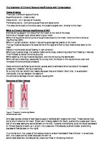
Pathophysiology The cerebral venous outflow system is particularly complex due to the large numbers of tributaries, and the marked calibre variations correlated to minimal variations in pressure. Moreover, to these should be added consistent variations related to postural conditions. Cerebral outflow from the skull occurs by means of two main systems: the jugular system and the vertebral system, both symmetrical and bilateral. These systems are subject to systemic and postural variation.12 It should also be noted that valve malfunction or malformation has been widely described for some time.13 Analogous vein alterations are detectable at lowerlimb level, where they are responsible for the occurrence of chronic venous insufficiency (CVI)14 (Figure 1). Given that histopathological lesions similar to those found in CVI have also been detected at cerebral and spinal system level, the hypothesis that a similar pathological process may occur at the superior cava level can be advanced.15 Similarly, the damage caused by an inefficient collateral pathway, which is able to determine further venous insufficiency in the lower limbs, may also cause damage to the cerebral and spinal system. The nervous system is known to be sensitive to all forms of stress, and particularly so to microcirculatory hypertension and to a volumetric increase in the circulatory system, which may exert extrinsic compression. As a consequence, nervous damage may derive either from a descending collateral pathway
Figure 1 Femoral vein valve. The valve leaflets are prolapsed determining valve incompetence
Phlebology 2012;27 Suppl 1:178–186
179
Original article
M Lugli et al. The hypothesis of patho-physiological correlation between CCSVI and MS
induced by jugular stenosis, or from an ascending one (May – Thurner syndrome, nutcraker syndrome, anomalies of the origin of the azygos vein). There are more doubts concerning the significance of cerebral reflux due to jugular valve incompetence, even if it seems to be correlated to neurological symptoms.5 It is not yet known whether obstruction or reflux is the more significant element, nor where respective responsibility lies in the case of their association. Nevertheless, reflux would appear to have a hierarchically inferior role.
Methods A series of 167 consecutive patients affected by clinically defined MS who underwent endovascular treatment for CCSVI, over the period February 2011 – June 2011, has been evaluated retrospectively. Ultrasound findings, venographic and intravascular ultrasound imaging have been compared.
Selection criteria Patient characteristics are provided in Table 1. Co-morbidities are shown in Table 2. Neurological assessment and cerebral magnetic resonance (MRI) no older than six months were required. Patients were selected for treatment on the basis of at least two positive CCSVI criteria present at ultrasound examination as per Zamboni et al.,3 and subsequent indications of the International Society for Neurovascular Disease (ISNVD).16 All patients provided their written consent to undergo the procedure.
Venography and venoplasty procedures Procedures were performed in an angiographic suite (Philips Allura XPER FD10). Patients were positioned supine, with a centralhole silicone pillow under their head in order to avoid neck rotation. Patient monitoring included conTable 1 Patient characteristics Patients ¼ 167 Age (years) Gender (M/F) Smoker or ex-smoker, n (%) Obesity, n (%) MS clinical course (RR/SP/PP), n (%)
46.5 (20/81) 72/95 76 (45.4) 38 (22.7) 79/62/26 (47.3/37.1/15.6)
RR, relapsing remitting; SP, secondary progressive; PP, primary progressive
180
Phlebology 2012;27 Suppl 1:178–186
Table 2 Patient co-morbidities Co-morbidity
n
%
Chronic renal insufficiency Diabetes Ischaemic heart disease Hypertension Thyroid disease Previous deep venous thrombosis Previus IJV ligation Presence of cava filter
4 12 5 27 17 12 1 1
2.4 7.2 3 16.7 10.2 7.2 0.6 0.6
IJV, internal jugular vein
tinuous electrocardiogram recording, pulse-oximetry, arterial pressure measurement in the arm (at 5-minute intervals) and breath-wave recording. All procedures were performed under local anaesthesia. The contrast dye used was Iodixanolum (Visipaque 300w, Amserham Health As, Oslo, Norway) and injections were performed mechanically with the ACISTTM injection system. Venographic access was the left common femoral vein in all cases. Ultrasound-guided catheterization with 5 Fr sheath (Terumow) was performed and replaced with a 9 Fr one. A hydrophilic wire (Radiofocus wire M angled 260 cm, Terumow) was inserted upwards into the right IJV, usually without the need of catheters to direct the tip. A straight angiographic catheter (Imager II 5 Fr, Boston Scientific, USA) was placed at the origin of the IJV and a first injection (20 mL – 10 mL/second) was performed to assess anatomy and possible activation of collateral pathways for drainage, intracranial or extracranial. The catheter was pulled back in the jugular bulb, below the confluence of the common facial vein and a second injection is performed (10 mL – 10 mL/second, ipsilateral oblique projection 30– 458, digital subtraction) to assess the junction anatomy, valve morphology and presence of anomalies. Venous pressure was recorded for at least three respiratory cycles and printed out. If required, selective catheterization of collateral veins was performed (e.g. external jugular vein, common facial vein, thyroid vein) to clarify anatomy and drainage patterns. The same procedure was extended to the left side. To engage the left IJV, a vertebral catheter was usually employed (Torque DX 5 Fr; AGA Medical Corp., USA). Before proceeding with venoplasty, systemic un-fractionated heparin was administered (depending on patient weight, routinely 5000 IU). A stiff wire (Amplatz Super StiffTM straight-tip short taper, Boston Scientific, USA) was put in place and intravascular ultrasound examination (IVUS) was performed. The scan was carried out by
M Lugli et al. The hypothesis of patho-physiological correlation between CCSVI and MS
means of a Volcano S5 imaging system equipped with a catheter Visionw PV 8.2. While at the beginning of our experience IVUS was applied only in situations requiring more information, nowadays it is routinely practised. The IVUS probe was inserted further up and ring-down calibration was performed, usually in the proximity of the right atrium. The probe was pushed into the IJV above the confluence of the common facial vein. The probe was slowly withdrawn and examination was recorded. Valve morphology was assessed and the jugular ostium was measured (longitudinal and transversal diameter): this is essential for sizing the angioplasty balloon precisely. The examination proceeds to the origin of the superior cava, on both sides. Inferior cava anatomy and the possible occurrence of iliac compression were also investigated.17 The choice of the balloon was based on 20% oversizing, preference being given to a high-pressure balloon (Atlas balloon 10– 16 mm × 40 mm, Bard Peripheral Vascular Inc., USA). In patients who present re-stenosis after previous venoplasty or who present small veins, smaller balloons and progressive diameters were utilized. After venoplasty, the IVUS scan was performed to record the post-dilation ostium diameter and to assess possible variations in valve morphology and function. Subsequently, the angiographic catheter was inserted into the IJV, the stiff wire was removed, and a control injection was performed (10 mL – 10 mL/second, ipsilateral oblique projection 30– 458, digital subtraction) to document the efficacy of the procedure and the absence of complications (dissection, rupture and thrombosis). Postprocedural venous pressure was recorded as described above. The use of stents in jugular veins was excluded a priori. The azygos vein study was then performed. Selective catheterization is normally performed using a headhunter catheter (Femoral-cerebral 5 Fr, Terumow). A hydrophilic wire was progressed up to L3 level, where the azygos vein usually is in continuity with the ascending right lumbar vein. The origin of the hemiazygos vein was identified. Selective catheterization of the left ascending lumbar vein may be required. Two or more injections (20 mL – 8 mL/second) were usually required to assess the azygos system anatomy. An oblique projection was required to assess arch and valve morphology, as well as adequate drainage. After the procedure is completed, the sheath was removed with manual compression. Ultrasound examination was performed to assess the absence
Original article
of haematoma, and eccentric compression was applied. Four hours after the conclusion of the procedure, a first low-molecular-weight heparin (LMWE) subcutaneous injection was administered, which continued daily for one month. Patients were discharged the day after the procedure. Ultrasound examination of the neck and groin was performed to check the possible occurrence of thrombosis and haematomas.
Follow-up Ultrasound follow-up was scheduled one month, six months and yearly after the procedure.
Results A total of 232 IJV venoplasties were performed. Only two azygos veins were treated. Procedural data are provided in Table 3. Complications are listed in Table 4. In one case a femoral cut-down was required to remove a broken balloon. The patient was discharged three days after the procedure without further complications. Jugular thrombosis was detected in three cases at the first ultrasound follow-up assessment; these patients were treated for re-stenosis, two of them presenting recanalized thrombosis at the cranial segment of the IJV and one having a permanent catheter in the homolateral subclavian vein. Concomitant occurrence of left iliac vein compression (May– Thurner syndrome) was detected Table 3 Procedural data Variable Procedures (n) Procedure duration (minutes) Contrast dye (mL) IVUS examination (n of patients) Treated vein (n) Right internal jugular vein Left internal jugular vein Azygos vein
167 66 (33/116) 170 (80/320) 43 234 97 135 2
IVUS, intravascular ultrasound examination
Table 4 Postoperative complications Complication
n
%
Open surgical balloon removal Vein thrombosis Transitory headache Contrast dye allergy
1 3 21 1
0.6 1.8 12.6 0.6
Phlebology 2012;27 Suppl 1:178–186
181
Original article
M Lugli et al. The hypothesis of patho-physiological correlation between CCSVI and MS
In 69% of cases, patients reported subjective clinical amelioration regarding non-specific symptoms such as headache, fatigue, rigidity and steadiness.
Discussion
Figure 2 Venographic image corresponding to nutcracker syndrome: abnormal collateral pathway efferent from left renal vein
in seven cases, confirmed by IVUS examination. Venographic images corresponding to nutcracker syndrome were recorded in one case, albeit asymptomatic (Figure 2). In 37% of cases there was no correspondence between preoperative ultrasound assessment and venographic findings, as described in Table 5. In the event of incongruity between venography and sonography, the IVUS investigation, when performed, confirmed ultrasound findings in 42% of cases and venography results in 58%, as detailed in Table 6 (Figures 3 and 4). At one month, in 12% of cases, ultrasound assessment showed the persistence of altered flow.
Table 5 Comparison between ultrasound/venography findings in the IJV of 167 patients Variable
n
%
IJV examined (n) Vein anomalies identified by ultrasound examination Vein anomalies identified by ultrasound examination and confirmed by venography Veins receiving a discordant definition (ultrasound false-positive) Vein normality identified by ultrasound examination
366 310
– 84.7
219
59.8
91
24.8
56
15.3
Vein normality identified by ultrasound examination and confirmed by venography Veins receiving a discordant definition (ultrasound false-negative)
13
3.5
43
11.9
Ultrasound anomalies were described according to CCSVI identifying criteria, as described above IJV, internal jugular vein
182
Phlebology 2012;27 Suppl 1:178–186
The intriguing hypothesis that many neurological diseases may trace their aetiopathogenic origins to a circulatory problem, and that this, given that it affects macrocirculation, may provide a point of correction, has stimulated research in various disciplines for over three decades. The correlation between MS and altered venous macrocirculatory function has received particular attention of late thanks to the non-invasive ultrasound-investigation protocols proposed by Zamboni et al.3 The macrocirculatory problems alluded to consist in structural, and hence also functional, alterations of the principal encephalic outflow pathways, which are able to exert a damaging effect not only by obstructing the flow but also by inducing a collateral pathway. The fact that a collateral pathway can exert a damaging effect at the level of the inferior cava system has been known for some time, and thus to exclude a priori that similar action can occur at other sites finds no rational justification. On the contrary, histopathological findings show that the physiopathological processes can be similar. In determining damage caused by haemodynamic action, multiple factors come into play, such that the same lesion may have various effects in different patients. This is due to pre-existing anatomical conditions, which can create an increase or decrease in the effect of an obstructive process, or reduce or intensify the damage caused by a compensatory collateral pathway. Thus, an azygos system of anomalous origin, a May– Thurner or nutcracker syndrome may well generate a variety of completely different haemodynamic scenarios, as demonstrated by Aboulker et al.4 If it seems difficult to classify clinical scenarios beyond single cases, establishing a causal nexus with a neuropathy is even more arduous. This is a crucial point, since the argument has moved from the hypothesis of causality, to a hypothesis of consequence (whereby the venopathy is thought to be a consequence of the disease), to a nexus of concausality, whereby the two elements may well aggravate each other in proportions, that cannot be defined. Not being able, on the basis of currently available observational studies, to distinguish between the hypotheses formulated above, it follows that any forecast regarding results of corrective treatment
M Lugli et al. The hypothesis of patho-physiological correlation between CCSVI and MS
Original article
Table 6 Findings of IVUS investigation in 18 IJV showing incongruity between venography and sonography N
Ultrasound findings
Venography
IVUS
1 2 3 4 5 6 7 8 9 10 11 12 13 14 15 16 17 18 Congruence IVUS/ultrasound Congruence IVUS/venography
IJV valve anomaly IJV reflux Normal IJV Normal IJV Normal IJV JJV stenosis JJV stenosis IJV reflux IJV reflux IJV valve anomaly IJV valve anomaly JJV stenosis IJV reflux JJV stenosis IJV flap IJV valve anomaly JJV stenosis IJV reflux
Normal valve IJV ostium stenosis Axial IJV stenosis Axial IJV stenosis Axial IJV stenosis Normal IJV Normal IJV IJV ostium stenosis IJV ostium stenosis Normal valve Normal valve Normal IJV IJV ostium stenosis Normal IJV IJV stenosis Normal valve Normal IJV IJV ostium stenosis
Congruence with Congruence with Congruence with Congruence with Congruence with Congruence with Congruence with Congruence with Congruence with Congruence with Congruence with Congruence with Congruence with Congruence with Congruence with Congruence with Congruence with Congruence with n ¼ 8 (42%) n ¼ 10 (58%)
US venography US US US venography venography venography venography US US venography venography venography US US venography venography
IVUS, intravascular ultrasound examination; US, ultrasound; IJV, internal jugular vein
performed on the macrocirculatory venous pathways is still unreliable. When correcting obstructions, angioplasty can prove decisive; however, it is well known from wide-ranging experience in treating obstructions to the lower limbs that the absence of stenting entails a high failure rate.18 Notwithstanding, our decision to exclude the implant of stents in the IJV is based on jugular physiology itself, which implies modified geometry during orthostatic and clinostatic variations. Inserting a stent in such conditions leads to loss of the vein’s physiological and haemodynamic features and may result in an
occlusion caused by the stent itself (Figure 5). Angioplasty alone, however, quite apart from the use of stenting, has unquestionable limits, in that the obstructed flow is frequently linked to a malfunctioning valve and this is a structure which is moved by the action of the balloon for no more than a highly transitory period. Removal of the valve using endoscopic techniques or by open surgery might be a better option, even if recourse to open surgery is less justifiable when the results are not predictable. The removal of a valve determines, as a direct consequence, the occurrence of a reflux. This
Figure 3 Left internal jugular vein: venography image showing stenosis of the medium third
Figure 4 Same vein. The intravascular ultrasound examination examination demonstrates regular anatomy of the vessel, with no significant stenosis
Phlebology 2012;27 Suppl 1:178–186
183
Original article
M Lugli et al. The hypothesis of patho-physiological correlation between CCSVI and MS
Figure 5 Intrastent non-occlusive obstruction of the left internal jugular vein
Figure 7 Recanalization of occluded left internal jugular vein by means of endophlebectomy. The anterior vein wall is reconstructed by means of bovine pericardium
reflux may cause encephalic damage19 and, consequently, in performing this action, we should admit the possibility that reflux can play a secondary haemodynamic role in obstructive lesions. The precise hierarchy of the relationship has not yet been demonstrated, bearing in mind that the reflux will be more effective in the supine position, due to the increased clinostatic role of the IJV.12 Pathologies deriving from obstructed encephalic outflow20 certainly produce significant symptoms which undergo a rapid regression following surgical or endoluminal correction, especially in acute scenarios (Figures 6 and 7). On the other hand, due to the fact that CCSVI is a chronic non-occlusive obstructive condition, the related
symptomatology is not so striking and expected outcomes after treatment are less objective. There should be much closer co-operation between neurologists, vascular surgeons and radiologists, since precise neurological evaluation is essential. Further studies and investigation are needed of those points that are susceptible to corrective surgery, leading to a standardized methodology so as to avoid the intrusion of subjective judgement. Venography, as is well known to those who have performed it for decades, provides many misleading images, such as the false impression of a stenosis created by the normal, semi-open position of the valve, presumed twists created by the flow of contrast dye through a major tributary, as occurs in the proximal portion of the azygos (Figure 8). Such images must be thoroughly investigated so as to remove any shadow of doubt, integrating venographic diagnosis with IVUS. The latter, which we initially applied only in selected cases, makes such a crucial contribution to diagnosis that we now use it routinely. It is able to confirm that a presumed stenosis is false, to differentiate between extrinsic compression from endoluminal elements, as well as reveal the presence of malformed valves or valves with rare conformations (monocuspid or tricuspid). Indiscriminate treatment of pseudo-lesions with angioplasty is not without risk for the treated vein, since parietal lesions induced by angioplasty can result in fibrosis and/or thrombosis that may require open surgery in symptomatic patients.
Figure 6 Right internal jugular vein – right atrial appendage bypass
184
Phlebology 2012;27 Suppl 1:178–186
M Lugli et al. The hypothesis of patho-physiological correlation between CCSVI and MS
Original article
Figure 8 (a) The arrow indicates a minus image mimicking a possible twist of the azygos vein. (b) An adequate contrast injection shows as the minus image is determined by an upper tributary vein inflow
Angioplasties of presumed twists are also unjustified since, quite apart from the fact that most of them are false images, angioplasty on a twist makes no sense at all, given that correction can only be carried out by intervening at both ends of the segment. Given the current state of play, we consider that research directed towards a possible improvement of neurological diseases – and in particular multiple sclerosis by means of correcting a microcirculatory lesion, where present – should proceed, in view of the available alternative therapies proposed to these patients. However, it is also necessary to establish in the briefest time possible protocols aimed at achieving predictable outcomes, since without such protocols any procedure in the field of medicine becomes a cause of frustration. Conflict of interest: None of the authors have any conflicting interests to declare.
2 3
4
5
6
7
8
9
Funding: None. 10
References 1
Ramagopalan SV, Dobson R, Meier UC, et al. Multiple sclerosis: risk factors, prodromes, and potential causal pathways. Lancet Neurol 2010;9:727 –39
11
Putnam TJ, Adler A. Vascular architecture of the lesions of multiple sclerosis. Arch Neurol Psychiat 1937;38:1 Zamboni P, Galeotti R, Menegatti E, et al. Chronic cerebrospinal venous insufficiency in patients with multiple sclerosis. J Neurol Neurosurg Psychiatry 2009;80: 392 –9 Aboulker J, Aubin ML, Leriche H, et al. L’hypertension veineuse intra-rachidienne par anomalies multiples du systeme cave. Acta Radiol 1975;S347:395 – 401 Schelling F. Damaging venous reflux into the skull or spine: relevance to multiple sclerosis. Med Hypotheses 1986;21:141– 8 Fog T. On the vessel-plaque relationships in the brain in multiple sclerosis. Acta Neurol Scand Suppl 1964;40: S10:9– 15 Doepp F, Paul F, Valdueza JM, et al. No cerebro-cervical venous congestion in patients with multiple sclerosis. Ann Neurol 2010;68:173– 83 Dolic K, Weinstock-Guttman B, Marr K, et al. Risk factors for chronic cerebrospinal venous insufficiency (CCSVI) in a large cohort of volunteers. PLoS ONE 2011;6:e28062 Zamboni P, Galeotti R, Menegatti E, et al. A prospective open-label study of endovascular treatment of chronic cerebrospinal venous insufficiency. J Vasc Surg 2009; 50:1348– 58 Petrov I, Grozdinski L, Kaninski G, et al. Safety profile of endovascular treatment for chronic cerebrospinal venous insufficiency in patients with multiple sclerosis. J Endovasc Ther 2011;18:314– 23 Ludyga T, Kazibudzki M, Simka M, et al. Endovascular treatment for chronic cerebrospinal venous insufficiency: is the procedure safe? Phlebology 2010;25:286– 95
Phlebology 2012;27 Suppl 1:178–186
185
Original article
12
13 14
15
16
M Lugli et al. The hypothesis of patho-physiological correlation between CCSVI and MS
Schaller B. Physiology of cerebral venous blood flow: from experimental data in animals to normal function in humans. Review. Brain Res Rev 2004;46:243– 60 Franklin DM. Valves in veins: an historical survey. Proc R Soc Med 1927;21:1 – 33 Maleti O, Lugli M, Perrin M. Chirurgie du reflux veineux profond. Encyclope´die Me´dico-Chirurgicale; Techniques chirurgicales – Chirurgie vasculaire. Paris: Elsevier Masson SAS, 2009: 43 – 163 Adams CW. Perivascular iron deposition and other vascular damage in multiple sclerosis. J Neurol Neurosurg Psychiatry 1988;51:260– 5 Zamboni P, Morovic S, Menegatti E, et al. Screening for chronic cerebrospinal venous insufficiency (CCSVI) using ultrasound – recommendations for a protocol. Int Angiol 2011;30:571– 97
186
Phlebology 2012;27 Suppl 1:178–186
17
18
19
20
Negle´n P, Raju S. Intravascular ultrasound scan evaluation of the obstructed vein. J Vasc Surg 2002;35: 694 – 700 Lurie F, Kistner R, Perrin M, Raju S, Neglen P, Maleti O. Invasive treatment of deep venous disease. A UIP consensus. Int Angiol 2010;29:199– 204 Nedelmann M, Eicke BM, Dieterich M. Increased incidence of jugular valve insufficiency in patients with transient global amnesia. J Neurol 2005; 252:1482– 6 Kalra M, Bjarnason H, Gloviczki P. Surgical and endovenous treatment of superior vena cava syndrome. In: Gloviczki P, ed. Handbook of Venous Disorders. 3rd edn. London: Hodder Arnold, 2009. Cap. 50: 553 – 73