Published September 1, 1913
T H E H I S T O G E N E S I S O F BLOOD P L A T E L E T S . * BY W A D E H. B R O W N , M.D.
(From the Pathologicat Laboratory of the University of North Carolina, Chapel Hill.) PLATE 9.
* Aided by a g r a n t f r o m The Rockefeller Institute for Medical Research. Received for publication, May 26, I913 . Wright, J. H., Boston Med. and Surg..four., I9O6, cliv, 643 ; Jour. Morphol., I91O, xxi, 263. _oSchridde, H., Anat. Hefte, Ite Abt., I9O7, xxxiii, I. a Bunting, C. H., Jour. Exper. Med., I9O9, xi, 54I. 4 0 g a t a , Beitr. z. path. Anat. u. ~. allg. Path., I912 , lii, I92.
278
Downloaded from on January 26, 2017
The problem of the histogenesis of blood platelets has been attacked from many points of view, but none of the theories advanced seems t'o be wholly satisfactory. Among the causes which have contributed to this confusion is the failure to recognize the unity in character of all blood platelets, and, until recently, the inability to reproduce the structural and tinctorial characteristics of blood platelets in section as they appear in smears stained by any of the stains of the Romanowsky type. The methods of staining blood platelets in section devised by Wright 1 and by Schridde 2 have obviated the technical difficulties of the problem and have served to emphasize the idea of the unity of blood platelets. By means of a special method of technique Wright succeeded in staining blood, platelets in section as characteristically as in smears, and he was further able to demonstrate the existence of a parent protoplasm with identical structure and staining characteristics in the megakaryocytes of the blood-forming organs. This theory of the origin of blood platelets from the megakaryocyte has been confirmed by Bunting, 3 0 g a t a , 4 and others. It is especially convincing on account of the ease and clearness with which the essential facts can be demonstrated. In tracing the development of platelets in embryonic and adult
Published September 1, 1913
Wade H. Brown.
279
TECHNIQUE.
Tissues.--Tissues for platele~s should be obtained as fresh as possible, but good granular stains of megakaryocytes and other Fells can be obtained from tissues not fixed for eight to ten hours after the death of the animal. Fixation.--The saturated solmion of bichloride of mercury in 0. 9 per cent. salt solution, recommended by Wright, is probably the best fixative for both the Wright and the Schridde stains.
Downloaded from on January 26, 2017
tissues, however, Wright refers to only two types of cells in mammals that possess a blue staining protoplasm with purple granules analogous to those of the platelet; namely, the megakaryocyte and their forerunners in embryonic tissues, or, as he calls them, "small megakaryocytes." One is left uncertain, therefore, as to whether other tissue cells possess a protoplasm of like character, or whether this type of protoplasm is restricted to the two cells named. W e know from smear preparations that this is not the case with the cells of the circulating blood, as the transitional leucocyte possesses a granular protoplasm quite like that of the platelet, and to a less degree azure granulations are occasionally observed in other mononuclear leucocytes. \ ¥ e might infer, therefore, that such narrow limitations as Wrigt{t's work implies could not be placed upon the distribution of protoplasm with the characteristic azure granulation of platelets, and the question naturally arises as to whether cells w i t h such protoplasm might not participate, to some degree, in the formation of blood platelets. It was with these points in mind that an investigation of the distribution of platelet-like protoplasm and the participation of this protoplasm in the formation of blood pla,telets was undertaken. The material used in the investigation was taken largely from the rabbit and the guinea pig, although some human tissue was used. The Schridde and the Wright methods of staining were used as the basis of the technique. Both methods, especially that of Wright, permit of considerable elasticity in their use. A brief note of some of the points that I have found of advantage in the application and modification of this technique is of impor.tance.
Published September 1, 1913
280
The Histogenesis of Blood Platelets.
Mallory, F. B., and Wright, J. H., Pathological Technique, 5th edition, Philadelphia and London, 191I, 373.
Downloaded from on January 26, 2017
Almost any other fixing fluid will give good results, except for the finer details. Formalin, methyl alcohol, grain alcohol, Orth's fluid, and even Zenker's fluid preserve both cell granules and platelets and permit of .clear, differential stains. Of these, methyl alcohol, Orth's fluid, and formalin can be recommended. Imbedding and Sectioning.--Any method of paraffin imbedding is permissible; there is little or no choice so far as the effect upon the staining is concerned. Sections should be as thin as possible. Staining.--This is the important feature of the technique, and with both the Wright and the Schridde stains it must be varied according to the fixation of the tissue and the results desired. The proportion of polychrome methylene-blue to eosin now recommended by Wright is I to Io. ~ I have found that it is essential to vary these proportions according to the fixation of the tissue in order to procure the best results, and suggest the following scheme of variations in the composition of the staining mixture with an average preparation of the polychrome methylene-blue, according to Wright, stated in volumes of the eosin solution to one volume of the polychrome methylene-blue: after fixation with bichloride of mercury, 8 to IO volumes of eosin; after fixation with methyl alcohol, io to i2 volumes of eosin; after fixation with formalin or Orth's fluid, I2, I4, or I6 volumes of eosin; after fixation with Zenker's fluid, 7 or 8 volumes of eosin. The mixtures are not stable and should be prepared and diluted with an equal volume of distilled water immediately before use in order to secure constan't results. With the Giemsa stain, used by Schridde, similar variations in dilution are advisable. The time of staining varies from ten to thirty minutes, as the proportion of eosin in the mixture is increased. The weaker methylene-blue mixtures with a longer time in a warm oven are preferable. The best results, considering all types o.f cells, are obtained with tissues fixed in Wright's bichloride of mercury salt solfftion and stained with Schridde's stain. Dehydrating, Clearing, and Mounting.--These steps are best carried out with pure acetone, oil of turpentine, and turpentine colophonium, as indicated by Wright.
Published September 1, 1913
Wade H. Brown.
281
OBSERVATIONS.
No exhaustive investigation of the occurrence of protoplasm with azure granulations has been attempted, but sufficient work has been done to show that it is not confined to platelets and megakaryocytes nor yet to blood or marrow cells. Such protoplasm has been found in the embryonic trophoblast, the foreign body giant cell with abundant protoplasm, .the well preserved tubercle giant cell, the giant cells of sarcomata, and the syncytial masses of chorionic epitheliomata. None of the cells shows a cytoplasm that could be regarded as identical w i t h t h e cytoplasm of the megakaryocyte, except ,the syncytial masses of trophoblast and the giant and small cells of the giant-celled osteosarcoma. The other cells mentioned show pale to dark blue cytoplasm and usually dust-like granulations that are easily overshadowed if the cytoplasm be stained too intensely blue. The fragments of such protoplasmic masses are usually abundant in the tissues and strongly resemble platelets. In fact, cell detritus and granular precipitates of many kinds may show ,the purple granular staining, but the absence of the hyaline blue cytoplasm usually distinguishes such detritus from blood platelets. It is obvious that such cells and cell detritus are in no way con-
Downloaded from on January 26, 2017
The adjustment of the proportions of the components of the staining mixture to t h e fixation or staining affinity of the tissue is of prime importance. Formalin-fixed tissue is difficult to s,tain with eosin, while tissue fixed in bichloride of mercury has a strong affinity for this stain, and this principle has been found applicable to the polychrome methylene-blue mixtures. The mixtures of Wright's stain with small amounts of methylene-blue are especially advantageous in demonstrating cell granules, but are apt to stain the cytoplasm of platelets extremely faintly or not at all. A stain that contains sufficient methylene-blue to impart the characteristic blue color to the platelet protoplasm will inevitably overshadow nmch of the finer granules of other elements, except the megakaryocytes. The staining of these cells is most brilliant with the Wright stain. The superiority of the Giemsa stain with tissues fixed in bichloride of mercury is that all elements are brought out clearly and characteristically.
Published September 1, 1913
282
The Histogenesis of Blood Platelets.
Downloaded from on January 26, 2017
cerned wi,th platelets, and attention is called to them merely to emphasize the fact that the protoplasm is not restricted to cells forming platelets, lest it be assumed that the possession of such protoplasm by cells of the marrow or blood implies a parental relationship to the blood platelet. On the other hand, it should be understood that all megakaryocytes do not necessarily show the same degree of granular staining or, in fact, any granulations at all. The number, size, and staining tone of megakaryocyte granules are extremely variable. The existence of a group of cells in the circulating blood with platelet-like granulations has been referred to. In the young rabbit these cells are more numerous than in the adult, and a study of the spleen and femur marrow of young rabbits showed that the cells were clearly demonstrable with dilute methylene-blue mixtures of Wright's marrow stain. In order to study these cells with greater advan,tage, procedures were adopted which destroyed the platelets in the circulating blood and caused proliferation of marrow cells. Injections of alkaline hematin were largely used for this purpose, as its effect upon the various elements of the blood and marrow had recently been worked out, and it gave promise of the type of reaction desired. In marrow ,thus stimulated to heightened activity, the endothelial cells covering the surface of the marrow begin to proliferate; they increase enormously in size and become nmltinucleated, their cytoplasm is vacuolated and stains blue with a rich purple granulation. The granules are irregular; some are extremely fine while others are comparatively coarse. The cell shown in figure I is such a cell in an early stage of hyperplasia. These cells continue to increase in size, becoming large multinucleated syncytial masses with pseudopods that burrow into the depths of the marrow. Occasionally one of these pseudopods penetrates the wall of a capillary vessel and segments into small round, oval, or irregular masses wi,th the typical structure and staining of blood platelets. The character and staining of the cytoplasm, the arrangement of granules, and mode of segmentation of the pseudopods of these cells are practically identical with those of the megakaryocyte, except that the processes are usually finer, shorter, and segment more rapidly. The platelets, therefore,
Published September 1, 1913
Wade H. Brown.
283
Downloaded from on January 26, 2017
are smaller and more irregular than those formed from the megakaryocy,te, but are identical in other respects. Figure 2 shows an endothelial cell mass of this nature, a polykaryocyte, and demonstrates conclusively the impossibility of confusing these structures with the megakaryocytes as well as the fact that they give rise to structures that, from all available criteria, we must regard as blood platelets. Most of the processes from these hyperplastic endothelial cells are h o t directly concerned with platelet formation, however, but, as they burrow into the depths of the marrow, segment off from the parent mass and form a new generation of round, oval, or irregular cells. Some of these are distinct myeloblasts with scant dark blue staining cytoplasm and dust-like pm:ple granules that are distinguished with difficulty, while others are larger and possess a more abundant dark blue to pale blue staining cytoplasm with coarse or fine purple granules. These cells are neither ordinary myeloblasts nor myelocytes. In rabbit marrow they are readily distinguished from myelocytes as there are no myelocytes that possess azure granulations. To bring out this distinction, marrow fixed with bichloride of mercury and stained with the Giemsa stain is of especial advantage. In rabbits that have received several large doses of alkaline hematin intravenously the platelets are reduced to a very low level and the megakary6cytes in the marrow of the femur show pyknotic nuclei and a hyaline cytoplasm with few or no granules. The megakaryocytes are necrotic and functionless. The marrow now shows a rapid and peculiar hyperplasia, the cells formed being mainly the large mononuclear or transitional cells with pale blue staining cytoplasm and fine purple granules. Various orders of cells that pass by slight gradations from the ordinary myeloblast to the megakaryocyte can be identified. Some are like the transitional leucocyte of the blood, and the entire group is comparable to the embrvonic forerunners of the megakaryocyte. The cytoplasm of the cells is frequently ragged or shows distinct pseudopods, sqme of which extend into adj.acent capillaries, and the cells themselves enter the blood stream in large numbers. Within the vessels of the marrow most of the cells preserve a comparatively clear cut outline, while only a few show fine, irregular streamers of
Published September 1, 1913
284
The Histogenesis of Blood Platelets.
Downloaded from on January 26, 2017
platelet-like protoplasm segmenting into platelets. In the splenic sinuses, where they collect in large numbers, the segmentation into platelets is more abundant. Figure 3 shows some of the more common appearances presented by the cells in the splenic sinuses. Examination of the blood shows an increased number of large mononuclear and transitional leucocytes and an exceptionally large number of these ceils with azure granulations. The morphology of the cells in smears is much the same as in sections from the marrow and spleen. As far as this group of cells is concerned, poisoning with saponin produces the same type of reaction. The myeloid metaplasia occurring in the spleen shows clearly the hyperplasia of premegakaryocytes and the formation of platelets directly from them. This mode of platelet formation has been observed once in the spleen of an old rabbit with a spontaneously developed aplastic condition of the marrow. The case shows that such a mode of platelet formation may exist under pathological conditions that are not of experimental origin. The morphological evidence from marrow, spleen, and blood points clearly to the formation of blood platelets from the group of marrow and blood cells described above under conditions of excessive demand and incapacity of the megakaryocytes. This evidence was further strengthened by parallel studies of blood and marrow which showed that, after destruction of platelets and megakaryocytes, regeneration of platelets to the normal might occur while there was yet but slight evidence of recovery of the megakaryocytes. Numerical changes in the elements of the circulating blood furnish still further evidence of a relationship between the mononuclear transitional cells with azure granulations and the blood platelets. The increase in absolute nmnbers of the cells and the increase in platelets after intravenous injection of hematin are practically parallel. As the normal platelet count is reestablished the cells gradually sink to their normal level. It is recognized that such changes as these may be only a matter of coincidence, and as the majority of blood platelets are probably formed in the marrow or spleen, under all conditions, evidence of this character is only suggestive. Still, there is at least one condition, Hodgkin's disease, in
Published September 1, 1913
Wade H. Brown.
285
which the high platelet and transitional leucocyte counts occur together as the most characteristic features of .the Mood picture. ° DISCUSSION.
Downloaded from on January 26, 2017
My investigations have confirmed much of the work of Wright, but they indicate that some modification or extension of his theory of the origin of blood platelets is necessary. The evidence of formation of blood platelets from the megakaryocyte is conclusive. Except for the origin of platele~s in the early embryo from a forerunner of the megakaryocyte, Wright recognizes no other source for blood platelets in mammals. Although the megakaryocyte is undoubtedly the principal source of blood platelets in normal adult life, there is sufficien~ evidence to indicate that the embryonic forerunner, or homologue, of the megakaryocyte, while progressively decreasing with age, never entirely disappears from either the bloodforming organs or the circulating blood, in the latter situation being represented by the transitional leucocyte. It is uncertain to what degree these cells participate in platelet production under normar_ circumstances. Although I believe that some platelets are normally derived from transitional leucocytes, the evidence to support this: belief comes largely from the embryological relations of the cells and their unmistakable participation in platelet formation under pathological conditions. The evidence, therefore, is not conclusive. Under experimental conditions it has been found possible to destroy the platelets in the circulating blood in large numbers and, at the same time, partially or completely to incapacitate the megakaryocytes. In such instances the regeneration of platelets from giant endothelial cells in the marrow and from premegakaryocytes and transitional cells in the marrow, spleen, and blood is sufficient to disprove the idea of the exclusive origin of blood platelets from the megakaryocyte, unless we accept for all of these cells the designation of small megakaryocytes, applied by Wright to the forerunners of the megakaryocyte in the embryo. Under conditions of such excessive demand for platelets as cannot be met by the normal mechanism of platelet production, there is undoubtedly a greater Bunting, C. H., Bull. Johns Hopkins Hosp., I9II , xxii, 369.
Published September 1, 1913
286
The Histogenesis of Blood Platelets.
o r less r e v e r s i o n to an e m b r y o n i c m o d e o f f o r m a t i o n in w h i c h cells o f a less h i g h l y specialized v a r i e t y a s s u m e t h e r61e o f platelet production. SUMMARY.
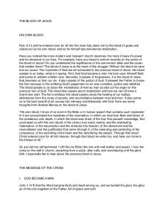
E X P L A N A T I O N OF P L A T E 9. The water color drawings were all made with the aid of a camera lucida, and with a Leitz No. 4 ocular and 2 mm. objective. All the figures are from formalinfixed tissue stained by Wright's method, except the central cell in figure 3, which is from tissue fixed in bichloride of mercury and stained with the Giemsa stain. The composition of the Wright stain used in figures I and 2 was methylene-blue part, eosin I2 parts; in figure 3, methylene-blue I part, eosin x4 parts. FIG. I. An endothelial cell from the surface of the femur marrow of the rabbit, illustrating an early stage of hyperplasia. Fro. 2. A cell of the same type and situation as that in figure i, showing marked hyperplasia. A stubby mass of protoplasm in the center is projecting into a capillary and breaking up into blood platelets. The more prolonged mass to the right shows no segmentation, while the process to the left is burrowing through the connective tissue covering the marrow. FIG. 3. Three types of cells that were numerous in the splenic sinuses of one of the rabbits injected with hematin. The presence of various forms of pseudopods and segmentation into platelets is shown. The platelet count in this animal rose to normal, while the megakaryocytes had regenerated to a very slight degree.
Downloaded from on January 26, 2017
I. U n d e r n o r m a l c i r c u m s t a n c e s blood platelets are l a r g e l y d e r i v e d f r o m the m e g a k a r y o c y t e o f the b l o o d - f o r m i n g o r g a n s . 2. T h e t r a n s i t i o n a l leucocyte, r e p r e s e n t i n g a persistent f o r m o f the e m b r y o n i c p r e m e g a k a r y o c y t e , is a c i r c u l a t i n g h o m o l o g u e o f the m e g a k a r y o c y t e a n d p r o b a b l y plays s o m e p a r t in n o r m a l platelet formation. 3. U n d e r conditions o f excessive d e m a n d f o r platelet p r o d u c t i o n , there m a y be a g r e a t e r o r less r e v e r s i o n to a n e m b r y o n i c m o d e o f platelet f o r m a t i o n in w h i c h less h i g h l y specialized cells t h a n the m e g a k a r y o c y t e participate in platelet p r o d u c t i o n . 4. I n a d d i t i o n to the m e g a k a r y o c y t e , the cells t h a t h a v e been o b s e r v e d to take p a r t in platelet f o r m a t i o n are hyperplas,tic e n d o thelial cells in the m a r r o w , a n d m o n o n u c l e a r a n d t r a n s i t i o n a l cells ( p r e m e g a k a r y o c y t e s ) in the m a r r o w , spleen, a n d Mood.
Published September 1, 1913
THE JOURNAL OF EXPERIMENTAL MEDICINE VOL. XVIII.
PLATE 9.
Downloaded from on January 26, 2017
(Brown: Histogenesis of Blood Platelets.)