CNS Drugs 2003; 17 (9): 641-652 1172-7047/03/0009-0641/$30.00/0
REVIEW ARTICLE
Adis Data Information BV 2003. All rights reserved.
The Glutamatergic System and Alzheimer’s Disease Therapeutic Implications D. Allan Butterfield and Chava B. Pocernich Department of Chemistry, Center of Membrane Sciences and Sanders-Brown Center on Aging, University of Kentucky, Lexington, Kentucky, USA
Contents Abstract . . . . . . . . . . . . . . . . . . . . . . . . . . . . . . . . . . . . . . . . . . . . . . . . . . . . . . . . . . . . . . . . . . . . . . . . . . . . . . . . . . . . 641 1. Alzheimer’s Disease . . . . . . . . . . . . . . . . . . . . . . . . . . . . . . . . . . . . . . . . . . . . . . . . . . . . . . . . . . . . . . . . . . . . . . 642 1.1 Overview of the Disease . . . . . . . . . . . . . . . . . . . . . . . . . . . . . . . . . . . . . . . . . . . . . . . . . . . . . . . . . . . . . . 642 1.2 Role of Oxidative Stress in Aetiology . . . . . . . . . . . . . . . . . . . . . . . . . . . . . . . . . . . . . . . . . . . . . . . . . . . 642 2. The Glutamatergic System . . . . . . . . . . . . . . . . . . . . . . . . . . . . . . . . . . . . . . . . . . . . . . . . . . . . . . . . . . . . . . . . 642 2.1 Role in Learning and Memory . . . . . . . . . . . . . . . . . . . . . . . . . . . . . . . . . . . . . . . . . . . . . . . . . . . . . . . . . 642 2.2 Changes in Alzheimer’s Disease . . . . . . . . . . . . . . . . . . . . . . . . . . . . . . . . . . . . . . . . . . . . . . . . . . . . . . . 643 3. Potential Therapies Directed at the Glutamatergic System . . . . . . . . . . . . . . . . . . . . . . . . . . . . . . . . . . . 645 3.1 The Two-Stage Model . . . . . . . . . . . . . . . . . . . . . . . . . . . . . . . . . . . . . . . . . . . . . . . . . . . . . . . . . . . . . . . . 645 3.1.1 Therapies Aimed at Glutamatergic Hyperactivity . . . . . . . . . . . . . . . . . . . . . . . . . . . . . . . . . . 645 3.1.2 Therapies Aimed at Glutamatergic Hypoactivity . . . . . . . . . . . . . . . . . . . . . . . . . . . . . . . . . . 647 4. Conclusion . . . . . . . . . . . . . . . . . . . . . . . . . . . . . . . . . . . . . . . . . . . . . . . . . . . . . . . . . . . . . . . . . . . . . . . . . . . . . . 648
Abstract
Alzheimer’s disease affects nearly 5 million Americans currently and, as a result of the baby boomer cohort, is predicted to affect 14 million Americans and 22 million persons totally worldwide in just a few decades. Alzheimer’s disease is present in nearly half of individuals aged 85 years. The main symptom of Alzheimer’s disease is a gradual loss of cognitive function. Glutamatergic neurotransmission, an important process in learning and memory, is severely disrupted in patients with Alzheimer’s disease. Loss of glutamatergic function in Alzheimer’s disease may be related to the increase in oxidative stress associated with the amyloid β-peptide that is found in the brains of individuals who have the disease. Therefore, therapeutic strategies directed at the glutamatergic system may hold promise. Therapies addressing oxidative stress induced by hyperactivity of glutamate receptors include supplementation with estrogen and antioxidants such as tocopherol (vitamin E) and acetylcysteine (N-acetylcysteine). Therapy for hypoactivity of glutamate receptors is aimed at inducing the NMDA receptor with glycine and cycloserine (D-cycloserine). Recently, memantine, an NMDA receptor antagonist that addresses the hyperactivity of these receptors, has been approved in some countries for use in Alzheimer’s disease.
642
Butterfield & Pocernich
1. Alzheimer’s Disease 1.1 Overview of the Disease
Alzheimer’s disease is the major dementing disorder of the elderly.[1] According to Katzman,[2] in the absence of any intervention, patients with Alzheimer’s disease will number 22 million worldwide in just over 2 decades. Current estimates suggest that the healthcare costs in the US in the nearly 5 million patients with Alzheimer’s disease approach $US100 billion annually.[1,2] In the absence of an effective therapy, the costs associated with 14 million Americans with Alzheimer’s disease in a few decades will be enormous. Alzheimer’s disease is associated with three major pathological hallmarks: loss of synapses and the presence of neurofibrillary tangles and senile plaques. Amyloid β-peptide (Aβ), in the form of insoluble fibril deposits, is the major component of senile plaques.[3,4] Senile plaques are surrounded by degenerated neurons,[1] and Aβ is toxic to neurons in culture.[5-19] Genetic studies of familial Alzheimer’s disease offer the strongest evidence for a central role of Aβ in the pathogenesis of the disease.[20] Several familial Alzheimer’s disease mutations have been found in the amyloid precursor protein (APP) and presenilin genes; these mutations invariably lead to increased Aβ deposition.[20,21] APP is expressed on chromosome 21, as is Down’s trisomy, and persons with Down’s syndrome have increased Aβ deposits[22] and develop Alzheimer’s disease eventually. APP-overexpressing mice exhibit some characteristics of Alzheimer’s disease pathology.[23-32] Increasing evidence suggests that the toxic species of the 42-mer Aβ(1-42) is small aggregates of the peptide.[33-35] 1.2 Role of Oxidative Stress in Aetiology
The Alzheimer’s disease brain is characterised by extensive oxidative stress manifested by increased protein oxidation, lipid peroxidation, free radical formation and DNA and RNA oxidation.[36] The Aβ – particularly Aβ(1-42), known to accumulate in the brains of individuals with Alzheimer’s disease – is Adis Data Information BV 2003. All rights reserved.
thought to be central to the pathogenesis of the disease.[37] Our laboratory combined these two concepts into a comprehensive model for neurodegeneration in Alzheimer’s disease based on oxidative stress associated with Aβ(1-42).[17,36,38,39] Aβ(1-42) induces protein oxidation, lipid peroxidation, reactive oxygen species formation, stimulation of nitric oxide synthase, alteration of mitochondria and many other markers of oxidative stress, all of which are inhibited by antioxidants.[17,36-40] In addition to the above effects, Aβ(1-42) also inhibits aspects of the glutamatergic system, including causing oxidative modification of glutamine synthetase (the enzyme that catalyses the conversion of glutamate, the excitatory neurotransmitter and activator of NMDA receptors, to glutamine) and the formation of small soluble, highly neurotoxic aggregates of Aβ(1-42).[18,40-42] Moreover, glutamate potentiates the toxicity of Aβ peptides.[43] Conceivably, this effect may reflect the increased free radical production observed following NMDA receptor stimulation.[44] Thus, although glutamate serves an important role in neurotransmission, excess glutamate-induced receptor stimulation can be toxic. The involvement of glutamatergic pathways in Alzheimer’s disease, the potential role played by Aβ(1-42) in the alterations in the glutamatergic system seen in the disease and possible therapies aimed at the glutamatergic system are addressed in this review. 2. The Glutamatergic System 2.1 Role in Learning and Memory
Activation of NMDA receptors in different ways can lead to either long-term potentiation (LTP) or long-term depression of synaptic strength. These forms of synaptic plasticity may represent ways of encoding memories in the brain. The size and nature of the changes in synaptic strength are highly regulated processes in learning and memory. The strength of the synapse can be altered in several ways. The end result can be affected by the probability of transmitter release from an activated presynaptic terminal, a change in the number of CNS Drugs 2003; 17 (9)
Glutamatergic System and Alzheimer’s Disease
receptors, a change in the size of the current produced by each receptor at a postsynaptic site, a change in the excitability of the dendritic membrane and/or changes in the cytoskeleton and membrane trafficking that ultimately produce a new connection.[45,46] Alzheimer’s disease is considered a synaptic failure.[47] The degree of cognitive decline in patients with Alzheimer’s disease has been correlated with synaptic loss.[48] A study of temporal and frontal cortical biopsies performed within an average of 2–4 years of the onset of clinical Alzheimer’s disease revealed a 25–35% decrease in the numerical density of synapses and a 15–35% decrease in the number of synapses per cortical neuron.[49] 2.2 Changes in Alzheimer’s Disease
The possibility that activation of glutamate receptors contributes to cell death in neurodegenerative disorders such as Alzheimer’s disease has been postulated. As mentioned in section 2.1, short-term release of glutamate is involved in important processes such as learning and memory,[50] possibly involving synaptic protein remodelling.[46] However, abnormally prolonged release of glutamate causes excitotoxicity and cell death and may play a role in the pathogenesis of chronic neurodegenerative disorders such as Alzheimer’s disease.[51] No enzymes exist in the synaptic cleft to degrade glutamate; thus, this neurotransmitter must be cleared from the synapse by high-affinity presynaptic and glial transporters. Five different forms of glutamate transporters have been identified: GLAST (EAAT1), GLT-1 (EAAT2), EAAC-1 (EAAT3), EAAT4 and EAAT5. GLAST and GLT-1 are restricted to astroglial cells, with GLAST expression observed during early stages of development and GLT-1 expression observed throughout maturity.[52] In the adult brain, GLAST and GLT-1 are differentially located in astroglia.[53] In particular, levels of GLT-1 expression are highest in the forebrain with moderate expression throughout the remaining neuroaxis, whereas expression of GLAST is highest in the cerebellum.[53] The neuronal glutamate transporters EAAC1 and EAAT4 are located outside of Adis Data Information BV 2003. All rights reserved.
643
and contribute relatively little to the clearance of glutamate from the synaptic cleft.[52,54] Glutamate can be neurotoxic through a stimulatory effect on NMDA, AMPA, kainate or group 1 metabotropic receptors (mGluR1), but selective neuronal death in Alzheimer’s disease appears to be dependent primarily on NMDA receptor activation.[55] Indeed, a recent study suggests that NMDA receptor activation stimulates APP processing to produce Aβ.[56] Consequently, overstimulation of the receptor by excess glutamate could lead to excess Aβ(1-42) production with consequent oxidative stress-induced neurotoxicity.[36,38,39] In turn, Aβ selectively depresses excitatory synaptic transmission onto neurons and is NMDA receptor activity dependent.[57] Nontoxic concentrations of Aβ can produce a rapid inhibition of LTP, although there is no longterm effect on normal synaptic transmission.[58] Aβ can also inhibit NMDA receptor-mediated synaptic potentials; however, results suggest that Aβ does not inhibit LTP via effects on NMDA receptors but rather interferes with a downstream pathway.[58] Synaptic depression from excessive Aβ could contribute to cognitive decline during early Alzheimer’s disease. Glutamatergic neurotransmission in neocortical regions and the hippocampus is severely disrupted in Alzheimer’s disease.[59-61] In addition, a reduction in the number of NMDA receptors is reported in individuals with the disease.[59] Once transported to glia, glutamate is converted to glutamine by glutamine synthetase. The glutamine is released, taken up into neurons and converted to glutamate by mitochondrial glutaminase. Noninvasive detection of glutamate + glutamine (GLX) in vivo showed a significant reduction in GLX in the cingulate cortex of patients with Alzheimer’s disease that strongly correlates with both their cognitive and functional status.[62] Two other in vivo reports of GLX in Alzheimer’s disease revealed no significant difference in the mid-frontal or temporoparietal GLX[63] but an increase in GLX in the occipital lobe[64] of patients with Alzheimer’s disease. One enzyme that is particularly sensitive to oxidative stress is glutamine synthetase.[18,41,42] Novel CNS Drugs 2003; 17 (9)
644
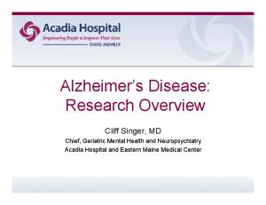
electron paramagnetic resonance (EPR) approaches suggested that glutamine synthetase isolated and purified from the brains of individuals who had Alzheimer’s disease was more oxidised than that isolated and purified from the brains of healthy agematched individuals.[42] A recent study has shown, using proteomics, that glutamine synthetase indeed is oxidised in the brains of individuals with Alzheimer’s disease relative to control individuals.[65] Consistent with these findings, a significant decrease in glia-resident glutamine synthetase activity in the hippocampus and neocortex of those with Alzheimer’s disease has been reported.[66] A decrease in glutamine synthetase activity could result in an increase in glutamate levels, prolonged NMDA receptor activation and neuronal injury in brain areas susceptible to glutamate toxicity. Aβ(1-42) significantly decreased the activity of the enzyme in cytosolic fractions of brain homogenates and cultured hippocampal neurons and astrocytes.[17,67] The GLT-1 glutamate transporter system in the brain is altered in Alzheimer’s disease. A significant reduction of glutamate transporter activity in the cortex has been reported.[68,69] GLT-1, the main astroglial glutamate transporter, is oxidatively modified by binding of 4-hydroxy nonenal (HNE), a lipid peroxidation product, in the inferior parietal region of the brains of individuals with Alzheimer’s disease.[40] This finding might explain the loss of activity of this transporter in the disease.[68] Aβ peptides added to synaptosomes lead to lipid peroxidation[39,40,70] and increased HNE binding to GLT-1,[40] suggesting that, in Alzheimer’s disease, where excess Aβ(1-42) deposition in the brain occurs, Aβ could account for the increased HNE binding to GLT-1. Inhibition of glutamate transport, coupled with decreased activity of glutamine synthetase in Alzheimer’s disease, would likely lead to increased extraneuronal glutamate with consequent stimulation of NMDA receptors and excitotoxic processes involving excess intraneuronal calcium accumulation and eventual cell death (figure 1). A recent study in Alzheimer’s disease reported that particular neurons that displayed cytoskeletal abnormalities Adis Data Information BV 2003. All rights reserved.
Butterfield & Pocernich
Glutamate Removal by GLT-1 and GS Excitotoxicity
Disruption of Ca2+ homeostasis; free radical formation
Cell death Fig. 1. Dysfunction in components of the glutamatergic system in Alzheimer’s disease leading to cell death in the brain. The glutamate transporter (GLT-1) and glutamine synthetase (GS) – both means of inactivating extraneuronal glutamate – are oxidatively modified and dysfunctional in Alzheimer’s disease. This leads to increased opportunity for excitotoxicity to occur, with subsequent cell death.
with abnormal tau-protein expressed GLT-1 or EAAT2, usually expressed in astrocytes.[71] Such a neuronal glutamate transporter is likely to be inhibited by HNE. The latter, as noted above, results from lipid peroxidation caused by Aβ(1-42) among other pro-oxidants.[39,40] The expression of EAAT2 in altered neurons in Alzheimer’s disease strongly supports the hypothesis that abnormalities in glutamate transport play an important role in the pathogenesis of Alzheimer’s disease. Glutamate is also converted to GABA, an inhibitory neurotransmitter in the brain, by glutamate decarboxylase. Messenger RNA for glutamate decarboxylase in patients with Alzheimer’s disease has been reported to be increased by 50% in the caudate nucleus and putamen.[72] In contrast, no alteration was found in the ventral striatum. These data suggest that, in Alzheimer’s disease, GABAergic neurotransmission may be increased in the dorsal striatum but not the ventral striatum. In patients with Alzheimer’s disease, the enzyme synthesising GABA is increased, potentially leading to higher levels of GABA,[73] even though glutamate is being removed from stimulating neurons. The elevated interstitial GABA acts on presynaptic GABAA receptors, causing their chronic depolarising block and dystropy, resulting in deafferentation and degeneration of neuronal systems (for review, see Marczynski[74]). As noted above, neurons produce glutamate from glutamine in a reaction catalysed by the enzyme CNS Drugs 2003; 17 (9)
Glutamatergic System and Alzheimer’s Disease
glutaminase. In the temporal cortex of individuals with Alzheimer’s disease, no significant changes were seen in the levels of phosphate-activated glutaminase,[75] suggesting that neurons in the cortex area in Alzheimer’s disease still have the ability to produce glutamate. In contrast, glutamate and glutaminase immunoreactive pyramidal neurons in the hippocampal dentate gyrus CA fields are decreased in number in patients with Alzheimer’s disease.[76] These same neurons were found to contain neurofibrillary tangles. Although speculative, this regional difference in glutamatergic neurons may reflect the role of the hippocampus in memory processing, a function severely compromised in patients with Alzheimer’s disease. 3. Potential Therapies Directed at the Glutamatergic System There are several subtypes of NMDA receptors. Stimulation of NMDA receptors leads to signalling events formulated by the protein-protein interaction among the receptor and other proteins.[46] As noted in section 1.2, free radicals can result from NMDA receptor stimulation,[44] and overstimulation of the receptor may be associated with oxidative stress and excitotoxicity. Therapies to alleviate the loss of memory associated with the dysfunctional glutamatergic system in Alzheimer’s disease vary greatly. There are two principal trends in potential Alzheimer’s disease therapy involving the glutamatergic system. One trend addresses the NMDA receptors in a hyperactive state, suggesting the blocking of the receptors and thus the blocking of excitotoxicity as a therapeutic strategy. The second trend argues that late in the disease, the NMDA receptor system is hypoactive and needs to be stimulated to avoid the cognitive decline seen with Alzheimer’s disease. Table I outlines agonists and antagonists of the NMDA receptor. 3.1 The Two-Stage Model
Olney and colleagues[77] have proposed a twostage mechanism encompassing both of the above hypotheses. The first stage entails the excess pro Adis Data Information BV 2003. All rights reserved.
645
duction and aggregation of Aβ in the brain and the interaction of Aβ(1-42) with NMDA receptors so that the neurons become hyperstimulated and degenerated by excess glutamate-induced calcium entry, free radical formation, membrane remodelling or Aβ(1-42) production. The second stage occurs when the loss of NMDA receptors is sufficient to produce hypoactivity. Coinciding with the hypoactivity of NMDA receptors, subtle changes in cognitive function become evident. This two-stage hypothesis for pharmacological intervention implies unique therapies for each stage. 3.1.1 Therapies Aimed at Glutamatergic Hyperactivity
Stage one most likely occurs in asymptomatic patients. Unfortunately, there is no current method for identifying Alzheimer’s disease at this stage. Therapies would include preventing excess Aβ peptide formation, energy deficits and oxidative stress while increasing glutamate uptake or blocking NMDA receptors. Oxidative stress can be addressed with high levels of antioxidants that can pass through the blood-brain barrier, such as thioctic acid (α-lipoic acid), glutathione ethylester or tocopherol (vitamin Table I. Agonists and antagonists of the glutamate NMDA receptor Drug/compound
NMDA receptor binding site
Agonists NMDA
NMDA/glutamate
Quinolinate
NMDA/glutamate
L-aspartate
NMDA/glutamate
Glycine
Glycine
Milacemide
Glycine
Cycloserine
Glycine
Spermidine
Polyamine
Antagonists Nimodipine
Calcium channel
Dizocilpine (MK801)
Calcium channel
AP-5; AP-7
Calcium channel
CHF 3381
Calcium channel
Ifenprodil
Calcium channel
Phencyclidine
Calcium channel
Memantine
Calcium channel
Huperzine A
Polyamine
Arcaine
Polyamine
CNS Drugs 2003; 17 (9)
646
E).[78-82] Yet, because free radicals may be important in cell signalling and other pathways,[81] blockade of all free radical processes may prove problematic. Antioxidant levels can be increased through diet[81] or supplementation. Several clinical trials in Alzheimer’s disease have addressed tocopherol supplementation[82-84] and indicate a retardation in the progression of cognitive decline. The increase in the antioxidant glutathione is another pathway for preventing oxidative stress. Glutathione can be increased many different ways.[79] Recently, a study in which glutathione levels were increased by supplementation with N-acetylcysteine has reported favourable outcomes on several cognitive battery tests in patients with Alzheimer’s disease.[85] Inhibition of the voltage-dependent calcium channels by glutamate receptor antagonists, such as nimodipine, dizocilpine (MK801), AP-5, AP-7, CHF 3381 [n-(2-indanyl)-glycinamide hydrochloride][86] and ifenprodil,[87] all NMDA receptor antagonists, can be achieved. In addition, arcaine[88] and huperzine A,[89] antagonists of the NMDA receptor polyamine binding site, and other anticonvulsant drugs aimed at NMDA receptors may be useful in Alzheimer’s disease. However, many NMDA receptor antagonists, such as dizocilpine, aptiganel (cerestat), licostinel and selfotel,[90-93] produced highly undesirable adverse effects in clinical trials at doses within their therapeutic range. The poor results obtained in clinical trials with dizocilpine and selfotel may have discouraged investigators from therapeutic approaches involving antagonism of NMDA receptors. However, memantine, a moderate-affinity noncompetitive NMDA receptor antagonist, has been reported to have therapeutic potential without the undesirable adverse effects of other antagonists at therapeutic doses.[90] Memantine is reported to prevent neurodegeneration and learning deficits in animal models of dementia.[94,95] This drug suppresses Aβ(1-42) formation in a transgenic animal model of Alzheimer’s disease.[96] In a double-blind, placebo-controlled clinical trial to assess the clinical efficacy and safety of memantine, 166 patients with moderately severe to severe Alzheimer’s disease and vascular dementia Adis Data Information BV 2003. All rights reserved.
Butterfield & Pocernich
were administered the drug for 12 weeks, starting at a dosage of 5 mg/day and then increasing to 10 mg/ day after the first week. This regimen reportedly led to adverse effect-free functional improvement and reduced care dependence in patients with severe dementia regardless of dementia type.[97] Recent studies have been reviewed evaluating the use of memantine in moderate to severe Alzheimer’s disease.[98] During a 28-week, double-blind treatment period, memantine 10mg twice daily was shown to slow the rate of deteriorisation as seen in Clinician’s Interview-Based Impression of Change (CIBIC)-Plus, Alzheimer’s Disease Cooperative Study Activities of Daily Living Inventory modified for more severe dementia (ADCS-ADLsev), Severe Impairment Battery (SIB), Functional Assessment Staging (FAST)[99] and Resource Utilization in Dementia (RUD) questionnaire endpoints.[100] At the end of the 28-week, double-blind phase, patients continued memantine 10mg twice daily for a 24-week open-label extension study.[101] Patients switching to memantine from placebo improved relative to the projected rate of decline established during the initial 28-week study.[101] Memantine is currently approved in the EU for the treatment of moderately severe to severe Alzheimer’s disease, and has been filed with the US FDA for this indication.[102] This pharmacological agent also could be combined with acetylcholinesterase inhibitors, which are the current standard treatment for Alzheimer’s disease.[103,104] A phase III, randomised, double-blind, 6-month study evaluated combination therapy of memantine 10mg twice daily with the cholinesterase inhibitor donepezil versus donepezil plus placebo.[105] The combined drug therapy showed a sustained improvement from baseline in cognitive function as assessed by the SIB, less decline in activities of daily living and a significant difference in CIBIC-Plus compared with donepezil plus placebo.[105] Recently, research has been directed towards developing amino-alkyl-cyclohexanes as new noncompetitive NMDA receptor antagonists based on the positive results of memantine for the treatment of neurodegenerative dementia.[106] CNS Drugs 2003; 17 (9)
Glutamatergic System and Alzheimer’s Disease
Studies to address the effects of estrogen on glutamate toxicity and uptake have been reported. Liang and colleagues[107] examined the effect of estrogen on glutamate uptake in astrocytes taken from individuals without dementia and patients with Alzheimer’s disease. Astrocytes from patients with Alzheimer’s disease that were treated with 17-βestradiol for 48 hours had significantly increased glutamate uptake and an increased level of protein expression of GLT-1 and GLAST.[107] GLT-1 expression was found to be sensitive to the estrogen receptor antagonist fulvestrant (ICI 182,780), whereas this drug had no effect on GLAST protein expression, possibly reflecting the differential location of each protein. In addition, protein synthesis was involved in the estrogen-induced upregulation of GLAST protein expression as well as glutamate uptake activity in astrocytes from patients with Alzheimer’s disease. GLAST may play a major role in the estrogen-induced increase of glutamate uptake in Alzheimer’s disease astrocytes since this transporter was estrogen receptor insensitive. Estrogen also has been shown to reduce Aβ production and increase the clearance of Aβ by enhancing microglia phagocytosis,[108-110] thus potentially decreasing the production of HNE by Aβ-induced lipid peroxidation[38-40] and inactivation of glutamate transporters by HNE.[40] Estrogen is reported to reduce the formation of free radicals and to protect neurons against oxidative damage induced by glutamate uptake impairment,[111,112] glutamate-induced cytotoxicity[113] and Aβ.[110,114] Clinical trials for estrogen replacement in the treatment of Alzheimer’s disease have produced ambiguous results. Initial, small, open-label and double-blind clinical trials indicated improved cognitive function in women with Alzheimer’s disease.[115,116] However, recent large trials failed to show a beneficial effect for long-term estrogen replacement for women with Alzheimer’s disease.[117-122] There are several variables that could affect these results, such as genetic factors, time between estrogen loss and replacement, route of estrogen administration, form of estrogen (conjugated estrogens vs estradiol), duration of treatment and Adis Data Information BV 2003. All rights reserved.
647
extent and type of Alzheimer’s disease pathology. Studies such as the Women’s Health Initiative Memory Study (WHIMS)[123] and Women’s International Study of long Duration Oestrogen after Menopause (WISDOM)[124] are currently underway to determine the exact timing and administration required and optimal dosage sufficient to slow the progress of dementia. Currently, estrogen replacement therapy remains a therapeutically unproven method of treating or delaying the onset of Alzheimer’s disease. 3.1.2 Therapies Aimed at Glutamatergic Hypoactivity
Second-stage therapies in the Olney et al.[77] hierarchy, addressing the stimulation of glutamate receptors, include selective agonists and drugs that prevent antagonist-induced damage to neurons. Such an approach may modulate the disease-induced damage to neurons, especially late in the disease. Glycine and polyamine stimulation of the NMDA receptor might be an effective treatment for Alzheimer’s disease. Large doses of glycine led to improvement in memory performance in various rodent studies, but also caused hind limb paralysis.[125,126] The pro-glycine drug milacemide and cycloserine (D-cycloserine), a partial glycine agonist, also reportedly improved memory in humans and rodents,[127-131] but clinical trials in patients with Alzheimer’s disease showed little therapeutic success.[127,128] In contrast, an improvement of implicit memory in patients with Alzheimer’s disease has been reported following administration of cycloserine.[132] More research is necessary to determine if this agent will prove useful as an Alzheimer’s disease therapy. The polyamine modulatory site agonist spermidine can potentate the learning impairment associated with dizocilpine treatment,[133] improve the performance of aged rats in the Stone maze[61] and cause an increase in test step-down latencies.[88] Ifenprodil dose dependently prevented cell death induced by glutamate or NMDA but did not affect that induced by kainate. The protective effects of ifenprodil against glutamate neurotoxicity were sigCNS Drugs 2003; 17 (9)
648
nificantly reduced by spermidine, but not by glycine.[134] The bis-ammonium adamantyl-containing compounds IEM-1460, a polyamine agonist, and IEM-1754, a polyamine antagonist/agonist, also can modulate the polyamine site of NMDA, AMPA/ kainate and nicotinic receptors.[135] These data suggest that polyamines may be involved in learning and memory. However, caution is urged in the use of polyamine-stimulating approaches, since Aβ(1-42) causes increased polyamine accumulation in neurons with subsequent increased neurotoxicity that can be prevented by the free radical scavenger tocopherol.[19,136] Synthesis of new glutamate receptor agonists is currently being addressed. Substitution on the carbon backbone of (S)-glutamic acid [(S)-Glu], as well as absolute stereochemistry, are structural parameters of key importance for the pharmacological profile of (S)-Glu receptor ligands.[137] Guldbrandt and colleagues[137] synthesised a series of methylsubstituted 2-aminoadipic acid (AA) analogues, 3-methyl-AA, 4-methyl-AA, 5-methyl-AA and (E)δ(4)-5-methyl-AA. Compounds with S-configuration at the α-carbon generally showed mGluR2 agonist activity of similar or slightly lower potency than (S)-AA. The compounds had different pharmacological effects at glutamate receptors. NMDA receptor agonists included (2S,5RS)-5-methyl-AA, and an NMDA receptor antagonist was (2R,4S)-4-methyl-AA. The two unsaturated analogues (S)- and (R)-(E)-δ(4)-5-methyl-AA turned out to be a weak AMPA receptor agonist and a weak mixed NMDA/AMPA receptor antagonist, respectively.[137] Whether these newer analogues will have therapeutic efficacy in Alzheimer’s disease will require further testing. Alzheimer’s disease is characterised by abnormalities in several neurotransmitter pathways.[1] Consequently, pharmacological treatment of the cognitive deficits in Alzheimer’s disease may require combinational therapy. Cholinergic hypofunction has been reported to correlate with the severity of dementia in Alzheimer’s disease, and this has led to the development of the current standard therapy for the disease, cholinesterase inhibitors.[61] In pa Adis Data Information BV 2003. All rights reserved.
Butterfield & Pocernich
tients with the disease, the activity of the enzyme synthesising GABA is increased, potentially leading to higher levels of GABA and cell death (see section 2.2).[73] Administration of glutamatergic and cholinergic agonists along with the use of serotonergic and GABAergic antagonists conceivably may be beneficial. 4. Conclusion Excitotoxicity in the brains of individuals with Alzheimer’s disease, resulting in neurodegeneration and stemming from excess stimulation of NMDA receptors, potentially could be avoided if the three main pathways for disposing of excess glutamate (glutamate transporters, glutamine synthetase and glutamate decarboxylase) were not oxidatively modified and therefore not of inhibited function. Aβ(1-42), which induces oxidative stress and may be central to the pathogenesis of Alzheimer’s disease, oxidatively modifies glutamine synthetase and GLT-1.[40-42,65] Other glutamate transporters conceivably may be similarly subject to Aβ(1-42)-induced oxidative modification. Consequently, selective glutamatergic antagonists that also possess antioxidant capabilities may be a novel approach to protect against both excitotoxicity and oxidative stress associated with excess Aβ(1-42) in Alzheimer’s disease. Studies to test these ideas are in progress. Memantine, a glutamatergic antagonist, is already being used for the treatment of Alzheimer’s disease. Acknowledgements This work was supported in part by grants from the National Institutes of Health (AG-10836; AG-05119). The authors have no conflicts of interest with regard to the contents of this manuscript.
References 1. Katzman R, Saitoh T. Advances in Alzheimer’s disease. FASEB J 1991; 5: 278-86 2. Katzman R. Epidemiology of Alzheimer’s disease. Neurobiol Aging 2000; 21 Suppl.: S1 3. Glenner GG, Wong CW. Alzheimer’s disease: initial report of the purification and characterization of a novel cerebrovascular amyloid protein. Biochem Biophys Res Commun 1984; 120: 885-90
CNS Drugs 2003; 17 (9)
Glutamatergic System and Alzheimer’s Disease
4. Masters CL, Simms G, Weinman NA, et al. Amyloid plaque core protein in Alzheimer disease and Down syndrome. Proc Natl Acad Sci U S A 1985; 82: 4245-9 5. Yankner BA, Dawes LR, Fisher S, et al. Neurotoxicity of a fragment of the amyloid precursor associated with Alzheimer’s disease. Science 1989; 245: 417-20 6. Frautschy SA, Baird A, Cole GM. Effects of injected Alzheimer beta-amyloid cores in rat brain. Proc Natl Acad Sci U S A 1991; 88: 8362-6 7. Kowall NW, Beal MF, Busciglio J, et al. An in vivo model for the neurodegenerative effects of beta amyloid and protection by substance P. Proc Natl Acad Sci U S A 1991; 88: 7247-51 8. Pike CJ, Walencewicz AJ, Glabe CG, et al. In vitro aging of beta-amyloid protein causes peptide aggregation and neurotoxicity. Brain Res 1991; 563: 311-4 9. Howlett DR, Jennings KH, Lee DC, et al. Aggregation state and neurotoxic properties of Alzheimer beta-amyloid peptide. Neurodegeneration 1995; 4: 23-32 10. Harris ME, Hensley K, Butterfield DA, et al. Direct evidence of oxidative injury produced by the Alzheimer’s beta-amyloid peptide (1-40) in cultured hippocampal neurons. Exp Neurol 1995; 131: 193-202 11. Aksenov MY, Aksenova MV, Butterfield DA, et al. Glutamine synthetase-induced enhancement of beta-amyloid peptide A beta (1-40) neurotoxicity accompanied by abrogation of fibril formation and A beta fragmentation. J Neurochem 1996; 66: 2050-6 12. Aksenov MY, Aksenova MV, Markesbery WR, et al. Amyloid beta-peptide (1-40)-mediated oxidative stress in cultured hippocampal neurons: protein carbonyl formation, CK BB expression, and the level of Cu, Zn, and Mn SOD mRNA. J Mol Neurosci 1998; 10: 181-92 13. Yatin SM, Aksenov M, Butterfield DA. The antioxidant vitamin E modulates amyloid beta-peptide-induced creatine kinase activity inhibition and increased protein oxidation: implications for the free radical hypothesis of Alzheimer’s disease. Neurochem Res 1999; 24: 427-35 14. Yatin SM, Varadarajan S, Link C, et al. In vitro and in vivo oxidative stress associated with Alzheimer’s amyloid betapeptide (1-42). Neurobiol Aging 1999; 20: 325-30 15. Yatin SM, Aksenova M, Aksenov M, et al. Effect of transglutaminase on Aβ(1-40) fibril formation and neurotoxicity. Alzheimer Rep 1999; 2: 165-70 16. Varadarajan S, Yatin S, Kanski J, et al. Methionine residue 35 is important in amyloid beta-peptide-associated free radical oxidative stress. Brain Res Bull 1999; 50: 133-41 17. Varadarajan S, Yatin S, Aksenova M, et al. Review: Alzheimer’s amyloid-peptide-associated free radical oxidative stress and neurotoxicity. J Struct Biol 2000; 130: 184-208 18. Aksenov MY, Aksenova MV, Harris ME, et al. Enhancement of beta-amyloid peptide A beta(1-40)-mediated neurotoxicity by glutamine synthetase. J Neurochem 1995; 65: 1899-902 19. Yatin SM, Yatin M, Aulick T, et al. Alzheimer’s amyloid betapeptide associated free radicals increase rat embryonic neuronal polyamine uptake and ornithine decarboxylase activity: protective effect of vitamin E. Neurosci Lett 1999; 263: 17-20 20. Selkoe DJ. Amyloid beta-protein and the genetics of Alzheimer’s disease. J Biol Chem 1996; 271: 18295-8 21. Scheuner D, Eckman C, Jensen M, et al. Secreted amyloid betaprotein similar to that in the senile plaques of Alzheimer’s disease is increased in vivo by the presenilin 1 and 2 and APP mutations linked to familial Alzheimer’s disease. Nat Med 1996; 8: 864-70
Adis Data Information BV 2003. All rights reserved.
649
22. Teller JK, Russo C, DeBusk L, et al. Presence of soluble amyloid beta-peptide precedes amyloid plaque formation in Down’s syndrome. Nat Med 1996; 2: 93-5 23. Games D, Adams D, Alessandrini R, et al. Alzheimer-type neuropathology in transgenic mice overexpressing V717F beta-amyloid precursor protein. Nature 1995; 373: 523-7 24. Hsiao K, Chapman P, Nilsen S, et al. Correlative memory deficits, Abeta elevation, and amyloid plaques in transgenic mice. Science 1996; 274: 99-102 25. Hsiao K. Transgenic mice expressing Alzheimer amyloid precursor proteins. Exp Gerontol 1998; 33: 883-9 26. Masliah E, Sisk A, Mallory M, et al. Comparison of neurodegenerative pathology in transgenic mice overexpressing V717F beta-amyloid precursor protein and Alzheimer’s disease. J Neurosci 1996; 16: 5795-811 27. Irizarry MC, McNamara M, Fedorchak K, et al. APPSw transgenic mice develop age-related A beta deposits and neuropil abnormalities, but no neuronal loss in CA1. J Neuropathol Exp Neurol 1997; 56: 965-73 28. Sturchler-Pierrat C, Abramowski D, Duke M, et al. Two amyloid precursor protein transgenic mouse models with Alzheimer disease-like pathology. Proc Natl Acad Sci U S A 1997; 94: 13287-92 29. Calhoun ME, Wiederhold KH, Abramowski D, et al. Neuron loss in APP transgenic mice. Nature 1998; 395: 755-6 30. Frautschy SA, Yang F, Irrizarry M, et al. Microglial response to amyloid plaques in APPsw transgenic mice. Am J Pathol 1998; 152: 307-17 31. Pappolla MA, Chyan YJ, Omar RA, et al. Evidence of oxidative stress and in vivo neurotoxicity of beta-amyloid in a transgenic mouse model of Alzheimer’s disease: a chronic oxidative paradigm for testing antioxidant therapies in vivo. Am J Pathol 1998; 152: 871-7 32. Smith MA, Hirai K, Hsiao K, et al. Amyloid-beta deposition in Alzheimer transgenic mice is associated with oxidative stress. J Neurochem 1998; 70: 2212-5 33. Lambert MP, Barlow AK, Chromy BA, et al. Diffusible, nonfibrillar ligands derived from Abeta1-42 are potent central nervous system neurotoxins. Proc Natl Acad Sci U S A 1998; 95: 6448-53 34. Walsh DM, Klyubin I, Fadeeva JV, et al. Naturally secreted oligomers of amyloid beta protein potently inhibit hippocampal long-term potentiation in vivo. Nature 2002; 416: 535-9 35. Drake J, Link CD, Butterfield DA. Oxidative stress precedes fibrillar deposition of Alzheimer’s disease amyloid β-peptide (1-42) in a transgenic Caenorhabditis elegans model. Neurobiol Aging 2003; 24: 415-20 36. Butterfield DA, Drake J, Pocernich C, et al. Evidence of oxidative damage in Alzheimer’s disease brain: central role for amyloid beta-peptide. Trends Mol Med 2001; 7: 548-54 37. Varadarajan S, Kanski J, Aksenova M, et al. Different mechanisms of oxidative stress and neurotoxicity for Alzheimer’s A beta(1-42) and A beta(25-35). J Am Chem Soc 2001; 123: 5625-31 38. Butterfield DA, Lauderback CM. Lipid peroxidation and protein oxidation in Alzheimer’s disease brain: potential causes and consequences involving amyloid-peptide-associated free radical oxidative stress. Free Radic Biol Med 2002; 32: 1050-60 39. Butterfield DA, Castegna A, Lauderback CM, et al. Evidence that amyloid beta-peptide-induced lipid peroxidation and its sequelae in Alzheimer’s disease brain contributes to neuronal death. Neurobiol Aging 2002; 23: 655-64
CNS Drugs 2003; 17 (9)
650
40. Lauderback CM, Hackett JM, Huang FF, et al. The glial glutamate transporter, GLT-1, is oxidatively modified by 4-hydroxy-2-nonenal in the Alzheimer’s disease brain: the role of Aβ1-42. J Neurochem 2001; 78: 413-6 41. Aksenov MY, Aksenova MV, Carney JM, et al. Oxidative modification of glutamine synthetase by amyloid beta peptide. Free Radic Res 1997; 27: 267-81 42. Butterfield DA, Hensley K, Cole P, et al. Oxidatively induced structural alteration of glutamine synthetase assessed by analysis of spin label incorporation kinetics: relevance to Alzheimer’s disease. J Neurochem 1997; 68: 2451-7 43. Mattson MP, Cheng B, Culwell AR, et al. Evidence for excitoprotective and intraneuronal calcium-regulating roles for secreted forms of the beta-amyloid precursor protein. Neuron 1993; 10: 243-54 44. Culcasi M, Lafon-Cazal M, Pietri S, et al. Glutamate receptors induce a burst of superoxide via activation of nitric oxide synthase in arginine-depleted neurons. J Biol Chem 1994; 269: 12589-93 45. Kennedy MB. Signal-processing machines at the postsynaptic density. Science 2000; 290: 750-4 46. Sheng M, Kim MJ. Postsynaptic signaling and plasticity mechanisms. Science 2002; 298: 776-80 47. Selkoe DJ. Alzheimer’s disease is a synaptic failure. Science 2002; 298: 789-91 48. Terry RD, Masliah E, Salmon DP, et al. Physical basis of cognitive alterations in Alzheimer’s disease: synapse loss is the major correlate of cognitive impairment. Ann Neurol 1991; 30: 572-80 49. Davies CA, Mann DM, Sumpter PQ, et al. A quantitative morphometric analysis of the neuronal and synaptic content of the frontal and temporal cortex in patients with Alzheimer’s disease. J Neurol Sci 1987; 78: 151-64 50. Collingridge GL, Singer W. Excitatory amino acid receptors and synaptic plasticity. Trends Pharmacol Sci 1990; 11: 290-6 51. Myhrer T. Adverse psychological impact, glutamatergic dysfunction, and risk factors for Alzheimer’s disease. Neurosci Biobehav Rev 1998; 23: 131-9 52. Furuta A, Rothstein JD, Martin JL. Glutamate transporter protein subtypes are expressed differentially during rat central nervous system development. J Neurosci 1997; 17: 8363-75 53. Sims KD, Robinson MB. Expression patterns and regulation of glutamate transporters in the developing and adult nervous system. Crit Rev Neurobiol 1999; 13: 169-97 54. Lehre KP, Levy LM, Otterson OP, et al. Differential expression of two glial glutamate transporters in the rat brain: quantitative and immunocytochemical observations. J Neurosci 1995; 15: 1835-53 55. Greenamyre JT, Young AB. Excitatory amino acids and Alzheimer’s disease. Neurobiol Aging 1989; 10: 593-602 56. Gordon-Krajcer W, Salinska E, Lazarewicz JW. N-methyl-daspartate receptor-mediated processing of beta-amyloid precursor protein in rat hippocampal slices: in vitro-superfusion study. Folia Neuropathol 2002; 40: 13-7 57. Kamenetz F, Tomita T, Hsieh H, et al. APP processing and synaptic function. Neuron 2003; 37: 925-37 58. Raymond CR, Ireland DR, Abraham WC. NMDA receptor regulation by amyloid-beta does not account for its inhibition of LTP in rat hippocampus. Brain Res 2003; 968: 263-72 59. Greenamyre JT. The role of glutamate in neurotransmission and neurologic disease. Arch Neurol 1986; 43: 1058-63
Adis Data Information BV 2003. All rights reserved.
Butterfield & Pocernich
60. Maragos WF, Greenamyre JT, Penney JB, et al. Glutamate dysfunction in Alzheimer’s disease: an hypothesis. TINS 1987; 10: 65-8 61. Palmer AM, Gershon S. Is the neuronal basis of Alzheimer’s disease cholinergic or glutamatergic? FASEB J 1990; 4: 2745-52 62. Antuono PG, Jones JL, Wang Y, et al. Decreased glutamate + glutamine in Alzheimer’s disease detected in vivo with 1HMRS at 0.5T. Neurology 2001; 56: 737-42 63. Moats RA, Ernst T, Shonk TK, et al. Abnormal cerebral metabolite concentrations in patients with probable Alzheimer disease. Magn Reson Med 1994; 32: 110-5 64. Ernst T, Chang L, Melchor R, et al. Frontotemporal dementia and early Alzheimer disease: differentiation with frontal lobe H-1 MR spectroscopy. Radiology 1997; 203: 829-36 65. Castegna A, Aksenov M, Aksenova M, et al. Proteomic identification of oxidatively modified proteins in Alzheimer’s disease brain. Part I: creatine kinase BB, glutamine synthase, and ubiquitin carboxy-terminal hydrolase L-1. Free Radic Biol Med 2002; 33: 562-71 66. Hensley K, Hall N, Subramaniam R, et al. Brain regional correspondence between Alzheimer’s disease histopathology and biomarkers of protein oxidation. J Neurochem 1995; 65: 2146-56 67. Butterfield DA. Beta-amyloid-associated free radical oxidative stress and neurotoxicity: implications for Alzheimer’s disease. Chem Res Toxicol 1997; 10: 495-506 68. Masliah E, Alford M, DeTeresa R, et al. Deficient glutamate transport is associated with neurodegeneration in Alzheimer’s disease. Ann Neurol 1996; 40: 759-66 69. Scott HL, Tannenberg A, Dodd PR. Variant forms of neuronal glutamate transporter sites in Alzheimer’s disease cerebral cortex. J Neurochem 1995; 64: 2193-202 70. Butterfield DA, Hensley K, Harris M, et al. beta-Amyloid peptide free radical fragments initiate synaptosomal lipoperoxidation in a sequence-specific fashion: implications to Alzheimer’s disease. Biochem Biophys Res Commun 1994; 200 (2): 710-5 71. Thal DR. Excitatory amino acid transporter EAAT-2 in tanglebearing neurons in Alzheimer’s disease. Brain Pathol 2002; 12: 405-11 72. Boissiere F, Faucheux B, Duyckaerts C, et al. Striatal expression of glutamic acid decarboxylase gene in Alzheimer’s disease. Neurochem 1998; 71: 767-74 73. Hertz L, Drejer J, Schousboe A. Energy metabolism in glutamatergic neurons, GABAergic neurons and astrocytes in primary cultures. Neurochem Res 1988; 13: 605-10 74. Marczynski TJ. GABAergic deafferentation hypothesis of brain aging and Alzheimer’s disease revisited. Brain Res Bull 1998; 45: 341-79 75. Haug LS, Ostvold AC, Cowburn RF, et al. Decreased inositol (1,4,5)-trisphosphate receptor levels in Alzheimer’s disease cerebral cortex: selectivity of changes and possible correlation to pathological severity. Neurodegeneration 1996; 5: 169-76 76. Kowall NW, Beal MF. Glutamate-, glutaminase-, and taurineimmunoreactive neurons develop neurofibrillary tangles in Alzheimer’s disease. Ann Neurol 1991; 29: 162-7 77. Olney JW, Wozniak DF, Farber NB. Excitotoxic neurodegeneration in Alzheimer’s disease: new hypothesis and new therapeutic strategies. Arch Neurol 1997; 54: 1234-40 78. Drake J, Kanski J, Varadarajan S, et al. Elevation of brain glutathione by glutamylcysteine ethyl ester protects against
CNS Drugs 2003; 17 (9)
Glutamatergic System and Alzheimer’s Disease
79.
80.
81.
82.
83.
84.
85.
86.
87.
88.
89.
90.
91. 92.
93.
94.
95.
peroxynitrite-induced oxidative stress. J Neurosci Res 2002; 68: 776-84 Butterfield DA, Pocernich CB, Drake J. Elevated glutathione as a therapeutic strategy in Alzheimer’s disease. Drug Dev Res 2002; 56: 428-37 Pocernich CB, Cardin AL, Racine CL, et al. Glutathione elevation and its protective role in acrolein-induced protein damage in synaptosomal membranes: relevance to brain lipid peroxidation in neurodegenerative disease. Neurochem Int 2001; 39 (2): 141-9 Butterfield DA, Castenga A, Pocernich CB, et al. Nutritional approaches to combat oxidative stress in Alzheimer’s disease brain. J Nutr Biochem 2002; 13: 444-8 Butterfield DA, Castegna A, Drake J, et al. Vitamin E and neurodegenerative disorders associated with oxidative stress. Nutr Neurosci 2002; 5: 229-39 Sano M, Ernesto C, Thomas RG, et al. A controlled trial of selegiline, alpha-tocopherol, or both as treatment for Alzheimer’s disease: the Alzheimer’s Disease Cooperative Study. N Engl J Med 1997; 336: 1216-22 Onofrj M, Thomas A, Luciano AL, et al. Donepezil versus vitamin E in Alzheimer’s disease. Part 2: mild versus moderate-severe Alzheimer’s disease. Clin Neuropharmacol 2002; 25: 207-15 Adair JC, Knoefel JE, Morgan N. Controlled trial of Nacetylcysteine for patients with probable Alzheimer’s disease. Neurology 2001; 57: 1515-7 Gandolfi O, Bonfante V, Voltattorni M, et al. Anticonvulsant preclinical profile of CHF 3381: dopaminergic and glutamatergic mechanisms. Pharmacol Biochem Behav 2001; 70: 157-66 Coughenour LL, Barr BM. Use of trifluoroperazine isolates a [(3)H]ifenprodil binding site in rat brain membranes with the pharmacology of the voltage-independent ifenprodil site on Nmethyl-D-aspartate receptors containing NR2B subunits. J Pharmacol Exp Ther 2001; 296: 150-9 Rubin MA, Stiegemeier JA, Volkweis MA, et al. Intra-amygdala spermidine administration improves inhibitory avoidance performance in rats. Eur J Pharmacol 2001; 423: 35-9 Zhang YH, Zhao XY, Chen XQ, et al. Spermidine antagonizes the inhibitory effect of huperzine A on [3H]dizocilpine (MK-801) binding in synaptic membrane of rat cerebral cortex. Neurosci Lett 2002; 319: 107-10 Parsons CG, Danysz W, Quack G. Memantine is a clinically well tolerated N-methyl-D-aspartate (NMDA) receptor antagonist: a review of preclinical data. Neuropharmacology 1999; 38: 735-67 Leppik IE, Marienau K, Graves NM, et al. MK-801 for epilepsy: a pilot study [abstract]. Neurology 1988; 38: 405 Sveinbjornsdottir S, Sander JWAS, Upton D, et al. The excitatory amino acid antagonist D-CPP-ene (SDZ EAA-494) in patients with epilepsy. Epilepsy Res 1993; 16: 165-74 Yenari MA, Bell TE, Kotake AN, et al. Dose escalation safety and tolerance study of the competitive NMDA antagonist selfotel (CGS 19755) in neurosurgery patients. Clin Neuropharmacol 1998; 21: 28-34 Barnes CA, Danysz W, Parsons CG. Effects of the uncompetitive NMDA receptor antagonist memantine on hippocampal long-term potentiation, short-term exploratory modulation and spatial memory in awake, freely moving rats. Eur J Neurosci 1996; 8: 565-71 Zajaczkowski W, Quack G, Danysz W. Infusion of (+)-MK-801 and memantine: contrasting effects on radial maze learning in
Adis Data Information BV 2003. All rights reserved.
651
96.
97.
98. 99.
100.
101.
102. 103. 104.
105.
106.
107.
108.
109.
110.
111.
112.
113.
114.
rats with entorhinal cortex lesion. Eur J Pharmacol 1996; 296: 239-46 Miguel-Hidalgo JJ, Alvarez XA, Cacabelos R, et al. Neuroprotection by memantine against neurodegeneration induced by beta-amyloid(1-40). Brain Res 2002; 958: 210-21 Winblad B, Poritis N. Memantine in severe dementia: results of the 9M-Best Study (benefit and efficacy in severely demented patients during treatment with memantine). Int J Geriatr Psychiatry 1999; 14: 135-46 Jarvis B, Figgitt D. Memantine. Drugs Aging 2003; 20: 465-76 Reisberg B, Doody R, Stoffler A, et al. Memantine in moderateto-severe Alzheimer’s disease. N Engl J Med 2003; 348: 1333-41 Wimo A, Winblad B, Stoffler A, et al. Resource utilisation and cost analysis of memantine in patients with moderate to severe Alzheimer’s disease. Pharmacoeconomics 2003; 21: 327-40 Ferris SH, Schmidt F, Doody R, et al. Long-term treatment with the NMDA antagonist, memantine: results of a 24-week, openlabel extension study in advanced Alzheimer’s disease [poster]. Annual Meeting of the American College of Neuropsychopharmacology; 2001 Dec 9-13; Waikoloa Village (HI) Kilpatrick GJ, Tilbrook GS. Memantine: Merz. Curr Opin Investig Drugs 2002; 3: 798-806 Jain KK. Evaluation of memantine for neuroprotection in dementia. Expert Opin Investig Drugs 2000; 9: 1397-406 Wenk GL, Quack G, Moebius HJ, et al. No interaction of memantine with acetylcholinesterase inhibitors approved for clinical use. Life Sci 2000; 66: 1079-83 Farlow MR, Tariot PN, Grossberg GT, et al. Memantine/ donepezil dual therapy is superior to placebo/donepezil therapy for treatment of moderate to severe Alzheimer’s disease [abstract no. 1035]. Neurology 2003; 60 Suppl. 1: A412 Danysz W, Parsons CG, Jirgensons A, et al. Amino-alkylcyclohexanes as a novel class of uncompetitive NMDA receptor antagonists. Curr Pharm Des 2002; 8: 835-43 Liang Z, Valla J, Sefidvash-Hockley S, et al. Effects of estrogen treatment on glutamate uptake in cultured human astrocytes derived from cortex of Alzheimer’s disease patients. J Neurochem 2002; 80: 807-14 Xu H, Gouras GK, Greenfield JP, et al. Estrogen reduces neuronal generation of Alzheimer beta-amyloid peptides. Nat Med 1998; 4: 447-51 Li R, Shen Y, Yang L-B, et al. Estrogen enhances uptake of amyloid protein by microglia derived from the human cortex. J Neurochem 2000; 75: 14447-54 Zheng H, Xu H, Uljon SN, et al. Modulation of A(beta) peptides by estrogen in mouse models. J Neurochem 2002; 80: 191-6 Keller JN, Germeyer A, Begley JG, et al. 17-Estradiol attenuates oxidative impairment of synaptic Na/K-ATPase activity, glucose transport, and glutamate transport induced by amyloid b-peptide and ion. J Neurosci Res 1997; 50: 522-30 Trotti D, Danbolt NC, Volterra A. Glutamate transporters are oxidant-vulnerable: a molecular link between oxidative and excitotoxic neurodegeneration? Trends Pharmacol Sci 1998; 19: 328-34 Singer C, Rogers KL, Strickland TM, et al. Estrogen protects primary cortical neurons from glutamate toxicity. Neurosci Lett 1996; 212: 13-6 Green PS, Perez EJ, Calloway T, et al. Estradiol attenuation of beta-amyloid-induced toxicity: a comparison of MTT and calcein AM assays. J Neurocytol 2000; 29: 419-23
CNS Drugs 2003; 17 (9)
652
115. Asthana S, Craft S, Baker LD, et al. Cognitive and neuroendocrine response to transdermal estrogen in postmenopausal women with Alzheimer’s disease: results of a placebo-controlled, double-blind, pilot study. Psychoneuroendocrinology 1999; 24: 657-77 116. Asthana S, Baker LD, Craft S, et al. High-dose estradiol improves cognition for women with AD: results of a randomized study. Neurology 2001; 57: 605-12 117. Yoon BK, Kim DK, Kang Y, et al. Hormone replacement therapy in postmenopausal women with Alzheimer’s disease: a randomized, prospective study. Fertil Steril 2003; 79: 274-80 118. Thal LJ, Thomas RG, Mulnard R, et al. Estrogen levels do not correlate with improvement in cognition. Arch Neurol 2003; 60: 209-12 119. Mulnard RA, Cotman CW, Kawas C, et al. Estrogen replacement therapy for treatment of mild to moderate Alzheimer disease: a randomized controlled trial: Alzheimer’s Disease Cooperative Study. JAMA 2000; 283: 1007-15 120. Henderson VW, Paganini-Hill A, Miller BL, et al. Estrogen for Alzheimer’s disease in women: randomized, double-blind, placebo-controlled trial. Neurology 2000; 54: 295-301 121. Rigaud AS, Andre G, Vellas B, et al. No additional benefit of HRT on response to rivastigmine in menopausal women with AD. Neurology 2003; 60: 148-9 122. Owens CT. Estrogen replacement therapy for Alzheimer disease in postmenopausal women. Ann Pharmacother 2002; 36: 1273-6 123. Shumaker SA, Reboussin BA, Espeland MA, et al. The Women’s Health Initiative Memory Study (WHIMS): a trial of the effect of estrogen therapy in preventing and slowing the progression of dementia. Control Clin Trials 1998; 19: 604-21 124. MacLennan AH, Paine BJ, Marley JE. WISDOM: will Australian women participate? Aust Fam Physician 2000; 29: 797-801 125. Myhrer T. Animal models of Alzheimer’s disease: glutamatergic denervation as an alternative approach to cholinergic denervation. Neurosci Biobehav Rev 1993; 17: 192-202 126. Steele JE, Palmer AM, Stratmann GC, et al. The N-methyl-Daspartate receptor complex in Alzheimer’s disease: reduced regulation by glycine but not zinc. Brain Res 1989; 500: 369-73 127. Herting RL. Milacemide and other drugs active at glutamate NMDA receptors as potential treatment for dementia. Ann N Y Acad Sci 1991; 640: 237-40
Adis Data Information BV 2003. All rights reserved.
Butterfield & Pocernich
128. Schwartz BL, Hashtroudi RL, Herting H, et al. Glycine prodrug facilitates memory retrieval in humans. Neurology 1991; 41: 1341-3 129. Flood JF, Morley JE, Lanthorn TH. Effect on memory processing by D-cycloserine, an agonist of the NMDA/glycine receptor. Eur J Pharmacol 1992; 221: 249-54 130. Monahan JB, Handelmann GE, Hood WF, et al. D-cycloserine, a positive modulator of the N-methyl-D-aspartate receptor enhances performance of learning tasks in rats. Pharmacol Biochem Behav 1989; 34: 649-53 131. Chessell IP, Proctor AW, Francis PT, et al. D-cycloserine, a putative cognitive enhancer, facilitates activation of N-methylD-aspartate receptor-ionophore complex in Alzheimer brain. Brain Res 1991; 565: 345-8 132. Schwartz BL, Hashtroudi S, Herting RL, et al. D-cycloserine enhances implicit memory in Alzheimer patients. Neurology 1996; 46: 420 133. Shimada A, Spangler EL, London ED, et al. Spermidine potentiates dizocilpine-induced impairment of learning performance by rats in a 14-unit T-maze. Eur J Pharmacol 1994; 263: 293-30 134. Zhang S, Kashii S, Yasuyoshi H, et al. Protective effects of ifenprodil against glutamate-induced neurotoxicity in cultured retinal neurons. Graefes Arch Clin Exp Ophthalmol 2000; 238: 846-52 135. Gmiro VE, Serdiuk SE. Bis-ammonium adamantane derivatives: novel modulators of polyamine binding sites. Eksp Klin Farmakol 2000; 63: 16-20 136. Yatin SM, Yatin M, Varadarajan S, et al. Role of spermine in amyloid beta-peptide-associated free radical-induced neurotoxicity. J Neurosci Res 2001; 63: 395-401 137. Guldbrandt M, Johansen TN, Frydenvang K, et al. Glutamate receptor ligands: synthesis, stereochemistry, and enantiopharmacology of methylated 2-aminoadipic acid analogs. Chirality 2002; 14: 351-63
Correspondence and offprints: Prof. D. Allan Butterfield, Department of Chemistry, Center of Membrane Sciences and Sanders-Brown Center on Aging, University of Kentucky, Lexington, KY 40506-0055, USA. E-mail:
[email protected]
CNS Drugs 2003; 17 (9)