European Journal of Cardio-thoracic Surgery 23 (2003) 794–798 www.elsevier.com/locate/ejcts
The efficacy of self-expanding metal stents for palliation of malignant esophageal strictures and fistulasq Alpay Sarper*, Necdet Oz, Cemalettin Cihangir, Abid Demircan, Erol Isin Department of Thoracic Surgery, Medical Faculty of Akdeniz University, Go¨gu¨s Cerrahisi Anabilim Dali, 07070 Antalya, Turkey Received 1 November 2002; received in revised form 26 December 2002; accepted 27 January 2003
Abstract Objectives: Esophageal strictures and esophagorespiratory fistulas are complications of malignant esophageal tumors, which are difficult to manage. The efficacy of self-expanding metal stents (SEMS) for palliation of malignant esophageal strictures and fistulas was investigated prospectively. Methods: Forty-three SEMS were inserted in 41 patients with malignant esophageal stricture or fistula. Our series included 32 men and nine women, of whom median age was 61.4 years. Twenty nine stents were inserted for stricture, ten for esophago-tracheal fistula, and four esophago-pleural fistula. Stents were inserted endoscopically under fluoroscopic control. Results: SEMS implantation was technically successful in 40 of 41 patients. A second stenting was needed in two patients. Median dysphagia score improved from 3.4 to 1.3. The covered SEMS was succesful in completely sealing 85.7% of the fistulas. Complication occurred in 11 (26.8%) patients. Especially in the case of tumor stenoses in the distal esophagus, complication rate was higher (44%). In total six patients (14.6%) died after stent placement during early postoperative period. Procedure-related mortality was 4.8% (2/41). Conclusions: We conclude that treatment of malignant esophageal obstructions, including esophagorespiratory fistulas, with SEMS is an alternative palliative procedure. Furthermore SEMS implantation seems more safe in the case of tumor stenoses locating in the middle esophagus. q 2003 Elsevier Science B.V. All rights reserved. Keywords: Malignant dysphagia; Esophagorespiratory fistula; Self-expandable metal stent
1. Introduction Tumors invading the adjacent structures from esophagus and cardia are rarely curable. Malignancies in which a fistula develops between the digestive and respiratory tract are associated with short survival time [1 – 3]. Palliation of malignant obstruction of the gastrointestinal tract and closure of fistula between digestive and respiratory fistulas are the primary goals of therapy, ideally in a single precedure involving minimal risk. Esophageal stenting is a valuable treatment in this purpose. Recently, self-expanding metal stents (SEMS) have been proved to be effective in reducing morbidity and mortality [1,2,4 – 13]. The aim of this study was to evaluate the results of esophageal SEMS in a series of 41 patients with malignant dysphagia and q Presented at the 10th Annual Meeting of the European Society of Thoracic Surgeons, Istanbul, Turkey, October 26–28, 2002. * Corresponding author. Tel.: þ 90-242-227-4343/21120; fax: þ 90-242227-8844. E-mail address:
[email protected] (A. Sarper).
esophago-tracheal fistula (ETF) or esophago-pleural fistula (EPF).
2. Materials and methods Between, September 1996 and February 2002, a total of 41 patients underwent esophageal stenting in our department. There were 36 patients with esophageal carcinoma, four patients with bronchial carcinoma and one patient with laryngeal carcinoma. Our series consisted of nine women and 32 men with a mean age of 62 years (range, 42– 75 years). Patients’ characteristics are included in Table 1. Twenty-seven patients had an esophageal stenosis alone. Eighteen tumors were non-resectable esophageal carcinoma for anatomic or functional reasons. Eight patients had an anastomotic recurrence after the esophagogastrectomy, and one patient with laryngeal carcinoma had a cervical recurrence 19 months after the laryngectomy. This patient had not tracheobronchial obstruction. The dysphagia was graded as follows: grade 0, normal
1010-7940/03/$ - see front matter q 2003 Elsevier Science B.V. All rights reserved. doi:10.1016/S1010-7940(03)00091-5
A. Sarper et al. / European Journal of Cardio-thoracic Surgery 23 (2003) 794–798 Table 1 Characteristics of 41 patients n (%) Mean dysphagia grade
3.4
Previous treatment Surgery (radically resection) Dilation Radiation Chemotherapy
14 2 25 15
(34.1) (4.8) (60.9) (36.5)
Stricture/fistula location Proximal esophagus Middle esophagus Distal esophagus Gastric cardia
1 27 10 3
(2.4) (65.8) (24.3) (7.3)
Tumor histology Squamous cell carcinoma Adenocarcinoma Pulmonary non-small cell Laryngeal carcinoma
23 13 4 1
(56.1) (31.7) (9.7) (2.4)
Mean stricture length (cm)
4.6 (3–7)
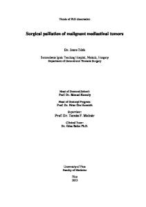
swallowing; grade 1, unable to swallow solids; grade 2, unable to swallow semisolids; grade 3, unable to swallow liquids; grade 4, unable to swallow own saliva. Ten patients had an ETF. The fistulas developed after the radiotherapy in seven patients with inoperable esophagus carcinoma. Three patients had a bronchial carcinoma. In one of these patients, mediastinal recurrence and esophagotracheal fistula occurred 3 years after lobectomy. Two patients had inoperable bronchial carcinoma, and fistula developed after the radiotherapy. Both of the patients had a central tumor and partial obstruction of main bronchus before the radiation therapy (Fig. 1).
Fig. 1. (A) Pre-stent esophagogram showing malignant obstruction with a fistula to the trachea. (B) After insertion of the covered stent, barium esophagram demonstrating complete occlusion of the fistula.
795
Four patients had an EPF. In two of them, fistula developed after the radiation therapy. Three of these patients with esophageal carcinoma had a fistula between anastomotic line and pleura after the operation (Fig. 2). In the other patient, who had undergone pneumonectomy because of bronchial carcinoma, fistula occurred due to mediastinal recurrence with esophageal invasion 2 years after the pneumonectomy. 2.1. Stent design We used 35 covered and eight non-covered SEMS, which are flexible in the longitudinal axis. The central portion of the covered stents has a polyurethane layer and aims at restricting tumor ingrowth. The inner diameter of the central portion of the stent is 17 – 18 mm when fully expanded. Its flanged ends measure 28 mm in diameter and facilitate anchoring of the stent to the esophageal wall. A 38F (13-mm) delivery system is used for insertion and consists of three coaxially arranged polypropylene tubes. The stent is preloaded on the inner tube while the outer tube compresses the stent. The central lumen of the inner tube allows guidewire insertion. This assists the introduction of the stent delivery system across an esophageal stricture. A 23% shortening occurs after full deployment. Two SEMS (100 or 120 mm in length) were used in our patients. This allowed for at least 2-cm portion of the stent beyond the proximal and distal margins of the gross tumor. 2.2. Stent insertion All patients were given general anesthesia for precise SEMS placement and patient comfort. Each lesion was assessed endoscopically, and the length of the stenosis was marked under fluoroscopic control using metallic markers
Fig. 2. (A) Barium esophagram demonstrating malignant stricture with a fistula to the pleura. (B) Repeat esophagram demonstrating complete occlusion of the fistula by the covered stent.
796
A. Sarper et al. / European Journal of Cardio-thoracic Surgery 23 (2003) 794–798
attached to the skin. To facilitate rapid expansion, all strictures were progressively dilated to 15 mm using flexible bougie dilators. The guidewire was inserted through the stricture via an endoscope, and the stent system was passed over it. The radiopaque markers of the delivery system were useful in the identification of the central part of the stent and allowed for accurate positioning of the stent across the structures. In patients with lesions near the gastroesophageal junction, the lower end of the stent was positioned in the fundus of the stomach, with the majority of the stent in the lower esophagus. Postoperatively, a chest roentgenogram was taken to exclude perforation and check the stent position.
development of symptoms and stenting was 14 days. The pneumonia could not be treated although the fistula was closed by a covered SEMS. In three out of four patients with EPF, fistula was closed successfully and stenting applied in proper position. But in one patient with mediastinitis, fistula repeated 6 days later. He died of sepsis 8 days after the stent placement. The patient with mediastinitis and EPF referred to our clinic 10 days after the development of symptoms. Although the fistula was treated by covered stent, this patient died of mediastinitis 17 days after the stenting. The median survival time of the patients with fistulas (ETF and EPF) was 49 days (range 5 – 186 days). 3.3. Overall complications and mortality
3. Results 3.1. Evaluation of the patients with esophageal stenosis alone Stent implantation was technically successful in 26 of 27 patients with esophageal stenosis alone. Malpositioning of the stent at deployment occurred in one patient, who had a malignant obstruction on the cardia. Dilation was needed. The median period of taking meals without the nutritional support after esophageal stent placements was 2.7 months (range, from 1 to 14 months). Mean dysphagia grade improved after stent placement (mean dysphagia grade before implantation was 3.4, mean dysphagia grade after implantation was 1.3, P , 0:05). In addition to this, dysphagia grade was gradually worse especially in the patients in whom a non-covered stent was used. A second stent was needed in two patients because of the stent obstruction caused by tumor ingrowth and overgrowth. In one of these patients, non-covered SEMS was used because covered SEMS were not commercially available at the time of the treatment. In the other patient, membrane separation from the stent was established. Both were treated by the insertion of a second stent. In two patients with distally malignant esophageal stenosis, perforation occurred during dilation. Covered SEMS was inserted. In one patient, perforation was healed after stent insertion. However, in the other patient, it failed, and mediastinitis and peritonitis developed. He died 8 days after the perforation. 3.2. Evaluation of the patients with fistula (ETF or EPF) The symptoms of fistula improved after the placement of covered stent in nine of the ten patients (90%) with ETF. In one patient, fistula recurred 2 days after the stent placement. Since fistula was just below the larynx, we did not prefer second stent placement. Gastrostomy was needed in this patient. He died of pulmonary insufficiency 22 days later. Another patient died 5 days after the stent placement. This patient had bilateral pneumonia and the time period between
Complications occurred in 11 (26.8%) patients (Table 2). Especially in the cases of tumor stenoses in the distal esophagus, complication rate was higher (44%, four of the nine patients). In total six patients (14.6%) died after stent placement during early postoperative period. Two patients had procedure-related mortality. In one patient massive hematemesis, and melana were noted, and he died 6 days after stent insertion. The second patient, in whom perforation occurred during dilation, died 8 days after the perforation. Remains four patients died because of natural progress or non-stent related complication in early postoperative period. In two patients, stenting could not treat the fistula. The fistula recurred and these patients died of complications of the fistula. Other two patients died of previously present overwhelming pneumonia and mediastinitis although the fistulas were closed by covered stent. The mean duration of hospital stay was 8.4 days (range, 21 – 23 days). During post-hospital period, all patients except two died as a result of the natural progression of the tumor. The median survival time was 94 days (range, 42– 431 days). Two patients are still alive and able to take a meal without nutritional support at 180 and 110 days after stenting.
Table 2 Complication in all patients Complications
n (%)
Chest pain lasting .24 h Regurgitation Hematemesis Aritmia Sensation of foreign body Malpositioning of the stent Incomplete stent expansion Tumor ingrowth and overgrowth Membrane separation Perforation Stent-related mortality
5 (12) 5 (12) 2 (4.9) 2 (4.9) 1 (2.4) 1 (2.4) 2 (4.9) 2 (4.9) 1 (2.4) 2 (4.9) 2 (4.9)
A. Sarper et al. / European Journal of Cardio-thoracic Surgery 23 (2003) 794–798
4. Discussion Palliation of malignant obstruction of the esophagus with endoscopically placed stent has been shown to improve patient quality of life by allowing restoration of oral alimentation [1,4]. Although plastic prostheses have been shown to be efficacious, gross dilatation of the tumor stenosis was necessary to enable the passage of these tubes [14 – 17]. Consequently, the rate of gross esophageal rupture at the tumor site, as well as aspiration, reflux, pneumonia, and sepsis with lethal complications was high, with a mortality rate of up to 42% [14 – 17]. SEMS have recently been introduced for this purpose [1,2,4 – 13]. Placement of the SEMS effectively relieved malignant dysphagia in 88.8% of patients treated in our series. The improvement in dysphagia grade after stent implantation was statistically significant. Tumor ingrowth and overgrowth is a significant problem with uncovered SEMS, occurring in 10– 35% of patients [1,2,4 –7]. One of the eight patients (12%) had a tumor ingrowth in our series. The addition of the covered membrane is successful in preventing tumor ingrowth. However, growth of the tumor above or below the stent may occur. The availability of longer stents may postpone this complication. In addition, covering of the SEMS (especially fully covered) may increase the rate of stent migration (2.5%) [4]. The SEMS used in our study are covered except at least 2 cm at least segment at each end. We did not encounter migration of the stent. SEMS are placed, they cannot be easily repositioned and are either very difficult or impossible to remove [5]. In some cases, malposition can be compensated by placing a second stent overlapping the first. Plastic and metal stents become dislocated in 5 – 10% of cases [7]. Malpositioning of the stent occurred in one patient. It was treated by dilation and restending did not require. Membrane separation from the SEMS observed in one patient has been addressed to laminating the membrane between two layers of wire mesh in a coaxial arrangement, which also increases the radial force exerted by the stent [1] ETF occur in approximately 5 – 15% of the patients with esophageal cancer [2,3,18,19]. The development of recurrent pulmonary infections secondary to aspiration and cachexia leads to short survival in patients with ETF, with most dying within 1 month [2,3,18]. Another form of fistula in these patients is EPF which is a very rare and fatal complication [20]. The surgical treatment of these fistulas has been associated with significant morbidity and an exceedingly high mortality rate [2,3,18]. The most reasonable palliation of the problems of malignant fistulas is an esophageal stenting. [2,3,5,6,14,18 – 20]. The covered SEMS was successful in completely sealing 80 –100% of the fistulas in literature [1 – 7,14,18 – 20]. In our patients with fistula (ETF and EPF) the rate was 85.7%. The median survival time of these patients in the present series was 49
797
days. This also compares favorably with literature, reporting median survival times of 31– 56 days [1,2,4,7,18 –20]. The timing for placement of stent is important. Stenting should be applied before complications of the fistula occurred. Especially in the patient with EPF, if the patients have a mediastinitis, morbidity and mortality rate is high. Complications of endoscopic stenting for malignant strictures have been well characterized [7]. Chest pain is a common complaint following stent insertion, with a reported incidence of up to 100% [7,8,10 –12]. In our series, 62% of patients complained of retrosternal pain (five of them lasting . 24 h and requiring narcotics), and this is most probably due to the dilation and stretching of the structures. Severe pain was related to the degree of stricture. Hematemesis is also a possible complication after SEMS insertion, and its incidence in our study is 5%. It is comparable with literature [4,7]. This complication could have been the result of pressure necrosis, the natural progress of the disease, or trauma from the sharp, uncovered end of the stent [8 – 10,12]. Food impaction is usually due to a lack of patient education or non-compliance with instructions for proper food selection, chewing, and swallowing. Stent diet instructions, verbal and written, should be given to patients and family members or caregivers prior to patients’ discharge from the hospital [19]. No food impaction occurred in our series. But regurgitation occurred in 38% patients with esophagogastric junction lesions. These patients were advised to sleep at 458 Fowler position to reduce reflux. Some were given H2-receptor blockers to relieve symptoms of reflux. Perforation is more serious complication in the patients with malignant dysfagia, and mortality rate is high. The incidence of perforations is more than 10% for plastic stents [14 –17] compared with less than 5% in SEMS [9,10]. Although this rate is 4.8% in our series, no perforations occurred in the last 30 patients, when our experience matured. As with other palliative therapies, increased risk and technical difficulties are the concerns for malignant stenoses in the cervical esophagus and at the gastroesophageal junction [21,22]. Lesions of the lower esophageal and gastric cardia can be effectively stented but do present potential technical problems [21]. Major complications (perforations, dislocation, hemorrhage) were higher than other location of the esophagus in our series. These were observed in four of the nine (44%) patients with distal esophageal stricture, and procedure related mortality rate was 23% in this group. In patients with poor general condition and more advanced tumors, rapid relief of dysphagia with minimum morbidity, enabling them to return home quickly and remain home during the terminal stage of their disease, is the ultimate goal. Although the SEMS are very expensive compared with the conventional plastic stents, palliation of malignant esophagotracheal or pleural fistulas and esopha-
798
A. Sarper et al. / European Journal of Cardio-thoracic Surgery 23 (2003) 794–798
geal stenoses using the SEMS may be alternative, and single treatment maintaining a patent esophageal lumen, especially in the case of tumor stenoses in the middle esophagus. But, the results of the patients with EPF are less enthusiastic in the presence of accompanying a mediastinitis. However, we think that if SEMS is applied as early as possible, the stenting may be more successful the in the patients with fistula.
References [1] Nelson DB, Axelrad AM, Fleischer DE, Kozarek RA, Silvis SE, Freeman ML, Benjamin SB. Silicone-covered Wallstent prototypes for palliation of malignant esophageal obstruction and digestiverespiratory fistulas. Gastrointest Endosc 1997;45:31 –7. [2] Raijman I, Lynch P. Coated expandable esophageal stents in the treatment of digestive-respiratory fistulas. Am J Gastroenterol 1997; 92:2188–91. [3] Duranceau A, Jamieson GG. Malignant tracheoesophageal fistula. Ann Thorac Surg 1984;37:346–54. [4] Wu WC, Katon RM, Saxon RR, Barton RE, Uchida BT, Keller FS, Ro¨sch J. Silicone-covered self-expanding metallic stents for the palliation of malignant esophageal obstruction and esophagorespiratory fistulas: experience in 32 patients and a review of the literature. Gastrointest Endosc 1994;40:22–33. [5] Ramirez FC, Dennert B, Zierer ST. Esophageal self-expandable metallic stents: indications, practice, techniques, and complications: results of a national survey. Gastrointest Endosc 1997;45:360–4. [6] Raijman I, Siddique I, Ajani J. Palliation of malignant dysphagia and fistulae with coated expandable metal stents: experience with 101 patients. Gastrointest Endosc 1998;48:172 –9. [7] Shields SJ. Esophageal self-expandable metallic stents. Gastrointest Endosc 1997;45:439–42. [8] Feins RH, Johnstone DW, Baronos ES, O’Neil SM. Palliation of inoperable esophageal carcinoma with the wallstent endoprosthesis. Ann Thorac Surg 1996;62:1603–7. [9] Watkinson AF, Ellul J, Entwisle K, Mason RC, Adam A. Esophageal carcinoma: initial results of palliative treatment with covered selfexpanding endoprostheses. Radiology 1995;195:821 –7.
[10] Ell C, Hochberger J, May A, Fleig W, Hahn E. Coated and uncoated self-expanding metal stents for malignant stenosis in the upper GI tract: preliminary clinical experiences with Wallstents. Am J Gastroenterol 1994;89:1496–500. [11] Song HY, Choi KC, Kwon HC, Yang DH, Cho BH, Lee ST. Esophageal strictures: treatment with a new design of modified Gianturco stent. Radiology 1992;184:729–34. [12] Vermeijden JR, Bartelsman JFWM, Fockens P, Meijer RCA, Tytgat GNJ. Self-expanding metal stents for palliation of esophageal malignancies. Gastrointest Endosc 1995;41:58–63. [13] Alfred M, Hans P, Gerhard BF, Heiko R, Freyja MSJ. Self-expandable coated stent after intraluminal treatment of esophageal cancer: a risky procedure? Ann Thorac Surg 1999;67:781– 4. [14] Stemerman DH, Caroline DF, Dabezies M, Mercader VP, Krevsky B, Gatenby RA. Non-expandable silicone esophageal stents for treatment of malignant tracheoesophageal fistulas: complications and radiographic appearances. Abdom Imaging 1997;22:14 –19. [15] Buess G, Schellong H, Kometz B, Gru¨bner R, Junginger TH. A modified prothesis for the treatment of malignant esophagotracheal fistula. Cancer 1988;61:1679–84. [16] Cusumano A, Rual A, Segalin A. Push through intubation: effective palliation in 409 patients with cancer of the esophagus and cardia. Ann Thorac Surg 1992;53:1010– 4. [17] Kratz JM, Reed CE, Crawford FA. A comparison of endoesophageal tubes. J Thorac Cardiovasc Surg 1989;97:19– 23. [18] Tomaselli F, Maier A, Sankin O, Woltsche M, Pinter H, Freyja MSJ. Successful endoscopical sealing of malignant esophageotracheal fistulae by using a coverved self-expandable stending system. Eur J Cardiothorac Surg 2001;20:734–8. [19] Boyce Jr HW. Palliation of advanced esophageal cancer. Semin Oncol 1984;11:186–95. [20] Watkinson A, Ellul J, Entwisle K, Farrugia M, Mason R, Adam A. Plastic-covered metallic endoprostheses in the management of oesophageal perforation in patients with oesophageal carcinoma. Clin Radiol 1995;50:304–9. [21] Spinelli P, Cerrai FG, Ciuffi M. Endoscopic stent placement for cancer of the lower esophagus and gastric cardia. Gastrointest Endosc 1994; 40:455–7. [22] Goldschmid S, Boyce Jr HW, Nord HJ. Treatment of pharyngoesophageal stenosis by polyvinyl prosthesis in. Gastrointest Endosc 1988; 83:513–8.