The effectiveness of syringe irrigation and ultrasonics to remove debris from simulated irregularities within prepared root canal walls
S.-J. Lee1, M.-K. Wu2 & P. R. Wesselink2 1
Department of Conservative Dentistry, Chonbuk National University, School of Dentistry, Chonju, South Korea; and 2Department of Cariology Endodontology Pedodontology, Academic Centre for Dentistry Amsterdam (ACTA), Amsterdam, The Netherlands
Abstract Lee S-J, Wu M-K, Wesselink PR. The effectiveness of syringe irrigation and ultrasonics to remove debris from simulated irregularities within prepared root canal walls. International Endodontic Journal, 37, 672–678, 2004.
Aim To compare the ability of syringe irrigation and ultrasonic irrigation to remove artificially placed dentine debris from simulated canal irregularities within prepared root canals. Methodology After canal enlargement, twelve canines were split longitudinally into two halves. On the wall of one half of each root canal a standard groove of 4 mm in length, 0.2 mm in width and 0.5 mm in depth was cut, 2–6 mm from the apex, to simulate uninstrumented canal extensions. On the wall of the other half, three standard saucer-shaped depressions of 0.3 mm in diameter and 0.5 mm in depth were cut at 2, 4 and 6 mm from the apex to simulate uninstrumented canal irregularities. Each groove and depression were filled with dentine debris mixed with 2% NaOCl to simulate a situation when dentine debris accumulates in uninstrumented canal extensions and irregularities during canal preparation. Each tooth was re-assembled by reconnecting the two halves, using
Introduction One of the most important procedures in root canal treatment is chemomechanical preparation of the canal
Correspondence: M.-K. Wu, Department of Cariology Endodontology Pedodontology, ACTA, Louwesweg 1, 1066 EA Amsterdam, The Netherlands (Tel.: +31 20 518 8367; fax: +31 20 669 2881; e-mail:
[email protected]).
672
International Endodontic Journal, 37, 672–678, 2004
wire and an impression putty material. Two per cent NaOCl was then delivered into each canal either using syringe irrigation (n ¼ 8) or using ultrasonic irrigation (n ¼ 8). Before and after irrigation, images of the two halves of the canal wall were taken, using a microscope and a digital camera, after which they were scanned into a PC as TIFF images. The amount of remaining dentine debris in the grooves and depressions was evaluated by using a scoring system between 0–3: the higher the score, the more the debris. The data were analysed by means of the Mann–Whitney U-test. Results Both forms of irrigation reduced the debris score significantly. The debris score was statistically significantly lower after ultrasonic irrigation than after syringe irrigation (P ¼ 0.002 for grooves, P ¼ 0.047 for depressions). Conclusion Ultrasonic irrigation ex vivo is more effective than syringe irrigation in removing artificially created dentine debris placed in simulated uninstrumented extensions and irregularities in straight, wide root canals. Keywords: dentine debris, irrigation. Received 15 September 2003; accepted 22 April 2004
system. Irrigation is complementary to instrumentation in facilitating removal of bacteria, debris and therapeutic materials such as gutta-percha, sealer and medicaments from root canals. The effectiveness of irrigation relies on both the mechanical flushing action and the ability of irrigants to dissolve tissue. Sodium hypochlorite (NaOCl) has been used as an endodontic irrigant for at least six decades (Walker 1936). Irrigation with NaOCl has been
ª 2004 International Endodontic Journal
Lee et al. Removal of debris from root canal
shown to dissolve organic tissues (Hand et al. 1978, Moorer & Wesselink 1982) and to be antibacterial in root canals (Bystro¨m & Sundqvist 1983). The flushing action of irrigants helps to remove organic and dentinal debris and microorganisms from the canal. Indeed, it has been suggested that the flushing action may be more important than the ability to dissolve tissue (Baker et al. 1975). The flushing action created by syringe irrigation is relatively weak and dependent not only on the anatomy of the root canal system but also on the depth of placement and the diameter of the needle (Abou-Rass & Piccinino 1982, Chow 1983). Increase in the volume of irrigant does not significantly improve its flushing action and its efficacy to remove debris (Walters et al. 2002). The flushing action of irrigants may be enhanced by using ultrasound because the directional flow from apical to coronal and eddies produced through the acoustic streaming created by the vibrating instrument is more intense in velocity and magnitude around its tip (Ahmad et al. 1987, Stock 1991). In a study by Cunningham et al. (1982a), the flushing action of hand syringe irrigation was compared with ultrasound using saline as the irrigant. Ultrasound removed 86% of the bacterial spores from the root canal while hand syringe irrigation removed 62%. Another potential methodology where the flushing action could be enhanced is the use of the noninstrumentation technique (NIT) (Lussi et al. 1995, 1997). Excellent cleaning was achieved after irrigation with NaOCl by generating alternating pressure fields within a reduced pressure environment thus creating hydrodynamic turbulence (Ardila et al. 2003). However, only limited clinical studies have been performed to date (Lussi et al. 1997). Various instruments are used to prepare root canals (Spa˚ngberg 1998). Rotary hand preparation techniques and rotary instruments with radial lands tend to produce round preparations (Vessey 1969, Hu¨lsmann et al. 2001). These round preparations may be produced in the centre or at one side of a long oval canal leaving uninstrumented canal extensions (Walton 1976, Gambill et al. 1996, Peters et al. 2001, Wu et al. 2001, 2003, Wu & Wesselink 2001, Ardila et al. 2003, Bergmans et al. 2003). In addition, many posterior teeth have complex isthmuses and other natural irregularities. Following conventional hand irrigation the uninstrumented canal extensions and irregularities may harbour debris and bacteria, thereby making thorough canal debridement difficult (Cunningham et al. 1982b,
ª 2004 International Endodontic Journal
Goodman et al. 1985, Wu et al. 2001, Wu & Wesselink 2001). If the dentine debris is not removed completely from the uninstrumented extensions, two unfavourable outcomes may result. First, calcium hydroxide and other medicaments that function only when in direct contact with the pathogens (Siqueira & Lopes 1999), cannot be placed in the space occupied by debris, and therefore could not be effective. Secondly, only those areas free of debris can be filled with gutta-percha and sealer, therefore, the debris-filled canal extensions may lead to leakage (Wu et al. 2001). Clearly, thorough removal of all debris is an essential part of root canal treatment. The purpose of this study was to evaluate and compare the ability of syringe irrigation and ultrasonic irrigation to remove artificial dentine debris from simulated canal extensions and irregularities.
Materials and methods The root canals in twelve maxillary and mandibular canines were used. The canals were accessed and prepared. The working length was established by deducting 1 mm from the actual canal length, which was determined by inserting a size 15 file into the canal until the tip of the file was just visible at the apical foramen. The coronal aspect of each canal was flared, using Gates Glidden drills (Dentsply Maillefer, Ballaigues, Switzerland); sizes 2–4, to a depth where resistance was met. Canals were then prepared using Flexofiles (Dentsply Maillefer) with a balanced-force technique (Roane et al. 1985) to size 50 at the working length and to sizes 55, 60, 70 and 80 to 1, 2, 3 and 4 mm from the working length respectively. Between files, each canal was irrigated with 2 mL of a freshly prepared 2% solution of sodium hypochlorite, using a syringe and a 27-gauge needle that was placed to 1 mm short of the working length, resulting in a total volume of 50 mL. The NaOCl solution was prepared by diluting a 10% NaOCl solution (Merck, Darmstadt, Germany). Its pH was adjusted to 10.8 with 1 N HCl. The concentration of the NaOCl solution was measured iodometrically (Moorer & Wesselink 1982). Two grooves were cut along the long axis of each tooth using a diamond disc (Horico, Berlin, Germany). All teeth were then split longitudinally using a chisel. The working portion of a hand spreader (A60, Dentsply Maillefer) was removed and the end of the shank sharpened to v shaped. Using this modified hand spreader, a standard groove 4 mm in length was created in the wall of one half of each root
International Endodontic Journal, 37, 672–678, 2004
673
Removal of debris from root canal Lee et al.
canal, 2–6 mm from the apex (Fig. 1a), to simulate uninstrumented canal extensions. The groove was 0.5 mm deep and 0.2 mm wide that was comparable with the diameter of narrow oval canals (Wu et al. 2000). The diameter of a round bur (006, Dentsply Maillefer) was reduced to 0.3 mm by grinding it on SiC-paper. Using this modified round bur, three standard saucershaped depressions, each 0.3 mm in diameter and 0.5 mm deep, were cut in the wall of the other half, at 2, 4 and 6 mm from the apex, in order to simulate uninstrumented canal irregularities (Fig. 1b). Dentine debris was produced by grinding dentine with round burs from the pulpal to the cementum side of a number of other teeth that had been split longitudinally. The dentine debris was mixed with 2% NaOCl 5 min before use; a wet sand-like mixture was prepared. Using a paper point, the grooves and depres-
sions were then filled with debris, taking care not to compact the debris. The two halves of each tooth were then re-assembled using wires and putty impression material (Provil novo, GmbH, Hanau, Germany). Either syringe irrigation or ultrasound irrigation was performed in the canal, using 2% NaOCl as irrigant. Ultrasound irrigation was performed in eight canines; each canal was irrigated with approximately 200 mL of a 2% NaOCl and a PMAX device (Satelec, Meriganc Cedex, France). After switching on the ultrasound device, an activated size-15 file was placed within 1 mm of the working length, thus, oscillation of the file and irrigation began almost at the same time. The oscillation in the direction of the groove at speed three lasted for 3 min. According to the manufacturer, the frequency employed under these conditions was approximately 30 kHz.
(a) Groove
Canal wall
0.2 mm
0.5 mm Cross section of the groove
2 mm 6 mm 0.3 mm
(b) Canal wall
Hole 0.5 mm Cross section of the hole
2 mm 4 mm 6 mm
674
International Endodontic Journal, 37, 672–678, 2004
Figure 1 Schematic representation of specimen preparation. On one half (a) of the instrumented root canal a groove was cut 2 to 6 mm from the apex. One the other half (b) three depressions were cut 2, 4 and 6 mm from the apex.
ª 2004 International Endodontic Journal
Lee et al. Removal of debris from root canal
Syringe irrigation was performed in four teeth. As the syringe irrigation procedure is not thought to damage the canal wall, it was deemed acceptable to use each tooth twice. Thus, a same-sample size of eight was used for the syringe irrigation. It has been reported that a suitable irrigation regimen would include 2 mL of sodium hypochlorite applied between each instrument (Walters et al. 2002) followed by a final flush of 5–10 mL (Santos et al. 1999). Occasionally 18 hand instruments may be used in each canal (Buchanan 2000). To simulate this situation, 50 mL of 2% NaOCl was injected into each canal for approximately 7 min using a 10-mL syringe and a 27-gauge needle, the tip of the needle was 1 mm short of the working length. The root halves were separated after the irrigation procedure in order to evaluate the removal of dentine debris. Images of the two halves of the canal wall were taken before and after irrigation, using a Photomakroskop M400 microscope with digital camera (Wild, Heerbrugg, Switzerland) at ·40 magnification; the pictures were scanned into a PC as Tagged Image File Format (TIFF) images. The amount of dentine debris in the groove and depressions was then scored by one investigator who did not know which irrigation technique had been performed. The first score (before irrigation) was used to assess whether the canals in the two groups (ultrasound and syringe) contained a comparable amount of debris before irrigation. A higher score indicated a larger amount of debris: score 0: the entire groove or depression was free of debris; score 1: less than half of the groove or depression was filled with debris; score 2: half or more than half of the groove or depression was filled with debris; and score 3: the entire groove or depression was filled with debris. The average score of the three depressions was used as the depression score for each specimen. Each specimen was finally re-examined by a second investigator. The difference in debris scores after the use of different irrigation techniques was analysed by means
Table 1 Debris scores after syringe irrigation and ultrasound irrigation Syringe irrigation n
Groove
Ultrasound irrigation
Depression* Groove
1 3.0 2.3 2 1.0 0.7 3 3.0 0.7 4 1.0 1.3 5 2.0 1.3 6 2.0 0 7 3.0 2.3 8 3.0 3.0 Sum 18.0 (25%) 11.6 (52%) (% reduction)
0 0 0 1.0 0 0 2.0 0 3.0 (88%)
Depression* 0.7 0 0 2.0 0.7 0.7 0.3 0 4.4 (82%)
*Because there were three depressions each root, an average depression score is presented for each root.
of the Mann–Whitney U-test. The level of significance was set at a ¼ 0.05.
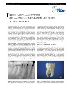
Results The two investigators differed in scoring five specimens; agreement was achieved following discussion. Before irrigation, the groove score and the average depression score were three for each specimen. The scores after irrigation are shown in Table 1. Overall less dentine debris remained in the grooves (P ¼ 0.002) and depressions (P ¼ 0.047) after ultrasonic irrigation (Figs 2 and 3); the summated score for grooves was reduced from 24 to 3 (88% reduction) while the summated score for depressions was reduced from 24 to 4.4 (82% reduction) (Table 1). Debris was completely removed after ultrasonic irrigation from grooves in six of the eight specimens (75%), and from depressions in three of the eight specimens (38%) (Table 1).
Discussion For the purposes of this study, a new model was designed to study the effectiveness of irrigation in
Figure 2 A specimen after syringe irrigation. The groove (a) and depressions (b) were still full of debris (score 3).
ª 2004 International Endodontic Journal
International Endodontic Journal, 37, 672–678, 2004
675
Removal of debris from root canal Lee et al.
Figure 3 A specimen after ultrasound irrigation. In one-half of the specimen (a) the groove was shown to be free of debris (score 0). In the other half (b) debris had been removed from the depressions, but each depression still contained a small amount of debris (score 1).
removing dentine debris in artificial irregularities and extensions of the root canal. In order to simulate uninstrumented extensions in the apical half, a groove was cut in the canal wall of each root 2–6 mm from the apex. Canines were used because they have wide canals (Kerekes & Tronstad 1977, Wu et al. 2000) and therefore, the roots could be easily split through the canals. The advantage of this model is that the amount of debris present both before and after irrigation can be compared, whereas in previous studies the amount of debris was evaluated only after preparation and irrigation (Abbott et al. 1991, Lumley et al. 1993, Wu & Wesselink 1995). Because it was not clear how much debris was present before irrigation in those studies, it could not be established how much was removed using the different irrigation procedures. This means that the ranking of the different techniques used to remove debris in these studies may not have been reliable. In the present study, the canals in the ultrasound and the syringe groups contained comparable amounts of debris before irrigation, as assessed by the initial scoring. A relatively small sample size (n ¼ 8) was applied but a statistically significant difference was indeed found, indicating that the model was sensitive, and a real difference existed between the two techniques. Application of suitable sample size is essential to minimize the risk of type I or II error (Schuurs et al. 1993). For either grooves or depressions the two mean scores significantly differed (Table 1, P < 0.05), indicating that the null hypothesis (H0), under which the two mean scores using the two different irrigation techniques did not differ, was rejected and the risk to mistakenly reject H0, i.e. type I error, was