Turkish Neurosurgery 2009, Vol: 19, No: 4, 360-366
The Effect of Nitrogen Mustard on the Rat Brain and the Therapeutic Value of Proanthocyanidin
Original Investigation
1
Ayhan TEKINER 2
Orhan YUCEL
3
Ayse KOSE SARGIN Onur GENC
4
5
Belgin CAN
6
Turhan KARAYILANOGLU
Nitrogen Mustard’›n Rat Beyni Üzerine Etkisi ve Proanthocyanidin’in Tedavi De¤eri
7
Jale KARAKAYA
8
Mehmet Akif BAYAR
1,8
ABSTRACT AIM: Nitrogen Mustard (NM) is an alkylating agent that damages cellular nuclear DNA after penetrating tissue. This results in cytostatic, mutagenic and cytotoxic effects. We used the electron microscope to investigate the effect of NM gas administered through the dermal and respiratory routes to rats on the brain cortex and also tried to show whether the antioxidant Proanthocyanidin (PC) could decrease this effect. MATERIAL and METHODS: A total of 32 rats were randomized into four groups: Group I: Control group, Group II: PC group, Group III: NM group, Group IV: NM + PC group. The rats were sacrificed 3 days after NM gas exposure. A segment of the cortical tissue was prepared for electron microscopy.
2,4
3,5
6
7
Ministry of Health, Training and Research Hospital, Department of Neurosurgery, Ankara, Turkey Gülhane Military Medical Academy, Department of Thoracic Surgery, Ankara, Turkey Ankara University School of Medicine, Department of Histology and Embryology, Ankara, Turkey Gülhane Military Medical Academy, Department of Nuclear, Biologic and Chemical Warfare, Ankara, Turkey Hacettepe University, School of Medicine, Department of Biostatistics, Ankara, Turkey
RESULTS: We used the electron microscope for cellular analysis of NM on cortical neural cells. These investigations revealed degeneration of the cortical neural cell nuclei together with oedema and axonal degeneration in the subcortical neural tissue. The group receiving antioxidants was found to have less oedema and degeneration. CONCLUSION: These findings imply that structural changes induced by mustard gas can be prevented and restored by proanthocyanidin treatment.
Received : 08.05.2009 Accepted : 31.08.2009
KEYWORDS: Nitrogen Mustard, Proanthocyanidin, Rat brain
ÖZ AMAÇ: Nitrogen Mustard (NM) dokuya penetre olduktan sonra hücresel nükleer DNA’ya hasar veren alkilleyici bir ajandır. Bu sitostatik, mutagenik ve sitotoksik etkilere neden olur. Cilt ve respiratuar yoldan uygulanan NM gazının beyin korteksine olan etkisini araştırmak ve aynı zamanda antioksidan Proanthocyanidinin (PC) bu etkiyi azaltıp azaltamayabileceğini göstermek için elektron mikroskobu kullandık. YÖNTEM ve GEREÇ: Toplam 32 rat dört gruba randomize edildi: Grup I: Kontrol grubu, Grup II: PC grubu, Grup III: NM grubu, Grup IV: NM+PC grubu. Ratlar NM gazı uygulamasından 3 gün sonra sakrifiye edildiler. Elektron mikroskop için kortikal bir doku segmenti hazırlandı. BULGULAR: NM’ın kortikal nöral hücreler üzerindeki hücresel etkisi için electron mikroskop kullandık. Bu incelemeler, ödemle birlikte kortikal nöral hücre nükleuslarının degenerasyonunu ve subkortikal nöral dokudaki aksonal degenerasyonu ortaya koydu. Antioksidan alan grupta daha az ödem ve degenerasyon gözlendi. SONUÇ: Bu bulgular mustard gazının indüklediği yapısal değişikliklerin proanthocyanidin tedavisi ile önlenebildiği ve onarılabildiğini ima etmektedir.
Correspondence address: Ayhan TEKINER E-mail:
[email protected]
ANAHTAR SÖZCÜKLER: Nitrogen Mustard, Proanthocyanidin, Rat beyni
360
Turkish Neurosurgery 2009, Vol: 19, No: 4, 360-366
INTRODUCTION Nitrogen Mustard (NM) is a structural analog of sulfur mustard, a potent warfare agent that affects the skin, eyes, lungs and the neuromuscular, hematological, gastrointestinal, endocrine and immune systems (3,5,7,10). It was first used as a chemical warfare agent during World War I and caused severe casualties. It was banned when the Geneva Convention signed in 1925 banned the use of all chemical gases in wars. It has not been used to a marked degree in World War II. Recent uses were during the Iran-Iraq war during 1982-1987 and against the civilian population in Halepce by the Iraqi government. NM is absorbed via the skin or the anterior surface of the eyes, and by inhalation (2). Its most common effect on the eye is conjunctival irritation with lacrimation (9). Although these agents have been studied for years, the main events that initiate cell death and the cytotoxic mechanisms induced by NM have still not been fully elucidated. The mechanism of mustard injury is thought to be associated with target alkylation (11). This results in cytostatic, mutagenic and cytotoxic effects. The interaction between NM and cellular structures is through ethylene imonium. The molecule creates a ring structure and binds to macromolecules. This ring alkaline structure affects the nucleophilic parts of intracellular macromolecules. The major alkylating reaction in nucleic acids is through the 7th nitrogen of guanine. It thus leads to the formation of cross-links between or within DNA helices. The 3rd nitrogen of adenine and the 6th oxygen atom of guanine are other regions where the alkylation reaction can be seen. It may also cause damage to RNA, proteins and the cell (3). These effects may lead to chromosomal aberrations in addition to inhibiting DNA, RNA and protein synthesis with the cells pausing at the G2-M phase of the cycle. The result is severe damage, especially in cells with high mitotic activity (3). The poly (ADP-ribose) polymerase (PADRPR) enzyme is activated on exposure to mustard gas, thus decreasing NAD+ levels. This causes cell death. A normal NAD+ concentration ensures continuity of the energy-providing system and prevents blister formation (3). Nicotinamide may be used as a reversible PADRPR inhibitor and this agent has been reported to decrease cytotoxicity when used within 24 hours of exposure to NM. DNA alkylation by mustard causes breaks in the DNA chain, stimulating the DNA repair mechanisms and
Tekiner et al: The Effect of Nitrogen Mustard on the Rat Brain
the activation of the PADRPR enzyme, which uses NAD+ as a substrate. Mustard also inhibits glycolysis and stimulates the NADP+-dependent hexose monophosphate shunt. These changes in cellular metabolism are reported to cause death especially in basal epidermal cells. The aim of this experimental study was to show the effects of mustard gas at the intracellular level and determine the effect of strong antioxidants such as PC in order to elucidate the diagnostic and therapeutic process to contribute to other studies aiming to decrease morbidity and mortality (1,13). The warfare agent mustard gas was administered to the subject via the transdermal and inhalation routes under conditions simulating the battlefield in this study. The combined effect of firearms and chemical weapons is usually made use of in current terrorist attacks. We investigated the harmful effect of mustard gas on neural tissue using an electron microscope in this study and also evaluated the efficacy of Proanthocyanidin (PC) as we thought it might be beneficial in subjects exposed to mustard gas. MATERIAL and METHODS I. Material: a- Animals: This experimental study took place at the Gulhane Military Medical Academy (GATA) Animal Experiments Laboratory. The Gulhane Military Medical Academy Animal Care and Usage Committee approved the experimental protocol. A total of 32 male Rattus Norvegicus weighing 140-160 g were used. These rats were randomized into four groups, each with eight rats. b- Chemicals: All chemicals were obtained from Sigma-Aldrich Chemie GmbH (Taufkirchen, Germany) and all organic solvents from Merck KGaA (Darmstadt, Germany). All reagents were of analytical grade, were prepared fresh each day (except the phosphate buffer) and stored in a refrigerator at +4 C. The reagents were equilibrated at room temperature for 0.5 h before use when the analysis was initiated or reagent containers were refilled. Phosphate buffers were stable at +4 C for 1 month. The animals were taken care of according to the relevant articles of the Helsinki Declaration and the guidelines of the U.S. National Institutes of Health. The mustard gas was administered to the subjects at the Nuclear Biologic and Chemical (NBC) laboratory and the sacrifice was 361
Turkish Neurosurgery 2009, Vol: 19, No: 4, 360-366
Tekiner et al: The Effect of Nitrogen Mustard on the Rat Brain
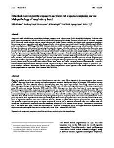
performed at the Experimental Animals Breeding and Research Laboratory. A microscope (Zeiss OpMi 99) and microsurgical instruments were used during the surgical procedures and decapitation. The electron microscopy investigation was performed at the laboratory of the Ankara University School of Medicine, Department of Histology and Embryology. II. Methods: Experimental design: A- Groups: These rats were randomized into four groups, each with eight rats as follows: Group I: Control group (no trauma and no NM). Group II: (PC group) The subjects were fed a diet including PC (100 mg/kg/day). Group III: (NM group) A toxic dose of vaporized 8 mg NM dissolved in 5 ml of distilled water was used for 10 minutes (800 mg/m3/min) on the NM group that was exposed to NM only. Group IV: (NM + PC group) These subjects were exposed to the same dose of NM and were fed a diet containing 100 mg/kg/day PC, administered orally via an orogastric injection. All exposures were carried out in a chamber 100 L in volume equipped with CBR filters. All rats were sacrificed with a lethal dose of xylazine and ketamine 72 hours later. B- Trauma induction by Mustard Gas administration and Perfusion-Decapitation-Obtaining tissue samples. 1. Anesthesia: Anesthesia was provided before the surgical procedure to the subjects in each group with an intramuscular injection of Ketamine hydrochloride (Ketalar 5% solution, Eczacıbaşı İlaç Sanayii, İstanbul with Parke-Davis license) - 35 mg/kg and Rompun (Xylazine 2% solution, Bayer, Istanbul) - 1.5 mg/kg. The rats in Group III and Group IV were administered 0.5 mg/kg NM gas. The subjects were placed in the chamber (Figure 1). The chamber was heated using a tungsten electric bulb (100/220V) with an average temperature of 22ºC +/- 2ºC. The subjects were directly exposed to vaporized NM. Rats in the PC Group were fed with PC beginning 8 h before the NM gas application and continuing 72 h afterwards. We assessed the rectal temperature, the number of breathing in one minute and the number of heart beats in one minute of the subjects twice a day for 72 hours. The activities and the neurological deficits of the subjects were also evaluated. The subjects were allowed to survive for an additional 72 h. All subjects were sacrificed on
Figure 1: The chamber
the third day (with a lethal dose of Ketamin + Xylazine). Buterphenol (0.5 mg/kg sc) was administered to the subjects to provide analgesia during this process. The parietal brain cortex (4x4x4 mm.) of the sacrificed subjects was removed for histological studies (macroscopic and electron microscopic). 2. Perfusion-Decapitation-Obtaining Tissue Samples: Subjects in all groups were put to sleep again with the same method following the specified duration (72 hours). A thoracotomy was performed and 1000 ml of 0.9% NaCl was administered at 100mmHg pressure through a catheter delivered from the left ventricle to the aorta. The right atrium was opened and the infused saline withdrawn. Perfusion was continued until the fluid was clear. The animals were then sacrificed by decapitation. The scalp was opened, craniotomy performed and the calvarium exposed in the middle. The brain and brainstem were removed intact. Tissue samples 4x4x4 mm in size were obtained from the hemisphere. A microscope and microsurgical instruments were used during these procedures. III. Tissue preparation: Procedure for Histological Examination Following a period of 72 hours, the animals were anesthetized with ketamine (85 mg/kg) and their craniums were opened by craniotomy. The brains were removed immediately, sectioned into small pieces (4x4x4mm) and fixed in 2.5% glutaraldehyde in 0.1 mol/l phosphate buffer, pH 7.2, at +4ºC for 2-4h and then post-fixed in 1% osmium tetroxide in phosphate buffer (pH 7). The materials were later dehydrated in serially increasing concentrations of alcohol. The tissues 362
Turkish Neurosurgery 2009, Vol: 19, No: 4, 360-366
Tekiner et al: The Effect of Nitrogen Mustard on the Rat Brain
were then washed with propylene oxide and embedded in Araldite 6005 (Ciba-Geigy, Summit, N.J., USA). Semi-thin sections of 0.8 μm were cut with a glass knife on a Leica Ultracut R ultramicrotome (Leica, Solms, Germany), stained with toluidine blue azur II and then examined under a Zeiss Axioscope photomicroscope (Thornwood, N.Y., USA). Ultrathin sections of 60 nm were cut with a glass knife on a Leica Ultracut R ultramicrotome, stained with uranyl acetate and lead citrate and examined on a LEO 906 E (LEO Elektronenmikroskopie, Oberkochen, Germany) transmission electron microscope. IV. Statistical Analysis: The Fisher-Freeman-Halton exact test was used to compare categorical variables among groups. p values less than 0.05 was evaluated as statistically significant. Statistical analysis was performed with SPSS 15.0 for Windows (SPSS Inc., Chicago, IL). RESULTS All slides obtained from all groups in this experimental study were studied with light and electron microscopy. These images obtaining from subjects of all groups were assessed for nucleus, mitochondria and perineural edema. The groups were compared in terms of pathological features. There were statistically significant differences between the groups (p