European Heart Journal (1998) 19, 1766–1775
Article No. hj981204
The Denolin Lecture The woman with congenital heart disease* J. Somerville GUCH Unit, Royal Brompton Hospital and Harefield NHS Trust, London, U.K.
Introduction Congenital heart disease, the main concern of paediatric cardiology, now presents problems for adult cardiologists, untrained and uninterested in such patients. Paediatric cardiology, stimulated by the challenge of cardiac surgery for congenital anomalies, has focused on anatomical disorders, changing nomenclature (often), embryological ‘myths’ and the results of various and many remarkable surgical feats. The latter’s success has created a new medical community — the ‘grown-up’ congenital heart patients — the GUCH. The patients’ gender has not concerned anyone. Whether it influences outcomes or natural history in congenital heart disease is neither known nor discussed. Since the opening of the adolescent cardiac unit in 1975 at the National Heart Hospital, which expanded into the GUCH unit now in the Royal Brompton, a database has been developed over the 20 years. This contains information on more than 5000 GUCH patients, and provides the basic data for this lecture. As a specialist unit within Cardiology there is bias for referral of complex problems which influence statistics, sometimes giving too gloomy a picture. Certainly the care of and provision for GUCH patients includes all teenagers (adolescents) and adults for their full life span, irrespective of gender. Whether gender influences outcomes and problems will be examined. The data on prevention and treatment of coronary heart disease and hypertension in men do not automatically apply to women. A woman is not an honorary man. By definition in the Shorter Oxford Key Words: Women, grown-up congenital heart disease. Manuscript submitted 14 June 1998, and accepted 3 July 1998. *Based on the European Society of Cardiology Lecture on Clinical Cardiology, The Denolin Lecture, given at the European Society of Cardiology, Stockholm, 26 August 1997. Correspondence: Dr Jane Somerville, GUCH Unit, Royal Brompton Hospital and Harefield NHS Trust, Sydney Street, London SW3 6NP, U.K. 0195-668X/98/121766+10 $18.00/0
English Dictionary (1956) a woman is ‘an adult female human being’, additionally with secondary definitions ‘a female servant’, ‘a mistress’, ‘a wife’ and ‘the reverse of a coin referring to Britannia’. A female belongs to the sex which bears offspring — it is this characteristic which makes a woman different from a man. A child becomes an adult when reaching a particular age, which varies from country to country, defined for the law, for health management, government, etc. Adolescence and adolescents are not recognised officially in the United Kingdom’s Department of Health; one is either a child or an adult at age 16 years. Since a female can and does bear children before the age of 16 years it is difficult to know when womanhood begins from the definitions.
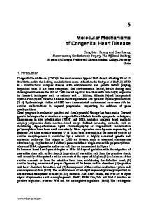
Gender incidence of congenital cardiac anomalies The old textbooks of paediatric cardiology and congenital heart disease, particularly Keith, Rowe and Vlad[1] provide us with knowledge of the incidence of the various anomalies. Four lesions appear to be more common in the female — mitral valve prolapse, secundum atrial septal defect, persistent duct (provided it is not caused by rubella when it is equally distributed between the sexes), and common atrium; all conditions affecting the inflow of the heart. Male dominance occurs in transposition of the great arteries, aortic valve stenosis, coarctation, univentricular hearts, Fallot’s tetralogy and total anomalous pulmonary venous drainage. Other lesions, including those that are rare, appear to be equally distributed between the sexes. Pulmonary valve stenosis has an increased incidence in males when born in the autumn (conceived in the winter) and an increase in females born in the spring (conceived in the summer)[2]. The result is that there are more males with congenital heart disease, in part explained by the relative frequency of the male dominated aortic valve disease and coarctation. � 1998 The European Society of Cardiology
The Denolin Lecture
(a)
1767
(b)
Res. coarctation
130
Fallot (98% TC)
269
Tricuspid atresia
50
One ventricle (DIV)
55
CPA/Pulm. atresia
101
Eisenmenger
112
0
10 20 30 40 50 60 70 80
PVS—valvotomy
96
TGA Mustard
54
Ebstein
41
Ostium primum ASD
79
Secundum ASD
200
A-V canal
26
0
20
40
60
Number of patients
67
Number of patients
AVS (valvotomy 60% systemic. Appeared pregnant and refused termination. Local obstetrician declined referral outside region. Special risks and needs explained by cardiologist. When 37 weeks pregnant the patient complained of tiredness. Labour was induced on a Friday evening. On Saturday morning, without consultant supervision, midwife and new junior registrar delivered a 7 lb baby. In third stage the cord pulled to remove the placenta. The patient collapsed and had a cardiac arrest. The cardiologist was called (for the first time!) but patient was unresuscitatable.
Aortic stenosis This is probably the commonest lesion in the pregnant ‘at risk’ congenital heart population, despite the condition being more common in the male. In the GUCH joint series[4] there were 58 patients with significant aortic stenosis who had 129 pregnancies: 50 (86%) had aortic valve stenosis, important in 14 with a resting gradient >50 mmHg at start or prior to pregnancy. This is the most important fact since the measured gradient doubles after the 13–15th week and is not a true reflection of the severity of the obstruction. Patients with gradients below 60 mmHg had no problems unless there was associated anaemia, infection, pre-eclampsia, or poor obstetrics. Those with severe aortic valve stenosis develop pulmonary oedema if not closely supervised. In three with pulmonary oedema after the 25th week, intravenous diuretics and rest controlled the situation until delivery by caesarean section when the infant was viable. Surgery is possible during pregnancy and avoids the dangers at delivery. Alternative therapy is catheter intervention, which can be performed by covering the abdomen all round with lead and has been successful in one patient at 32 weeks whose valve was domed and pliable without aortic regurgitation. Subaortic stenosis, where the gradients vary in the same patient, had no problems in pregnancy, except one with very severe concentric hypertrophy who developed runs of ventricular ectopy at 34 weeks and breathlessness responding to rest and small doses of diuretics; she was delivered by caesarean section at 37 weeks. Coarctation of the aorta before and after resection does not usually have a problem. If antihypertensive therapy is used it is important not to reduce pressure beyond the coarctation excessively, otherwise miscarriage may occur. There is a small risk of aneurysm formation and rupture during pregnancy. Note must be taken of new upper thorax back pain. One difficulty is that the definitive investigation, MRI, may not be possible because of the size of the patient and inability to lie on back in the scanner.
Fontan Although Canobbio and Mair[9] in a series collected from several centres reported good outcomes are Eur Heart J, Vol. 19, December 1998
1772
J. Somerville
possible with small babies, the author has some reservations as pregnancy and Fontan are prothrombotic. In two pregnancies from a series of 26 patients both had thromboembolic complications from right atrial thrombus and one died from embolic obstruction of the connection two weeks after delivery.
Valve replacements Pregnancy in patients with prosthetic and mechanical valves is more common in patients with rheumatic heart disease. The problems relating to anticoagulants, which are potentially teratogenic, cause haemorrhage and if reduced may permit thromboembolism, have not been solved. Heparin throughout pregnancy, even if controlled by KCT estimation, which is usually not done, is not a sufficient anticoagulant to prevent thromboses on valves and the dose of warfarin needs to be lessened to prevent flagrant embryopathy, although probably milder effects still occur. The accelerated disintegration of bioprostheses in pregnancy is concerning. The question of which valve to use in a young female child/ adolescent must include consideration of the risks for future pregnancy. When the congenital mitral valve, or in Marfan’s syndrome needs replacement, a valve requiring anticoagulants cannot be avoided. However when the aortic valve is replaced there are other possibilities such as the aortic homograft and the pulmonary autograft. The homograft in the right ventricular outflow tract, used widely in many complex anomalies, is not affected in pregnancy. In a study by Dore and Somerville on 27 females who had pulmonary autograft replacement, only eight had pregnancies and were normal in all respects[10]. Possibly there was minimal increase in mild right homograft stenosis and calcification, but no changes occurred in the autograft replacing the diseased aortic valve. Since the valve is ‘living’, this is not surprising. This would appear to be the ideal aortic valve replacement for the young female but one needs the ideal surgeon since this is technically demanding surgery requiring experience and dexterity. The choice of valve replacement and the effects of pregnancy must be taken into consideration in female children. Autografts and homografts have an immediate advantage of not needing anticoagulants, and degeneration is not accelerated. All patients with a systemic right ventricle may show a tendency to fail during pregnancy, with pulmonary oedema during delivery. This can be treated and usually pre-empted by careful pre-delivery and last trimester management. Those at special risk are patients who have transposition treated by Mustard/Senning and those with corrected (congenital) transposition, operated and unoperated, in whom the systemic ventricular (right) function may worsen. After the pregnancy is completed, the clinical responsibility is not finished for the GUCH patient. Sometimes in certain conditions the cardiac function does not recover to its pre-pregnancy state and may even Eur Heart J, Vol. 19, December 1998
Table 5 Hormone treatment sometimes required in female GUCH patients; these may have deleterious effects in certain diseases Danazol Oestrogen contraception Progesterone Clomiphene Hormone replacement therapy Thyroid (for ‘hypo’ and ‘hyper’, particularly with chronic amiodarone therapy)
worsen after the ‘stretching’. The new GUCH mother has more responsibility now the child is born and demanding, and this contributes to deterioration of a diseased heart. Thus the cardiologist must anticipate this and initiate appropriate therapy early. Deterioration occurs, particularly in those with impaired function before, as in systemic right ventricle, dilated cardiomyopathy, and always in those with pulmonary hypertension.
Gynaecological surgery Termination, hysterectomy and sterilization may be required. Gynaecologists tend not to seek advice about the care unless the patient is blue, symptomatic or insistent. Deaths do occur unnecessarily and there have been thromboembolic complications in the cyanotics who, like the pulmonary hypertensives and heart failure patients, require cardiology first, familiar anaesthetists and fragmin for the period of immobilization and at least 2 weeks after.
Hormone therapy Problems arise from hormone administration particularly in the cyanosed, the pulmonary hypertensive, borderline heart failure and those with predisposition to arrhythmia. Hormones of various sorts are needed by the female GUCH (Table 5). Besides contraception, the problem of in-vitro fertilization (IVF) and variations for egg harvesting are arising now with greater frequency, added to by the desire for surrogate pregnancies. Clomiphene is a danger for the cyanotic and pulmonary hypertensive, and has precipitated problems such as atrial fibrillation, heart failure and increasing cyanosis in six consecutive patients. Such patients should be strongly counselled against embarking on these activities. Danazol, used for endometriosis, may make the patients vomit and feel unwell. It is important to check electrolytes, particularly if the subjects are taking diuretics, as hypokalaemia with resultant arrhythmias causes severe problems in a number of conditions. There is nothing known about post-menopausal hormone replacement in the ‘at risk’ group of patients
The Denolin Lecture who have survived to the age where they might need it. Without doubt osteoporosis occurs particularly in small slender women and protection from this would be desirable. However, whether the increased risks of thrombosis and fluid retention should be taken in Eisenmenger patients and others with chronic cyanosis remains to be shown. Problems of physical and sexual immaturity are probably commoner than thought in GUCH and may require hormone help but much more investigation is needed. The giving of growth hormones to change the shape may also change the heart. Usually dwarfism is part of syndromes rather than attributable to the heart alone. However children in chronic heart failure are small and might be helped by growth hormone. Besides the female sex hormones, thyroid disease is important, affecting women more than men. Amiodarone-induced thyrotoxicosis also affects women more than men. If only specialists in other fields could be persuaded to consult and share the GUCH problems there would be fewer unnecessary upsets in GUCH lives.
Lifestyles and responsibility Inevitably and correctly responsibility for home and family is the woman’s. This, together with women’s constant feeling of guilt (part of the gender), imposes further pressures on the GUCH woman already afflicted with reduced effort tolerance. A study of habits in complex GUCH patients shows that smoking is uncommon, particularly in the female GUCH, and drug and drink abuse are rare, although 15% had taken recreational drugs; of these, the majority experimented with cannabis. Data from a Toronto General Hospital survey showed more female GUCHs remained unmarried compared with male GUCHs and normals, and there was a higher incidence of divorce in female GUCHs compared to male GUCHs. The males tend to leave the female GUCH, particularly when cyanotic, and when multiple hospital admissions and problems with pregnancy occur, although there are some incredible exceptions. In a study of major concerns of GUCH patients with complex heart disease comparing both genders, the females most fear deteriorating health and sudden death whereas the males rarely worry about sudden death or anything in relation to their cardiac health. It is possible that they will not admit anxieties and that this is male bravado. Concerns which the GUCH population voice to the GUCH departmental secretaries (via the telephone) are more frequent in the female compared with the male about every feature of life (Fig. 6). Such concerns should be channelled to the GUCH Patients’ Association.
Appearance Major problems in relation to appearance are voiced by females. Cardiologists should be sensitive to these. Con-
Males
1773
Females
Adoption Employment Mortgage/life ins. DHSS/disability Innoculations Housing General problems Extrac. surgery Pregnancy Extrac. problem Card. drugs/tests Contraception Holidays/fit to fly Dental
25 20 15 10 5 0 5 10 15 20 25 30 Number of queries
Figure 6 Queries/worries telephoned to the departmental secretary related to gender of the GUCH patient. Total queries=340 (male=134, female=206). Card.=Cardiac; DHSS=Department of Health & Social Security; Extrac.=extracardiac; Ins.=insurance. cerns are about physical immaturity, spotty cyanotic skin, abnormal breast development, badly placed scars, kyphoscoliosis, obesity (rare) and use of slimming tablets. The skills of the plastic surgeon are often needed and should be encouraged if the ugly residuum of life saving surgery can be improved. Much of this could have been avoided with more consideration of the future needs of the female child. Scoliosis in the general population is commoner in the female and much commoner in the female GUCH. This is an important cause of distressing deformity and many need help, despite the frequent association of serious congenital heart disease. The taking of slimming pills is a habit of females (rarely of males) and is dangerous for those who have potential to develop pulmonary vascular disease. It may be the cause of irreversible pulmonary vascular disease, complicating a simple atrial septal defect after taking it for only a month. The question of whether slimming pills have been taken should always be explored in GUCH patients. It is appropriate to remind physicians that the body mass of females is less than the male and considerably less in many female GUCHs. Thus, drug dosages must be adjusted — female GUCHs are often intoxicated by the ‘adult’ dose of drugs because of thoughtless prescriptions.
Advantages Having considered some of the difficulties and disadvantages of being a GUCH woman, it is important to consider possible advantages. Coronary artery disease causing symptoms and occasionally needing surgery is now manifesting in old Eur Heart J, Vol. 19, December 1998
J. Somerville
Table 6 Aortic valve replacement for aortic stenosis. The results of re-analysis of the age of adults operated on for replacement of calcified aortic valve. Some with tricuspid valves may not be congenital. In bicuspid and tricuspid valves the females are older Gender/ number of patients
Age range at operation (years (mean))
Bicuspid aortic valve Female=110 Male=205 Tricuspid aortic valve Female=66 Male=53
60 50 % Patients
1774
From the paper of Davies MJ et al. Br Heart J 1996; 75: 174–8.
35
30 20 10
35–93 (67) 31–84 (64) 60–95 (75) 49–88 (72)
40
0
6–10 11–15 16–20 Time post-valvotomy (years)
0–5
21–25
Figure 8 Time in years after aortic valvotomy in childhood and adolescence when patients needed re-operation for valve degeneration (from increasing calcific stenosis not endocarditis) in males and females. Delay is longer in the females. =females; =males.
25
100
20
80 % Survival
% Patients
30
15 10 5 0