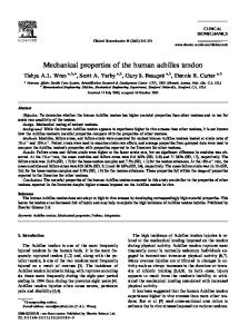
Send Orders for Reprints to
[email protected] The Open Orthopaedics Journal, 2014, 8, 135-139
135
Open Access
Tendon-Holding Capacities of Two Newly Designed Implants for Tendon Repair: An Experimental Study on the Flexor Digitorum Profundus Tendon of Sheep *,1
2
3
4
4
İsmail Ağır , Mahmut Nedim Aytekin , Onur Başçı , Barış Çaypınar and Bülent Erol 1
Department of Orthopedics and Traumatology, Adıyaman University Training and Research Hospital, Adıyaman, Turkey 2
Department of Orthopedics and Traumatology, Atatürk Training and Research Hospital, Ankara, Turkey
3
Department of Orthopedics and Traumatology, Dokuz Eylul University School of Medicine, Izmir, Turkey
4
Department of Orthopaedics and Traumatology, Marmara University School of Medicine, Pendik, 34662, Istanbul, Turkey Abstract: Background: Two main factors determine the strength of tendon repair; the tensile strength of material and the gripping capacity of a suture configuration. Different repair techniques and suture materials were developed to increase the strength of repairs but none of techniques and suture materials seem to provide enough tensile strength with safety margins for early active mobilization. In order to overcome this problem tendon suturing implants are being developed. We designed two different suturing implants. The aim of this study was to measure tendon-holding capacities of these implants biomechanically and to compare them with frequently used suture techniques Materials and Methods: In this study we used 64 sheep flexor digitorum profundus tendons. Four study groups were formed and each group had 16 tendons. We applied model 1 and model 2 implant to the first 2 groups and Bunnell and locking-loop techniques to the 3rd and 4th groups respectively by using 5 Ticron sutures. Results: In 13 tendons in group 1 and 15 tendons in group 2 and in all tendons in group 3 and 4, implants and sutures pulled out of the tendon in longitudinal axis at the point of maximum load. The mean tensile strengths were the largest in group 1 and smallest in group 3. Conclusion: In conclusion, the new stainless steel tendon suturing implants applied from outside the tendons using steel wires enable a biomechanically stronger repair with less tendon trauma when compared to previously developed tendon repair implants and the traditional suturing techniques.
Keywords: Early mobilization, implant, tendon repair, tendon ruptures. INTRODUCTION After tendon repair, early mobilization is necessary in order to prevent loss of motion [1, 2]. Verdan suggested that passive mobilization at four weeks after tendon repair improved the results by tearing fresh adhesions in 1960 [3]. Ten years later, Kleinert et al. and Lister et al. reported the results of tendon repair with a bunnel suture technique followed by immediate active extension and passive flexion [2-4]. Later studies confirm those findings and it is now widely accepted that the repaired tendons should be mobilized early to prevent contractures. In order to expose a repaired tendon tension force should be applied and the early repair should grant maximal strength. Two main factors determine the strength of tendon repair which are tensile strength of repair material and *Address correspondence to this author at the Department of Orthopedics and Traumatology, Adıyaman University Training and Research Hospital, Adıyaman, Turkey; Tel: +90(416)2161015; Fax: +90(416)2162659; E-mail:
[email protected] 1874-3250/14
gripping capacity of suture configuration [5-7]. These two variables should be maximum for maximum strength. Many studies were performed to demonstrate the tensile strengths of suture materials or tendon-holding capacities of different suture configurations [6, 8-10]. Different repair techniques and their modifications were developed to increase the strength of repairs. However, none of these techniques and suture materials seemed to provide enough tensile strength with large safety margins for early active mobilization [11, 12]. In order to overcome this problem tendon suturing implants are being developed. An implant applicable to a tendon should have an easy to apply design and is preferably economically advantageous. We designed two different suturing implants. The aim of this study was to measure the biomechanical characteristics of our newly designed implants and to compare them with commonly used suture techniques. MATERIALS AND METHODS In this study, we used 64 flexor digitorum profundus tendons of fore extremities of sheep. The tendons were 2014 Bentham Open
136
The Open Orthopaedics Journal, 2014, Volume 8
harvested in two hours after the sheep were slaughtered. After the paratenon was disected, a 15 cm piece of tendon was excised. The tendons were held in a wet gauze (isotonic 0.9% NaCl solution) during the 2 hour period in which the implants were applied to the tendon and the 3 hour period in which the measurements were taken.
Ağır et al.
testing machine was programmed to exert a 20-mm/sec tensile force. The repair construct was loaded to failure. The data were recorded automatically (Fig. 4).
There were four groups with 16 tendons in each group. We applied our newly designed model 1 and model 2 tendon holding implants to the first 2 groups respectively. Classical Bunnell and locking-loop techniques were applied to the 3rd and 4th groups respectively by using 5 Ticron sutures (Tyco, Waltham, MA) (Table 1). Design of the Implants (Model 1 and Model 2) Two metal implants were produced by using a 0.8 mm stainless steel wire. Both implants had a loop like shape with 2 identical prongs. In model 1, on each prong, holes with 0.7 mm diameter were formed with 2.1mm intervals and in model 2, on each prong, holes with 0.7 mm diameter were formed with 7.7 mm intervals (Fig. 1). In both models approximately 5 mm after the last hole of one prong, wires were bended in a demilune manner and on the opposite prong, after 5 mm, the holes were placed exactly opposite, exact alignment and exact number (Fig. 2). Model 1 had 10 holes and model 2 had 5 holes at each side. Two ends of the wire (8 cm long) were twisted from 7.5 mm distal to the last hole to form the tail of the implant (Fig. 2). Model 1 was applied to group 1, model 2 was applied to group 2. Tendon Repair Protocol and Biomechanical Testing The proximal end of the tendon was passed through a wire loop and turned over and sutured to itself using polyglactin sutures (Fig. 3). The proximal nonimplanted part of tendon was holded by aid of that circle shaped wire. Models 1 and 2 were applied to 16 tendons in groups 1 and 2, respectively. Each tendon was first placed between the prongs of the implant. Then 0.6 mm stainless steel pins were sent through the holes. The pins were paralel to each other and were perpendicular to the longitudinal axis of the tendon. Ten pins were used for fixation in model 1 and 5 pins in model 2. The most distal pin was placed 1cm above the distal end of the tendon (Fig. 3). Biomechanical tests were performed with the Instron device (electrohydrolic model 1321B; Instron, Canton, MA). The proximal wire loop and the distal tail of each implant were attached to the Instron clamps of the test device. The Table 1.
Fig. (1). Designing of holes in two implants.
Statistical Tests Kruskal-Wallis ( Nonparametric ANOVA) test was made with SSPE for statistical analysis. P